A successful undergraduate science education includes teaching students science process skills, including critical analysis of primary scientific literature (PSL). Unfortunately, the use of PSL in the classroom remains limited due to several barriers. These include students struggling with the actual practice of science (as opposed to the purely linear scientific method presented in textbooks), the scientific jargon found in articles, and an inability to connect PSL to the broader context of the discipline. Additionally, most novice students are still developing the critical thinking skills needed to interpret the results and conclusions found in PSL. To complicate matters, some educators may be uncomfortable using PSL themselves due to concerns about student pushback or their sense that the material is too complex for their students.
Despite these barriers, a growing body of literature shows that PSL is a valuable and useful tool for STEM education. For example, closely analyzing PSL in a classroom setting engages students in discussion and debate around interpretations of experimental data while building their insight into both the nature of science and researchers themselves (Hoskins, Stevens, & Nehm, 2007). Teaching with scientific research papers has been shown to promote critical thinking, experimental design ability, and epistemological maturation as well as improve students’ positive attitudes to science and (Gottesman & Hoskins, 2013; Hoskins, Lopatto, & Stevens, 2011; Kenyon, Onorato, Gottesman, Hoque, & Hoskins, 2016; Murray, 2014; Round & Campbell, 2013; Stevens & Hoskins, 2014). PSL can also show students that even after a study is completed and published, there are still many unanswered questions for future scientists, and therefore promote the development of creativity through study design assignments (Gottesman & Hoskins, 2013; Hoskins et al., 2007).
Despite the diversity of positive outcomes resulting from incorporating PSL into classrooms, this method of teaching science remains relatively uncommon. Some reasons for this include: the amount of educator time spent in developing PSL lessons; difficulty with sharing and/or re-using PSL curricula because topics are often specific to particular scientific disciplines; and difficulty evaluating the higher order thinking skills involved with the use of PSL.
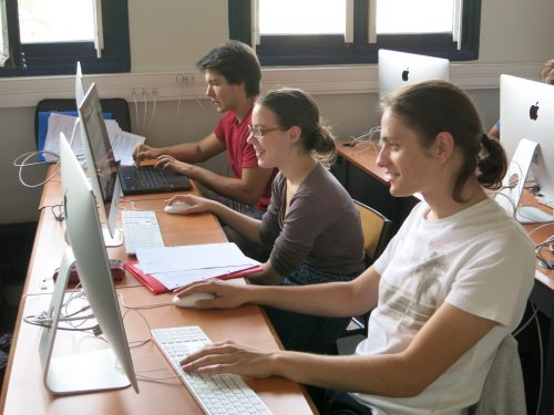
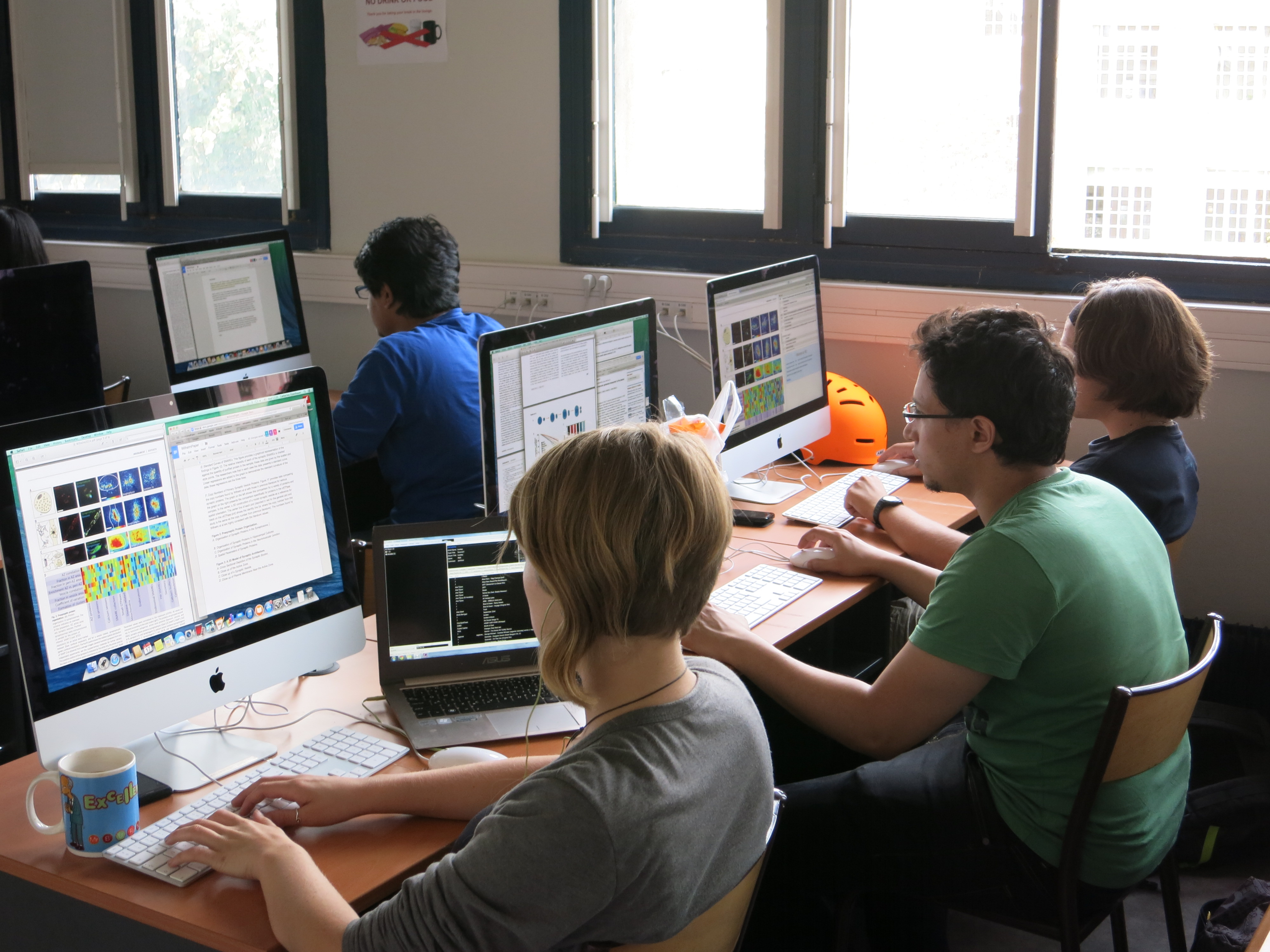
Annotated PSL is a promising example of a less-intensive intervention for including PSL into introductory biology courses at the undergraduate level. Annotated PSL is designed to help readers interpret complex science by overlaying additional information on top of the original PSL text: there is no modification or re-writing of the text itself. The “Learning Lens” tool is then used to selectively highlight different parts of the original text, and, when the highlighted text is clicked on, a pop-up box containing additional information will appear. Check out an example annotated PSL resource and try out the Learning Lens tool for yourself here: https://tinyurl.com/k7m329g
(Over 100 additional annotated papers covering a variety of scientific topics and disciplines can be found as part of the Science in the Classroom, a project supported by AAAS and NSF: http://scienceintheclassroom.org/)
Our lab has three main goals for incorporating annotated PSL into introductory biology courses:
- Minimize the time investment for instructors
- Minimize alteration to existing courses and plans of study
- Increase student confidence in being able to deconstruct future PSL
In our first pilot study, published recently in PLOS Biology, we addressed goals 1 and 2 and aimed to develop an implementation protocol. For this study, we collaborated with seven different biology courses at Florida International University. We used the same annotated PSL for all in-class activities described in this study: “Caffeine in floral nectar enhances a pollinator’s memory of reward” (https://tinyurl.com/k7m329g, the same example we encouraged you to try out above). We chose this article because incorporated many different aspects of biology, including evolution, ecosystem interactions, basic botany, learning and memory, and animal behavior in a single study, making this paper applicable in a wide variety of undergraduate courses (goal #2).

Because this was the first time annotated PSL was examined as part of a scientific study, our methods were exploratory in nature. We started at the beginning with collecting baseline data on how students interacted with the annotations themselves. Specifically, we measured average time students spent interacting with the annotated PSL, asked students what they liked about using annotated PSL, and whether or not the topic of the annotated PSL related to their course. We are currently working on analyzing these data.
Regarding implementation, we had the most success when instructors introduced the annotated PSL to their class and allowed for ~20 minutes of class time for reading followed by a few clicker questions related to the science in the article. We consider a total of ~40 minutes to be a minimal time investment for instructors with minimal alterations to existing courses and plans of study.
Moving forward, we are currently analyzing data collected from annotated PSL given as homework assignments with accompanying iClicker questions given at the start of the next class. We are also collecting student self-efficacy relating to reading PSL as a part of this study.
We are always looking for more collaborators to expand the reach of annotated PSL beyond FIU. If you and your students would like to join one of our studies please let us know!
Melissa McCartney
mmccartn @ fiu.edu
References
Gottesman, A. J., & Hoskins, S. G. (2013). CREATE Cornerstone: Introduction to scientific thinking, a new course for STEM-interested freshmen, demystifies scientific thinking through analysis of scientific literature. CBE Life Sciences Education. https://doi.org/10.1187/cbe.12-11-0201
Hoskins, S. G., Lopatto, D., & Stevens, L. M. (2011). The C.R.E.A.T.E. approach to primary literature shifts undergraduates’ self-assessed ability to read and analyze journal articles, attitudes about science, and epistemological beliefs. CBE Life Sciences Education. https://doi.org/10.1187/cbe.11-03-0027
Hoskins, S. G., Stevens, L. M., & Nehm, R. H. (2007). Selective use of the primary literature transforms the classroom into a virtual laboratory. Genetics. https://doi.org/10.1534/genetics.107.071183
Kenyon, K. L., Onorato, M. E., Gottesman, A. J., Hoque, J., & Hoskins, S. G. (2016). Testing CREATE at Community Colleges: An Examination of Faculty Perspectives and Diverse Student Gains. Cell Biology Education, 15(1), ar8-ar8. https://doi.org/10.1187/cbe.15-07-0146
Murray, T. A. (2014). Teaching students to read the primary literature using pogil activities. Biochemistry and Molecular Biology Education. https://doi.org/10.1002/bmb.20765
Round, J. E., & Campbell, A. M. (2013). Figure facts: Encouraging undergraduates to take a data-centered approach to reading primary literature. CBE Life Sciences Education. https://doi.org/10.1187/cbe.11-07-0057
Stevens, L. M., & Hoskins, S. G. (2014). The CREATE Strategy for Intensive Analysis of Primary Literature Can Be Used Effectively by Newly Trained Faculty to Produce Multiple Gains in Diverse Students. CBE Life Sciences Education, 13(2), 224–42. https://doi.org/10.1187/cbe.13-12-0239
 (4 votes)
 Loading...
 The BSDB Archive covers 70 years of our society’s history, providing deep insights into its early years, its long trail of scientific conferences, workshops and committee meetings; it includes an almost complete collection of the many newsletters that have been published since issue 1 came out in 1979. A year ago, many of the archive’s documents were made digitally available (see box below) and described in a dedicated blog post by Andreas Prokop (


 (No Ratings Yet)
 (16 votes)