Jamie: A bit of a mad mixture. The lab has always worked on organogenesis and self-organization in development, at the bench and at the computer. For around 20 years, we have combined natural development with tissue engineering, with a special interest in 3Rs applications as well as eventual clinical use. We were among the founders of the now burgeoning field of synthetic morphogenesis (our first publication, 2008). Many in the lab want to use it to make useful living materials; the PI has an interest in using synthetic biology ‘re-creating’ developmental mechanisms that we think we understand, in a very simple form, to check that we really do understand them (a bit like testing understanding of aerodynamics by making a paper ‘plane’). We began this by making synthetic patterning systems, and then added creation of shape. The next step is engineering agency and purpose. In addition to this, and almost by accident, we have a strong interest in pharmacoinformatics and run the global drug database for IUPHAR, the International Union of Basic and Clinical Pharmacology, and the antibiotic database for the Global Antibiotic Research and Development Partnership. Sadly, the IUPHAR meetings, which I have to attend, almost always clash with the BSDB spring conference.
From left to right: Wentong, Jane, Sneha, Elena, Natalia, Louise, Rhiannon, Jamie, Hüseyin, Simon.
Roll call (in order of length of time in the lab):
The lab gremlin, in the lab from time immemorial; spends its time corroding electrical connections, moving critical antibodies to the wrong fridge, and emptying CO2 cylinders overnight. Like magnetism, it is invisible but we know it to be present by its observable effects.
Jamie Davies, Professor and anyone’s-technician-in-a-crisis (the jobs go together); now grey-haired enough to have made most of the mistakes people make in research, so is able to help young scientists avoid them and instead discover new and exciting mistakes of their own.
Jane Armstrong, Senior Curator, who joined as a wet-lab post-doc over 25 years ago and later discovered the joy of informatics.
Simon Harding, Senior Developer, a molecular biology post-doc turned software wrangler, with the group for over a decade now.
Elena Faccenda, Curator, a pharmacology post-doc turned informatician, also in the group for more than a decade. As well as managing the database, Elena, Simon and Jane write high-profile papers together, and have each acquired h-indices higher than many full professors.
Rhiannon Beadman, a post-doc working on homeorhesis and error-correction in organ development, uses a mix of classic 1920s-style cut-and-paste embryology and up-to-date molecular techniques.
Hüseyin Gül, a PhD student of this lab turned post-doc; works on a new drug target he identified for polycystic kidney disease.
Natalia Penar, a PhD student; combines optogenetics and morphogenetic control to test hypotheses about symmetry-breaking in branching morphogenesis.
Sneha Ravi, a PhD student; uses our new patented biofabrication technique to engineer ureters and similar tubular tissues.
Louise Goossens, a PhD student who will use Sneha’s ureters to model bacteria-tissue interactions, and how they affect antibiotic resistance, in urinary tract infections.
Nick Younger, a post-doc just joining us to work on optogenetics and axioloids.
Wentong Fu, an MSc project student working on dynamics of pattern coarsening following wounding of a synthetic patterning system.
Favourite technique
Jamie: Reading developmental literature from a century or more ago; researchers then did not have our fancy tools, but they had observation, insight and imagination and their work is an unfailing source of inspiration.
Outside your own work, what are you most excited about in developmental or stem cell biology?
Jamie: Evo-devo generally. I have never worked in this area, but seeing links develop between understanding of developmental opportunities and constraints, and the areas of morphospace visited and avoided over phylogenetic evolution, is amazing and is a ‘new’ new synthesis.
How do you manage your group and your various tasks?
Jamie: I ‘manage’ with as light a touch as possible. If you give bright young people the right encouragement, they will ask interesting questions and do interesting things, many of which I would never have imagined. In recruitment, I value diversity, not just in terms of ethnicity, gender and religion, but also in terms of academic culture; my lab has included people with first degrees in philosophy, anthropology, mathematics, and engineering as well as in biological subjects, and has benefited from the different ways of thinking they bring. As for managing my own tasks, the golden rule is not to procrastinate, and to reserve time just to think; I mean actually reserve it in a calendar, with the same non-negotiability as one marks out a timetabled lecture or conference. Thinking is part of a scientist’s job, and it is entirely reasonable to demand time in which it can be done.
What’s the best thing about where you work?
Answers given by members of the lab in general include: “I like how everyone in the building helps each other…. I love my daily commute to work through the Meadows.” “It’s a space where complex problems turn into exciting challenges that motivate me to learn, experiment and grow. It’s a mix of curiosity, teamwork, and the occasional lab triumph—it’s never dull!” “The people! As a biocurator and office-based scientist, it is great to still be part of an academic research laboratory, especially one that is so supportive. Being based on a campus right in the heart of Edinburgh is pretty special as well.” “The lab is friendly and supportive — and seems to benefit from lots of different experiences.” “I enjoy the intellectual stimulation that researching for the Guide to Pharmacology brings. A huge bonus is working with competent and friendly colleagues who help you through the days when things aren’t quite going to plan.” “The best part about working here is the friendly and supportive lab environment, where you can always count on others and never feel like you are asking too much.” “The best thing is friendly atmosphere and cooperation in our lab. It makes dealing with setbacks easier, celebrating successes even more pleasant, and keeps us motivated.” “The people—everyone is approachable, motivated, and genuinely passionate about their work”.
What’s there to do outside the lab?
Answers given by members of the lab in general include: “Dancing! Edinburgh has a vibrant community for various dancing styles (my pick is Latin dances). It’s amazing for meeting new friends and staying healthy.” “I like to go for walks in the Pentland Hills followed by a well-deserved beer in one of the many pubs.” “The Meadows is a great spot to unwind and soak in some greenery or a hike up Arthur’s seat offers stunning views of the city. Plus, the area is filled with great eateries, making it easy to grab a bite between experiments” “The Scottish Highlands and Islands are all within a half-day of travel. Fantastic if you enjoy epic scenery and being outdoors.” “Being based in the centre of Edinburgh is great – there’s usually lots going on, especially during the festival. Lots of nice green spaces close by is another positive.” “There is no shortage of historical, cultural, sporting, entertainment and foody places and events to explore in the heart of Scotland’s capital city. What makes it really special is that beaches, hills and the great outdoors are within very easy reach, whether you’re a thrill seeker or just want to chill.” “I love exploring Scotland, with its endless hikes and breathtaking scenery, though it is most enjoyable on the rare occasion when the weather is not cold and wet!” “The best place to go if you find yourself outside the lab is back inside the lab again!”
This is part of the ‘Lab meeting’ series featuring developmental and stem cell biology labs around the world.
Where is the lab?
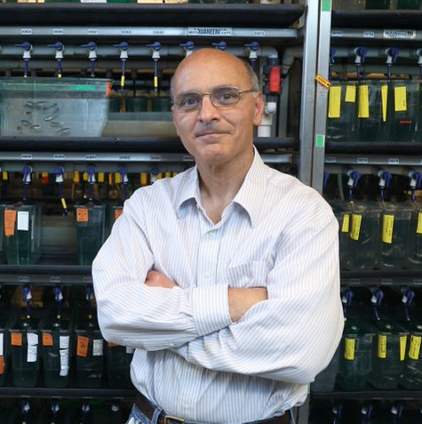
Li-Kun: My lab is located in Kobe, Japan. My lab initially belonged to RIKEN Center for Developmental Biology, which subsequently reformed to become the RIKEN Center for Biosystems Dynamics Research.
Li-Kun: My lab aims to understand how blood vessels are built and shaped into a hierarchical tubular network of arteries, veins and capillaries of optimal connections and size. We are particularly interested in endothelial cell mechanobiology – how physical forces and mechanical properties govern their shapes and behaviours to drive distinct steps of vessel morphogenesis, from sprouting angiogenesis to vessel remodelling. We are also keen to elucidate the interplay between endothelial cells and the perivascular environment such as haemodynamic forces (shear stress and pressure) at the apical surface and mural cells on the abluminal surface. In the long term, we aim to uncover whether altered endothelial cell mechanobiology contributes to endothelial dysfunction and vascular anamolies. To address these questions, we employ a spectrum of methodologies such as zebrafish genetics, high resolution time-lapse confocal microscopy, optogenetics, pharmacology, quantitative image analyses, scRNA sequencing and mathematical modelling.
Group photo of the lab
Lab roll call
Li-Kun Phng: We are an internationally diverse lab, and the current lab members are listed from oldest to newest here.
Emi Taniguchi: I have been a lab assistant since 2016. I am doing administrative work and zebrafish system care in the lab. Also, I check the condition of our zebrafish daily to keep them healthy.
Igor Kondrychyn: I am a Research Scientist and am currently working on a project investigating the role of aquaporins in embryonic hematopoiesis.
Yan Chen: I am a postdoctoral researcher investigating vascular remodelling during zebrafish development, with a focus on how endothelial cells regulate vessel size and structure. My research explores how endothelial cells undergo rearrangement and shape changes, particularly through actin-driven constriction, to fine-tune vessel dimensions. Using zebrafish models, molecular genetics, and live imaging, I aim to uncover the cellular mechanisms underlying vascular remodelling.
Haymar Wint: I am a postdoctoral researcher, and my research interest lies in investigating the role of microtubules in mediating endothelial cell responses to fluid shear stress, unveiling its role in vascular development and remodeling.
Mingzhao Hu: I am a PhD student enrolled at the Department of Biological Sciences at Osaka University. I am interested in understanding how mural cells impact vascular remodeling using genetics and high-resolution time-lapse imaging.
Jason da Silva: I am a research technician. I carry out general lab duties and am working on creating knock-in zebrafish lines.
Rajrishi Kumar: As a technical staff member in the lab, I contribute to research on developmental biology and vascular morphogenesis, with a focus on sprouting angiogenesis and mechanobiology using zebrafish as a model organism. My work involves maintaining zebrafish lines, performing genotyping, and assisting with experiments that dive into the molecular and biophysical aspects of blood vessel formation. Along the way, my science toolbelt’s been getting a glow up with hands-on experience in genetic manipulation, molecular biology, and advanced imaging techniques.
Favourite technique, and why?
Li-Kun: Undoubtedly light microscopy – the higher the resolution the better – as it allows us to peer into the dynamics of vascular cell behaviour, enabling us to understand the process of vessel morphogenesis.
Apart from your own research, what are you most excited about in developmental and stem cell biology?
Li-Kun: I am presently fascinated with the growing prominence of ion channels, osmotic gradient and hydrostatic pressure in regulating tissue morphogenesis.
How do you approach managing your group and all the different tasks required in your job?
Li-Kun: My job entails juggling research, administrative work and some teaching. It was a steep learning curve starting my lab in Japan without knowing the language. Thankfully, I am lucky to have a very capable assistant who helps me deal with work-related matters in Japanese within and outside the institute, allowing me to focus on the science. A major goal is to provide a good research environment (in terms of infrastructure and opportunities) that is conducive to scientific excellence and efficiency. I firmly believe that communication is crucial not only in keeping up-to-date with scientific progress and shaping research direction, but also in identifying and tackling problems (be it scientific or personal) that surface. As such, I meet researchers every two weeks, encourage open communication in our weekly lab meetings and foster team spirit over meals and lab outings. I also like to enliven the lab atmosphere by hosting visitors from abroad, which at the same time promotes exchange of ideas and technology as well as fostering collaborations. Another important aspect of my job is to ensure that we stay abreast with current developments in our field and to disseminate our work to the scientific community. In Japan, the JSPS-funded ‘Mechanical Self-transformation of Living Systems’ consortium (https://multicellular-mechanics.org/research) has been a vital platform for networking with scientists in the fields of biomedical sciences, engineering and physics. Beyond Japan, I attend internationally conferences and meetings but this is limited by family commitments. The plus side is that I spend more time with my lab members to develop and complete projects!
What is the best thing about where you work?
LKP: Core funding from RIKEN! And being able to pursue curiosity-driven research.
EM: I enjoy working in multi-cultural environment with the members come from different countries. So far, I have met the lab members from 15 different countries.
IK: Friendly atmosphere.
YC: One of the best aspects of working in our lab is the supportive and collaborative environment. Communication flows easily within our team and across research groups. We also have access to an excellent microscopy facility, where the friendly staff provide invaluable support.
HW: One of the best things about working at RIKEN BDR is its highly international environment, where researchers from diverse countries come together to collaborate. And, our lab’s warm, supportive, and well-structured team environment accelerates scientific progress, and makes work both productive and enjoyable.
MH: I’m the only student in our lab, so that I often receive helpful suggestions and feedback from other members, who have full experience in this field.
JDS: Everyone is nice and the atmosphere is very relaxed.
RK: The best part? The people and the place. Sure, RIKEN’s state-of-the-art facilities and cutting-edge instruments are great for high quality research, but it’s the environment and community that make it truly exceptional. From lab members to admin staff, everyone is incredibly friendly, communicative, and genuinely helpful. And then there’s our team leader, high energy, inspiring, and somehow always present when you need a hand, a nudge, or a fresh perspective. Oh, and bonus points for the stunning sea view from the lunch area. Yes, science with a seaside vibe!
What’s there to do outside of the lab?
LKP: I would typically like to recharge my batteries by not doing anything, but this, unfortunately, is not possible because I have two young ones at home to entertain. Kobe is sandwiched between the mountain and the sea, and so we often venture to the beach or forests to appreciate nature. In Spring, we enjoy hanami (cherry blossom viewing); in Autumn, momijigari (viewing of the vibrant autumn colours of maple leaves); in Winter, warming ourselves up by going to onsen (hot springs). And of course, we love trying out new cafes and restaurants.
EM: I like to travel not only in Japan but also to other countries to learn about cultures and ideas created by each region that has original geographical and historical factors.
IK: Relaxing outdoors (if no rain), cycling and listening to music while drinking wine.
YC: Outside the lab, Japan offers a perfect blend of nature, culture, and city life. Depending on the season, I enjoy hiking in the nearby mountains, visiting historical temples and shrines, or exploring the diverse food options. There are also many festivals throughout the year, offering a chance to experience traditional culture. Within the lab, we often celebrate small victories, birthdays, and milestones with group dinners and relaxed afternoon tea gatherings.
HW: Outside the lab, I enjoy exploring Japan’s rich culture and natural beauty. I visit historical sites, experience seasonal festivals, and try different local foods. Sometimes, I join social gatherings with colleagues or participate in local events to experience Japanese traditions firsthand.
MH: Outside of the lab, we often have a food-themed adventure to explore the local food, from Ramen restaurant to open-air bar. I also spend time in nature to increase feelings of happiness and pay attention to the changing seasons. I really enjoy cherry blossom in spring and Momiji viewing in autumn!
JDS: Kobe is close to the sea and to mountains so there are a lot of scenic places to visit. There are hundreds of restaurants to try around Kobe too. It’s also easy to visit other cities in Japan by public transport.
RK: I am new to the area, but outside of the lab I usually go for a run or play basketball. Here in Kobe, there are great cafes, cozy izakayas, and lively karaoke spots where we can relax after a long day. The city is also surrounded by mountains, offering plenty of hiking spots with scenic views of both the city and the ocean. And of course, we are in Japan, a hub for shopping and entertainment, so there is always something to do. Also, if you are feeling too adventurous, there are plenty of options for onsens to enjoy a hot spring experience.
The Company of Biologists is organising an essay competition entitled “Innovative Ideas for the Future of Sustainable Events”.
Participants are invited to write an essay of maximum 1000 words to details how your idea will change the way we organise scientific events and will open the door to a new concept of organising events more sustainably in the next 10 years.
Prizes:
1st place: £250
2nd place: £150
3rd place: £100
The winning essays will be selected by the Sustainability Committee and featured on The Company of Biologists’ website.
This is a fantastic opportunity for anyone passionate about sustainability and writing. For more details, please visit our post here.
In this ‘Featured image’ post, we find out more about the story behind Allan Carrillo-Baltodano’s image, which was one of the runners-up in the competition.
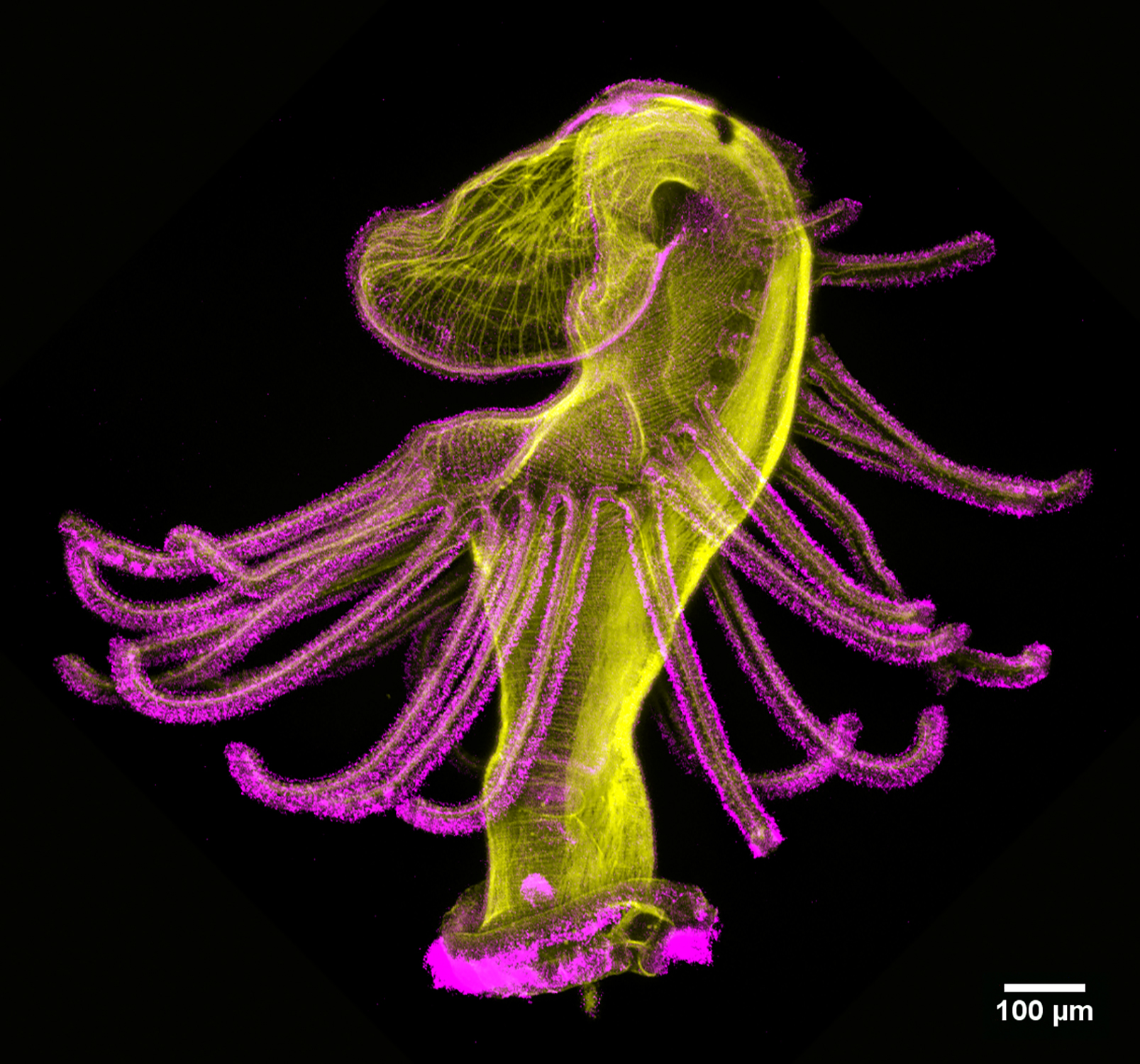
Dancing actinotroch Allan Carillo-Baltodano Actinotroch larva of a phoronid worm with phalloidin shown in yellow and acetylated tubulin in magenta. Imaged with a Zeiss LSM 800 at 10 x magnification.
What is your background?
I did my undergrad at the University of Costa Rica, in Costa Rica. Early on, I biased my interests towards invertebrate zoology, and ended up doing an undergrad thesis on marine zooplankton of coral reefs. It was back then when I saw the beauty of marine invertebrate larvae. I have been studying evolution and development (EvoDevo) of marine invertebrates since — first, during my PhD in Néva Meyer’s lab at Clark University in Massachusetts (USA), and currently as a postdoc in Chema Martín-Durán’s lab at Queen Mary University of London in UK.
What are you currently working on?
I study how body plans of marine annelids develop, and try to understand how different modes of development in these and other animals could have evolved.
Can you tell us more about the story behind the image that you submitted to the image competition?
The image shows a larva of a phoronid worm. The larva is commonly known as actinotroch, and it is very conspicuous among other members of the zooplankton community because of the tentacles surrounding the head. To swim the larva propels itself using cilia (shown in the image in magenta) concentrated in a ciliary band on the posterior part of the larva. The big hoodie you see at the top will open slightly so that food can be captured in the mouth. One of the wonderful things of the plankton is that if you are lucky, you can find any stage of development of these and many other amazingly weird invertebrates.
What is your favourite technique?
I like mostly confocal microscopy, although if a sample is nice enough, you can get some very beautiful images with DIC microscopy as well.
What excites you the most in the field of developmental and stem cell biology?
The breakthrough in technologies has really opened the door for many of us who like to study development in unusual research organisms. In combination with the EvoDevo field being more open and the growing interest in comparative biology across many species and many scales, we are creating a great environment for anyone to make new discoveries.
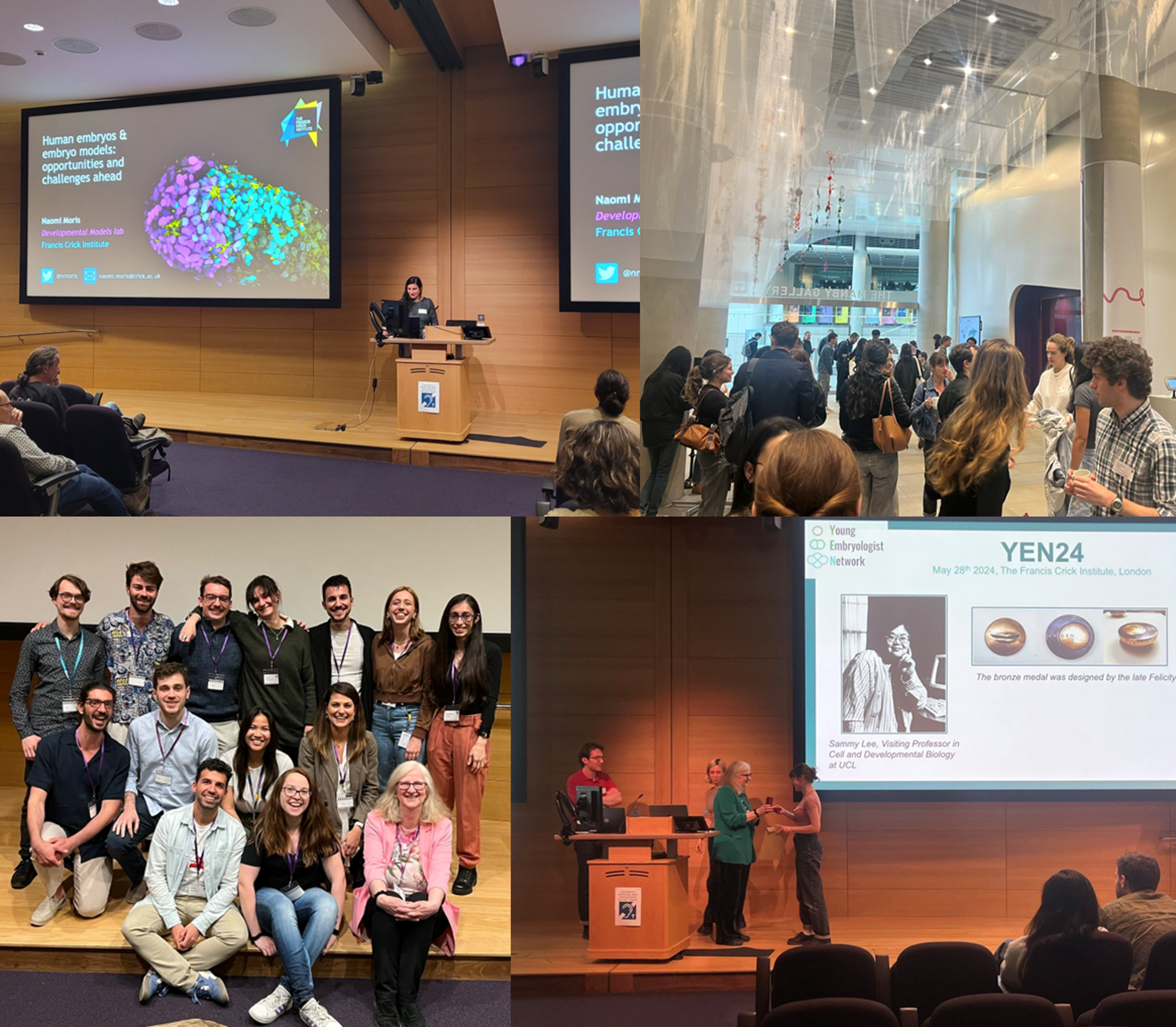
Abstract submission to the Young Embryologist Network Conference closes this week on the 13th April 2025. Please share!
This is a great opportunity for PhD students and postdocs working on differentiation, stem cells, embryonic development, IVF or organoids to present a short talk or a poster. YEN2025 will take places at the Francis Crick Institute on the 19th May and is free to attend.
Exiting news! We have revived our tradition of the YEN Image Competition. Please submit your science images and art by the 1st May 2025 and follow us to get updates on where to vote!
Special thanks to Maddie Ryan, Charli Corcoran & Michaela Noskova Fairley for putting this digest together! If you would like to thank the Zebrafish Rock! team for their time & effort, you can buy us a strong cuppa at the link below. Every little bit keeps us caffeinated and motivated! We appreciate your support 🙂
Spotted a preprint in this list that you love? If you’re keen to gain some science writing experience and be part of a friendly, diverse and international community, consider joining preLights and writing a preprint highlight article.
Christina McNerney, Clayton P. Santiago, Kiara C. Eldred, Ian Glass, Tom A. Reh, Arturo Hernandez, Seth Blackshaw, Nathan D. Lord, Robert J. Johnston Jr
Giulia Di Muzio, Sarah Benedetto, Li-Chin Wang, Lea Weber, Franciscus van der Hoeven, Brittney Armstrong, Hsin-Jui Lu, Jana Berlanda, Verena Körber, Nina Claudino, Michelle Krogemann, Thomas Höfer, Pei-Chi Wei
Christopher Chan Jin Ji, Daniel Santos-Olivan, Marie-Christine Ramel, Juliana Sanchez-Posada, Priscilla Paizakis, Toby GR Andrews, Emily S Noel, Alejandro Torres-Sanchez, Rashmi Priya
Binbin Ma, Guanghui Yang, Jonathan Yao, Charles Wu, Jean Pinckney Vega, Gabriel Manske, Saher Sue Hammoud, Satrajit Sinha, Abhyudai Singh, Haiqing Zhao, Xin Chen
Tsz Long Chu, Ostap Dregval, Farasat Zaman, Lei Li, Xin Tian, Xin Liu, Dana Trompet, Baoyi Zhou, Jussi O Heinonen, Claes Ohlsson, Lars Sävendahl, Igor Adameyko, Andrei S Chagin
Tina Balayo, Sharna Lunn, Pau Pascual-Mas, Ulla-Maj Fiuza, Amruta Vasudevan, Joshua D. Frenster, Hannah Y. Galloon, Raquel Flores Peirats, Alfonso Martinez Arias, André Dias, David A. Turner
Beatriz Gomes-Silva, Marta Furtado, Marta Ribeiro, Sandra Martins, Maria Teresa Carvalho, Henrike Maatz, Michael Radke, Michael Gotthardt, Rosina Savisaar, Maria Carmo-Fonseca
Michelangelo Corcelli, Ellen Petzendorfer, Kate Hawkins, Filipa Vlahova, Catherine Caruso, Mehedi Mohammad Hasan, Katie Durrant, Anna David, Fleur S van Dijk, Pascale V Guillot
Jengmin Kang, Abhijnya Kanugovi, M. Pilar J. Stella, Zofija Frimand, Jean Farup, Andoni Urtasun, Shixuan Liu, Anne-Sofie Clausen, Heather Ishak, Summer Bui, Soochi Kim, Camille Ezran, Olga Botvinnik, Ermelinda Porpiglia, Mark Krasnow, Antoine de Morree, Thomas A. Rando
Caramai N. Kamei, William G. B. Sampson, Carolin Albertz, Oliver Aries, Amber Wolf, Rohan M. Upadhyay, Samuel M. Hughes, Heiko Schenk, Frederic Bonnet, Bruce W. Draper, Kyle W. McCracken, Denise K. Marciano, Leif Oxburgh, Iain A. Drummond
Marie Alonso, Pierre Prévost, Aline Potier, Pascal GP Martin, Yves Caraglio, Michael Nicolas, Michel Hernould, Christophe Rothan, Béatrice Denoyes, Amèlia Gaston
Ioannis Sarropoulos, Mari Sepp, Tetsuya Yamada, Philipp S. L. Schäfer, Nils Trost, Julia Schmidt, Céline Schneider, Charis Drummer, Sophie Mißbach, Ibrahim I. Taskiran, Nikolai Hecker, Carmen Bravo González-Blas, Niklas Kempynck, Robert Frömel, Piyush Joshi, Evgeny Leushkin, Frederik Arnskötter, Kevin Leiss, Konstantin Okonechnikov, Steven Lisgo, Miklós Palkovits, Svante Pääbo, Margarida Cardoso-Moreira, Lena M. Kutscher, Rüdiger Behr, Stefan M. Pfister, Stein Aerts, Henrik Kaessmann
Dang M. Nguyen, Sarah K. Monroe, Danielle N. Rendina, Kevin S. Boyd, Erika D. Rispoli, Olivia M. Wirfel, A. Brayan Campos-Salazar, Anna R. Araujo, Trisha V. Vaidyanathan, Virginia L. Keziah, Benjamin A. Devlin, Caroline J. Smith, Staci D. Bilbo
N. Zollo, G. Zaffagnini, A. Canette, G. Letort, C. Da Silva, N. Tessandier, J. Dumont, C. Blugeon, S. Lemoine, B. Wattellier, E. Böke, M. Almonacid, M.-H. Verlhac
Ruta Meleckyte, Wazeer Varsally, Jasmin Zohren, Jerry Eriksson, Tania Incitti, Linda Starnes, Amy Pointon, Ryan Hicks, Benjamin E. Powell, James M.A. Turner
Katelyn M. Cooper, Carly A. Busch, Alice Accorsi, Derek A. Applewhite, Parth B. Bhanderi, Bruno da Rocha-Azevedo, Abhijit Deb Roy, Joseph P. Campanale, Fred Chang, Jerry E. Chipuk, Lee A. Ligon, G.W. Gant Luxton, Austin J. Graham, Camila Hochman-Mendez, Imge Ozugergin, Zachory M. Park, Claire M. Thomas, Alex M. Valm, Hongxian Zhu, Rebecca S. Alvania
In this ‘Featured image’ post, we find out more about the story behind Julia Peloggia de Castro’s image, which was one of the runners-up in the competition.
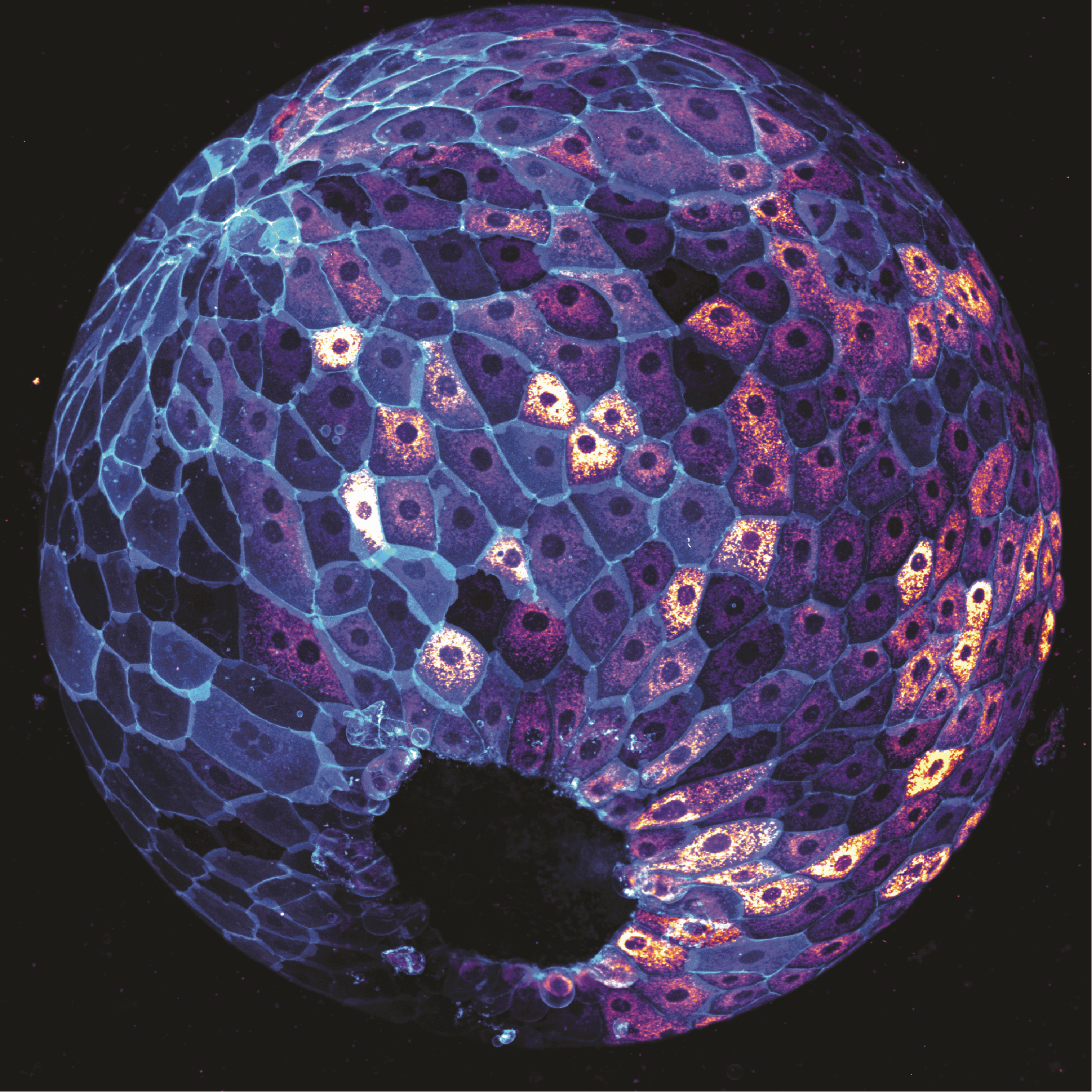
Who’s active? The image depicts a zebrafish embryo at 9 hours post-fertilisation on a lateral view. Cells are stained with MitoTracker, which labels active mitochondria, and cell membranes are labelled in cyan with a EGFP transgenic membrane tag. Image was taken using a 20x objective on a spinning disk confocal microscope.
What is your background?
I got my bachelor’s degree at the University of São Paulo, in Brazil. I majored in Biological Sciences with a focus on genetics and molecular biology. I then pursued my PhD at the Stowers Institute for Medical Research in Kansas City (USA), where I studied zebrafish development and phenotypic plasticity under the mentorship of Dr. Tatjana Piotrowski. I am now a postdoctoral fellow at University of California, Los Angeles (USA) working with Dr. Heather Christofk.
What are you currently working on?
My current interests are in developmental metabolism, and how maternal metabolic states impact mouse embryonic development.
Can you tell us more about the story behind the image that you submitted to the image competition?
This image shows a zebrafish embryo after a few hours of development. One of my favorite things about zebrafish is how fast they develop, giving us the opportunity to watch it —and image it— in real time. This particular image was taken late at night in the laboratory while I was testing a reagent and optimizing imaging parameters before running a larger experiment later that week. The result was definitely worth staying late for!
What is your favourite technique?
I really appreciate any type of microscopy. From the foldscopes to confocal and electron microscopes. Looking at cells never gets old!
What excites you the most in the field of developmental and stem cell biology?
I think this is a very exciting moment in developmental biology research. The last decade has brought many technical revolutions and more fields are bridging towards developmental biology, generating new questions and new ways to approach them. This makes me think we are poised to discover new biological principles soon.
This is part of the ‘Lab meeting’ series featuring developmental and stem cell biology labs around the world.
Where is the lab?
Jennifer: You can find the Zenker Lab at the Australian Regenerative Medicine Institute (ARMI) at Monash University. We are super lucky to call Australia, to be exact Melbourne, our home. More information is available on our Zenker lab webpage or follow us on X (formerly Twitter) @LabZenker or on LinkedIn @JennyZenker.
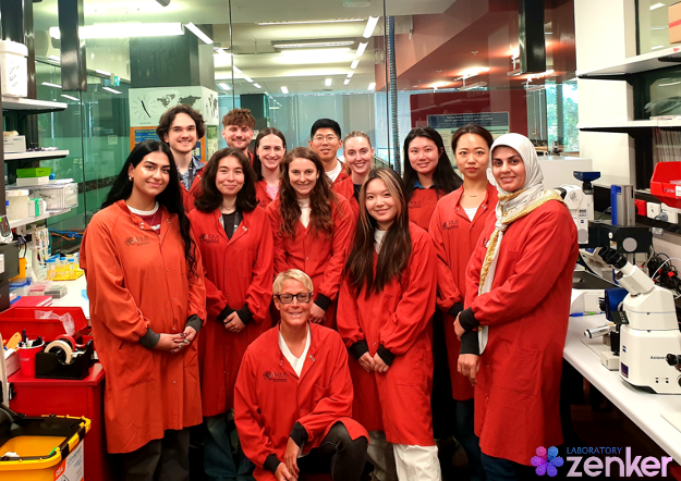
Zenker lab group photo
Research summary
The transformation of an early mammalian embryo from a tiny soccer ball-like structure into a newborn with four limbs, a beating heart, and big bright eyes is one of the most remarkable and fundamental processes of life. Errors in these early steps in development can profoundly shape a person’s lifelong ability to carry out essential tasks for everyday living. Improving the efficiency of in-vitro fertilisation (IVF) treatments 1, 2, which are expensive, physically demanding and emotionally challenging, could set up parents-to-be for the best chance of a successful cycle when it is right for them. Our research vision is to implement a new cell biological paradigm for reproductive and regenerative medicine by uncovering the real-time movements inside single cells of the living embryo and human induced pluripotent stem cells (hiPSCs).
Inside the soccer ball-like embryo resides a handful of “all-rounder” cells, known as pluripotent cells, which can give rise to any type of cell in the adult body. Using 4D (3D plus time) live-cell imaging, our research is the first in the world to conclusively demonstrate that pluripotent cells have a highly organised internal road map composed of the microtubule cytoskeleton. This road map guides the asymmetric transport of RNAs and organelles in single cells of the living mammalian embryo as a decision-making process of their future fate, becoming either part of the embryo proper or extraembryonic tissues, for instance placenta 3, 4, 5, 6, 7. Together with national and international collaborators, we have been developing innovative tools and technologies. We generated the first in vitro model of human embryos, termed iBlastoids 8, producing the most spectacular images about this new Frontier in human stem cell biology, which will continue to be a world-leading approach for many years to come 9. Furthermore, we have been pioneering the application of innovative light-switchable regulators in living 3D physiological systems to allow the spatiotemporal manipulation of the microtubule network to control the potency and function of pluripotent cells non-invasively 3, 10.
Can you give us a lab roll call?
Jennifer: I will have to start here with Louise, who is our research assistant and keeps the lab running smoothly and always stocked up. Every other lab member has their own research project. I like to design them so that team members can work easily together. There is a group of people working on the living mouse embryo. Postdoc Hongbin and two undergraduate students, Tia and Ava, are unravelling how the localisation of RNA subtypes inside single cells of the embryo contributes to the development of a healthy embryo. The project of PhD student Manlin is closely linked to it, but instead of looking at RNA subtypes, she is looking at various organelles. PostDoc Jess and undergraduate student Aya are developing new tools to interfere with the transport of RNAs or organelles by manipulating the microtubule cytoskeleton with spatial and temporal precision. PhD student Tracey and undergraduate student Zhi Bie aim to understand how similar (or not) such processes are between mouse and human model systems. Then we have PhD student Oliver, graduate student Adam and undergraduate student Micaela who are all working with human induced pluripotent stem cells (hiPSCs) to translate our discoveries in reproductive biology to regenerative medicine. This area is now even more strengthened by our newest member PhD student Minoo who is working with both systems to understand how one cell can have the capacity to give rise to all cell types an adult body is made of.
Favourite technique, and why?
Jennifer: There is only one answer — live imaging. Being able to see in real-time how a new life starts developing is simply mind-blowing, better than any science-fiction movie or so you can watch at the movies.
Apart from your own research, what are you most excited about in developmental and stem cell biology?
Jennifer: Developmental and stem cell biology is the foundation for regenerative medicine, which is one of the newest fields of research. It is an amazing time to be involved in such a rapidly evolving field. it has an exceptional potential to help the wider society worldwide by improving our capabilities to more effectively treat a wide range of diseases. I might be bias but that’s the future.
How do you approach managing your group and all the different tasks required in your job?
Jennifer: There is one quote that says it all: “Fail to prepare, prepare to fail”. It is a lot indeed, but I love to plan and prepare, being on an exact time schedule and writing to-do lists sorted by priority and time requirements. But all this only works if there is an open, clear and effective communication across all lab members.
What is the best thing about where you work?
Oliver: The best thing about where I work is being part of a great team that works on interesting and important research. We are all working together to produce cutting-edge discoveries and there is a huge amount of satisfaction that comes from that. I am also lucky that working here gives me access to high-quality facilities and instruments, as well as interactions with exceptional colleagues.
Zhi Bie: For me, it’s the opportunity to attend various internal and external seminars hosted by ARMI. Although all the research groups belong to the same institution and share a general goal in regenerative medicine, I still have a limited understanding of areas outside my own group. The weekly institute seminars provide a welcoming, knowledgeable, and meaningful platform where staff and students can engage, share ideas, and gain novel perspectives from researchers with similar backgrounds.
Hongbin: I enjoy the active exchange of scientific ideas and findings here at ARMI, through weekly internal seminars, as well as regular external seminars that invite researchers from other universities or even other countries. These seminars not only provide insights into their scientific work but also offer opportunities to discuss career paths and professional development.
Adam: In my opinion, one of the best things about working at ARMI is the people I work with. Not only are the people friendly and enthusiastic to help you however they can, but I think it is really beneficial to share a workspace that is rich in diversity because you gain valuable insight into various cultural heritages and research backgrounds.
Tia: The community, both in the institute and in the lab, is extremely special. Specifically, I like how the community continuously views science in an almost child-like wonder whilst trying to do the best we can to discover new ways to help people. Furthermore, outside of the laboratory and seminars, there is a strong sense of support that extends past just science.
Micaela: The best thing about where we work is the community among the different labs, and the always smiling faces in the tea room.
What’s there to do outside of the lab?
Louise: I started playing badminton again when I joined the lab, encouraged by my supervisor Jenny Zenker with her attitude to sport and exercise. Now I am putting more effort into this sport outside the lab with more training and I am feeling much better physically and mentally. I think that is what Jenny wants to deliver via her non-scientific role outside the lab —triathlete.
Manlin: Melbourne is one of the best student cities in the world (QS World University Rankings). As the cultural and educational hub of Australia, it offers a vibrant academic environment, diverse communities, and a high quality of life. I truly enjoy studying and living here!
Zhi Bie: Melbourne is surrounded by the sea, and I love driving along the coastal roads at sunset. Chasing the light as it fades over the ocean gives me a sense of peace and clarity—it’s windy, cozy, and feels incredibly freeing. I’m also passionate about playing squash and badminton. The time I spend on the court brings a deep sense of focus and an exhilarating sensation of explosive muscle power.
Hongbin: Outside of the lab, I enjoy Melbourne’s beautiful mountains and beaches, which help me relax, recharge, and gather fresh energy for the work ahead.
Adam: As someone who has recently joined the lab from the UK, I enjoy spending my time outside of work indulging myself in everything Australia has to offer. Whether this be early morning runs along the Yarra River trying to spot wild kangaroos, road trips to explore the Great Ocean Road and stunning rainforest or simply lazy days at the beach enjoying the Melbourne sun — no two weekends are the same!
Tia: On site, the campus is filled with many spots to explore nature while you eat lunch that are truly stunning and relaxing. Off-site, there are some amazing beaches just a short drive from the lab that are great for decompressing after a day of running experiments.
Micaela: Outside of work there are beautiful coastlines to explore where you can go swimming, surfing or hiking. At the right time of the year you can even watch humpback whales migrating up the southeast coast of Australia, which is one of the most extraordinary things to witness.
References
[1] Jin H, Han Y, Zenker J. Cellular mechanisms of monozygotic twinning: clues from assisted reproduction. Hum Reprod Update. 2024;30(6):692-705. doi:10.1093/humupd/dmae022
[2] Jin H, Zenker J (2024) Seeing is believing: Visualising the Formation of Monozygotic Twins in IVF. J Reprod Med Gynecol Obstet 9: 180.
[3] Zenker J, White MD, Templin RM, et al. A microtubule-organizing center directing intracellular transport in the early mouse embryo. Science. 2017;357(6354):925-928. doi:10.1126/science.aam9335
[4] Zenker J, White MD, Gasnier M, et al. Expanding Actin Rings Zipper the Mouse Embryo for Blastocyst Formation. Cell. 2018;173(3):776-791.e17. doi:10.1016/j.cell.2018.02.035
[5] Hawdon A, Geoghegan ND, Mohenska M, et al. Apicobasal RNA asymmetries regulate cell fate in the early mouse embryo. Nat Commun. 2023;14(1):2909. Published 2023 May 30. doi:10.1038/s41467-023-38436-2
[6] Hawdon A, Aberkane A, Zenker J. Microtubule-dependent subcellular organisation of pluripotent cells. Development. 2021;148(20):dev199909. doi:10.1242/dev.199909
[8] Liu X, Tan JP, Schröder J, et al. Modelling human blastocysts by reprogramming fibroblasts into iBlastoids. Nature. 2021;591(7851):627-632. doi:10.1038/s41586-021-03372-y
[9] Palacios Martínez S, Greaney J, Zenker J. Beyond the centrosome: The mystery of microtubule organising centres across mammalian preimplantation embryos. Curr Opin Cell Biol. 2022;77:102114. doi:10.1016/j.ceb.2022.102114
[10] Greaney J, Hawdon A, Stathatos GG, Aberkane A, Zenker J. Spatiotemporal Subcellular Manipulation of the Microtubule Cytoskeleton in the Living Preimplantation Mouse Embryo using Photostatins. J Vis Exp. 2021;(177):10.3791/63290. Published 2021 Nov 30. doi:10.3791/63290
A critical function of blood vessels is the transportation of plasma, blood cells, nutrients, and metabolites efficiently across the body. The formation and maintenance of blood vessels as hollow tubes is thus important for their function and is a research interest in our lab. One long-standing question in this subject is how apical membranes expand at luminal patches, which form between endothelial cells, at the initial phase of lumen formation. We questioned whether this could occur through physical forces generated from within the luminal patch that would drive the expansion of apical membranes to form a bigger lumen. This led to the hypothesis that endothelial cells may expel water at its apical surface into the luminal space to build up hydrostatic pressure that would consequently inflate the lumen. While we were exploring this idea, an scRNAseq screen in my lab identified two endothelial water channels, aqp1a.1 and aqp8a.1, to be differentially expressed within blood vascular networks of the developing zebrafish embryo. While aqp1a.1 mRNA is ubiquitously expressed in all blood vessels, aqp8a.1 mRNA expression is confined to the posterior trunk vessels and in the ventral regions of intersegmental vessels (ISVs). We therefore speculated that these two Aquaporins may have distinct roles in blood vessel formation and function.
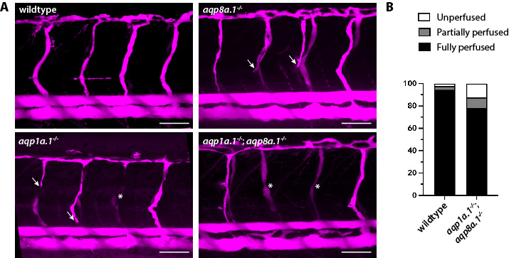
Igor, the first author of the paper, subsequently generated zebrafish mutants using CRISPR/Cas9 to investigate the function of Aqp1a.1 and Aqp8a.1. Our initial analysis was focused on determining whether blood vessel lumens formed normally in aqp1a.1 and aqp8a.1 mutants. To address this, we performed microangiography experiments to examine lumen patency at 2 and 3 days post fertilisation (dpf), after trunk vessels have lumenised. This analysis showed a decrease in the number of ISVs that are fully perfused and an increase in the number of partially perfused ISVs (Fig. 1), which supported our hypothesis that Aquaporin-mediated water flow may have a role in lumen formation. However, we needed to exclude the possibility that the decrease in fully perfused ISVs may have arisen from a defect in an earlier phase of ISV formation. We therefore examined sprouting angiogenesis in aqp1a.1 and aqp8a.1 mutants. This took a long time as we needed to cross three mutant zebrafish lines (aqp1a.1-/-, aqp8a.1-/-and aqp1a.1-/-;aqp8a.1-/-) to a transgenic endothelial reporter line to visualise endothelial cell shape and behaviours. Eventually, through time-lapse live imaging, we discovered that endothelial tip cells lacking endothelial Aquaporins take longer to emerge from the dorsal aorta (or not at all), generate fewer membrane protrusions and migrate more slowly, thus impairing sprouting angiogenesis1. This was an unexpected finding since it is a commonly held perception that endothelial cell migration depends predominantly on the remodelling of actin cytoskeleton. Our findings instead implicate Aquaporin-mediated water flow as an alternative mechanism of endothelial cell migration in vivo, and the defects in lumen perfusion observed is secondary to incomplete ISV formation.
Figure 1. Deletion of endothelial aqp1a.1 and aqp8a.1 decreases the number of fully perfused intersegmental vessels (ISVs). (A) Microangiography was performed in 2 dpf wildtype, aqp1a.1-/-, aqp8a.1-/-or aqp1a.1-/-;aqp8a.1-/- zebrafish to fill and visualise lumenised blood vessels with dextran tetramethyl rhodamine (2000 kDa). Arrows, partially perfused ISVs. *, unperfused ISVs. Scale bar, 50µm. (B) Quantification of ISV perfusion in wildtype and aqp1a.1-/-;aqp8a.1-/- embryos at 3 dpf.
How do we demonstrate that Aquaporins mediate water flow in endothelial tip cells in vivo?
Aquaporins are widely known to increase water permeation by allowing water to flow across the plasma membrane up an osmotic gradient2. However, we did not know in which direction water flows as endothelial tip cells migrate. This was (and still is) a technical challenge since there is no available method to track water flow inside cells in a living organism. We thus attempted to address whether the loss of Aquaporin function would lead to changes in cytoplasmic viscosity by tracking (Video 1) and calculating the diffusion coefficient (Fig. 2) of genetically encoded multimeric nanoparticles, which self-assemble into spherical particles with a diameter of 50nm (50nm-GEMs)3. Unfortunately, we were unable to observe a difference in 50nm-GEM mobility between wildtype and aqp1a.1-/-;aqp8a.1-/- endothelial tip cells. Still, this did not necessarily mean that Aquaporins do not mediate water flow in endothelial cells since the diffusion coefficient of particles depends on their size4. In our experiment, the 50nm-GEMs may not be sensitive enough to detect small changes in cytoplasmic viscosity that is altered by water influx or efflux, or that the changes in viscosity may be confined to local regions of the cell. We next resorted to measuring tip cell volume, with the assumption that cytoplasmic volume would increase if Aquaporins mediate water influx or decrease if they mediate water efflux. Using this approach, we found that the cytoplasmic volume of tip cells decreased when both Aqp1a.1 and Aqp8a.1 were depleted; conversely, tip cell volume increased when Aqp1a.1 was overexpressed. Coupled with a reduction in tip cell elongation in aqp1a.1-/-;aqp8a.1-/- embryos, we concluded that Aquaporins mediate water influx into tip cells and in doing so, increase cytoplasmic hydrostatic pressure.
Video 1. Single particle tracking of 50nm-GEMs expressed in endothelial tip cells of wildtype and aqp1a.1-/-;aqp8a.1-/- embryos during ISV formation at 1 dpf. This experiment was done in collaboration with Liam J. Holt (NYU), Sameer Thukral (RIKEN BDR) and Yu-Chiun Wang (RIKEN BDR).Figure 2. Diffusion coefficient of 50nm-GEMs expressed in endothelial tip cells of wildtype and aqp1a.1-/-;aqp8a.1-/- embryos during ISV formation at 1 dpf.
What controls the direction of water flow?
That there is directed water flow into tip cells points towards the generation of an osmotic gradient by endothelial tip cells. Upon one of the reviewers’ suggestions, we investigated whether the anion channel, SWELL1 (also known as LRRC8A), may generate an osmotic gradient across the cell membrane to control water flow. To our surprise, chemical inhibition of SWELL1 phenocopied many of the defects found in aqp1a.1-/-;aqp8a.1-/- zebrafish including decreased membrane protrusions, migration velocity and ISV length. All in all, our work on endothelial Aquaporin function uncovered a novel role of osmotic pressure-driven water inflow and increased hydrostatic pressure in driving endothelial tip cell migration and sprouting angiogenesis in vivo1.
These new findings took me back to my post-doctoral work more than 10 years ago, when I discovered that endothelial tip cells continued to migrate, albeit more slowly, after the inhibition of actin polymerisation and in the absence of filopodia5. (This finding was another surprise as it challenged the previously held dogma that filopodia are necessary for polarised tip cell migration.) Curiously, tip cells were able to generate lamellipodia-type membrane protrusions under the low dose of Latrunculin B (Lat. B) treatment used to inhibit filopodia formation. Back then, I had assumed that the low dose of Lat. B used did not inhibit all actin polymerisation events so that some actin-based membrane protrusions could still be generated to drive the slower migration observed. However, our new results suggested that local increases in hydrostatic pressure in the cytoplasm could instead be the driving force for the observed residual migration. To support this, we inhibited both actin polymerisation and hydrostatic pressure by treating aqp1a.1-/-;aqp8a.1-/- embryos with Lat. B and discovered that tip cell migration and ISV formation were more severely impaired compared to their individual inhibition. This final experiment cemented the conclusion that endothelial tip cells employ two modes of migration – actin polymerisation and hydrostatic pressure – to ensure robust sprouting angiogenesis in physically confined tissues.
In sum, the journey behind our paper is one of twists and turns with a revisit of a past observation. Such discovery-based science filled with unexpected findings is what makes research exciting, and I look forward to uncovering more surprises in endothelial cell behaviours!
References:
1. Kondrychyn, I., He, L., Wint, H., Betsholtz, C. & Phng, L.-K. Combined forces of hydrostatic pressure and actin polymerization drive endothelial tip cell migration and sprouting angiogenesis. eLife13, RP98612 (2025).
2. Agre, P. et al. Aquaporin water channels – from atomic structure to clinical medicine. J. Physiol.542, 3–16 (2002).
3. Hernandez, C. M., Duran-Chaparro, D. C., Eeuwen, T. van, Rout, M. P. & Holt, L. J. Development and Characterization of 50 nanometer diameter Genetically Encoded Multimeric Nanoparticles. bioRxiv 2024.07.05.602291 (2024) doi:10.1101/2024.07.05.602291.
4. Sakai, K., Kondo, Y., Goto, Y. & Aoki, K. Cytoplasmic fluidization contributes to breaking spore dormancy in fission yeast. Proc. Natl. Acad. Sci.121, e2405553121 (2024).
5. Phng, L. K., Stanchi, F. & Gerhardt, H. Filopodia are dispensable for endothelial tip cell guidance. Development140, 4031–4040 (2013).
 (1 votes)
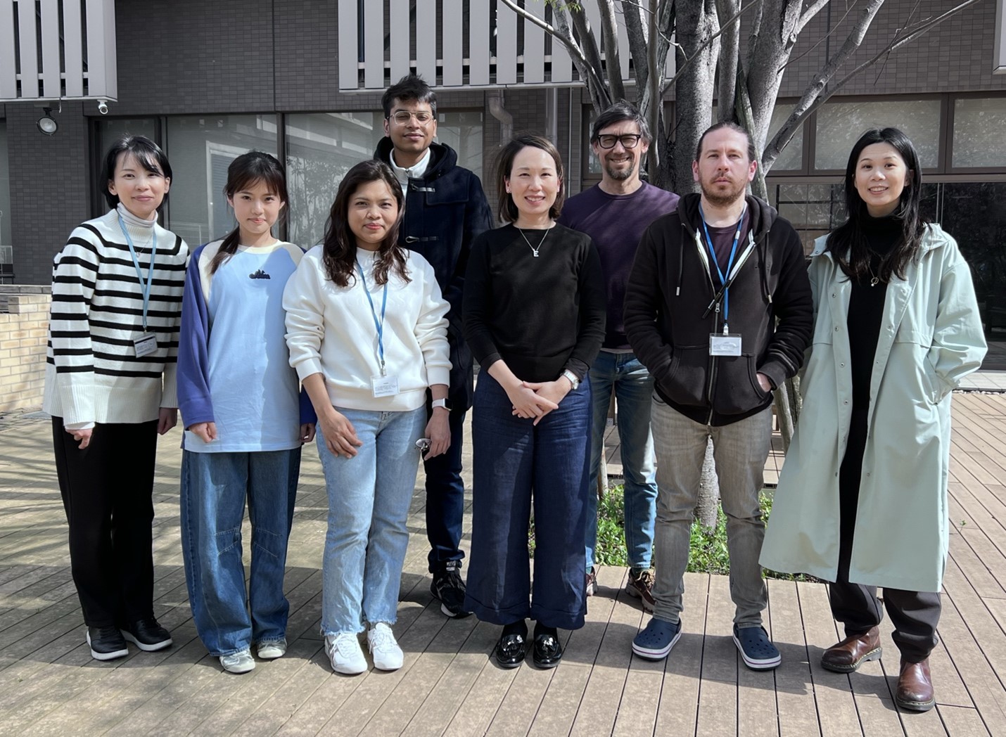


 (No Ratings Yet)