BSDB Report co-written by Simran Singh and Renato Duarte Dos Santos
We are extremely grateful to the BSDB for giving us the opportunity to attend the Biologists @ 100 conference in Liverpool. As PhD students investigating spinal cord injury and regeneration, this experience was invaluable. It allowed us to connect with scientists, gain insights into diverse fields and explore potential collaborations.
Simran Singh
The meeting started off with an exciting early career research session, offering a unique chance to interact with individuals who have pursued various scientific career paths. A highlight for me was the keynote talk by Dr. Richard Server, co-founder of bioRxiv and medRxiv. He shared his career trajectory, discussed the impact of bioRxiv on publishing – especially during the Covid-19 pandemic –and highlighted the numerous transferable skills gained from an academic career.
The following three days were filled with inspiring and thought-provoking scientific talks. One of the first talks was by Professor Muzlifah Haniffa, recipient of the Cheryll Tickle Medal. She described herself as “born into immunology, married into developmental biology, and became best friends with single-cell omics”. Her research focuses on decoding the human immune system, particularly the spatial and temporal composition of immune cells and their roles beyond immunology, such as in development. Additionally, her work on the Human Developmental Cell Atlas, integrating developmental disorders, has had a profound translational impact. Throughout her talk, she emphasised the importance of interdisciplinary approaches and collaboration in science. She is also a strong advocate for women in STEM and leadership. I particularly liked her powerful statement “Women should continue to thrive in science not despite but because of the system”.
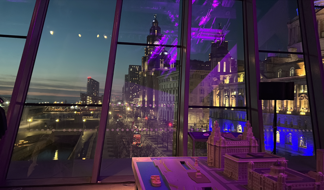
Fantastic end to day 1 of the conference with the welcome reception at Museum of Liverpool
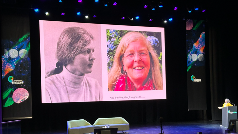
Another talk I enjoyed was by Professor Helen Skaer, winner of the BSDB Waddington Award for her outstanding research, contributions to the developmental biology community and excellent mentorship. Her research explores how cells work together to make an organ of the right shape, size and in the right place, with a focus on renal tubules in fruit flies, which are highly consistent. She eloquently described her research journey from studying Mercierella Enigmatica (reef building tubeworms) to fruit flies and shared some of her “most exciting moments” in the lab. One such moment was the identification of a ‘tip cell’, the master regulator that when ablated would arrest cell division of the renal tubules. My favourite part was due to the lack of a laser ablation machine, Professor Helen Skaer had to come up with a creative solution to manually “suck up” the tip cell. It reminded me the importance of being creative in research and not being afraid to think outside the box.
Winner of the BSDB Waddington Award (Professor Helen Skaer)
Overall, I had a great time at the Biologists@100 conference. It was a fantastic opportunity to hear talks from scientists across the world. I am now ready to go back into the lab feeling more inspired than ever!
Renato Duarte Dos Santos
Spreading science for 100 years, The Company of Biologists and BSDB have delivered an amazing event full of opportunities to learn more about the current work in developmental biology, but also in environmental awareness and career pathing in biology. This year, the developmental biology showcase at the conference had a clear focus on the role of signal patterning and mechanical signaling, which has been shown to affect multiple processes that we tend to view as solely based on biochemical reactions.
I had a special interest in the work done by Dr. Muzlifah Haniffa, awarded the BSDB Tickle Medal for her involvement in the Human Development Cell Atlas, a project aiming to incorporate single-omics from all human cells that intervene in human development. I believe this tool will become intemporal for the world of science, with applications for all the multitude of biomedical-related fields.
Another work that caught my eye was the development of a new barcoding method and bioinformatic processing capable of improving the output of single-cell expression analysis, increasing the sampling and reducing substantially the price in comparison with the current market offer (Maizels et al., 2024). This amazing work has been done by Dr. Rory Maizels while as a PhD student. His brilliance and achievements have led him to be awarded the well-deserved BSDB PhD student Beddington Medal. Another approach that I found very interesting was the use of the cell shape to determine the cell type and cell fate, like a pseudo-time analysis (Pönisch et al., 2024 preprint). This innovative work was made by Ewa Paluch from the University of Cambridge.
Besides development talks, there were also some morning plenary talks about climate change and biodiversity loss to help spread awareness about this urgent global matter that hasn’t been handled so far as it should.
Jane Francis talk – British Antarctic Survey
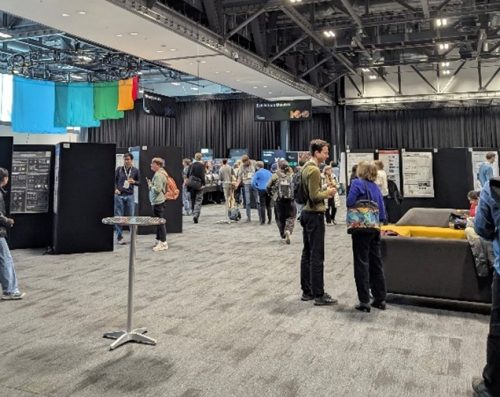
Lunch, posters and exhibition
I also found the early-career researcher career session quite insightful. It helped me and most likely many to gain a realistic view of the current scientific paradigm and the many options we biologists possess in terms of profession. The chance to have a one-to-one talk with a professional of each career path was for sure one of the most useful experiences. I also have to mention the gala dinner, which was of the highest luxury in a mouth-dropping location, the St George’s Hall. Overall, the conference was amazing, full of great talks, opportunities to network with high-tier researchers, and to enjoy the scientific community at its best.
One of my favourite uses of social media is sharing (and discovering) new preprints that seem relevant for my field. Rewardingly, posting preprints is usually well-received by followers. Since there used to be a button on the bioRxiv website for posting preprints on twitter/X, this was a relatively simple thing to do. However, twitter/X is abandoned and the majority of folks that I follow are now on Bluesky. So, now I’m posting preprints (among other sciency content) on BlueSky. Unfortunately, at the time of writing this piece, there are no buttons on the bioRxiv page that facilitate this process.
The information that I collect for posting the preprint are the title, an author and the link to the preprint, so that involves quite some copy-pasting between different browser windows. To simplify this process, I looked into web extensions (footnote 1). The web extensions that I needed was not there, so I decided to explore how easy it is to make one. It turns out that web extensions are written in Javascript and I have zero experience with this language. So, I turned to AI for help.
The prompt that was used to code the first web extension prototype
First, I tried ChatGPT, which wasn’t very successful. Then, I turned to Claude.ai and I got a working prototype after my first prompt. After a second prompt I already had something quite decent. Then, I decided to polish the web extension, e.g. add an icon, add only the name of the last author to the post and extend the functionality to medRxiv. This process involved several iterations of prompts and tests. The main issue is that new functionality has to be added without breaking the existing functions. So there were several rounds where the code stopped working, and I had to feed the error to Claude.ai and ask for repair. I also asked several times to ‘simplify the code’, to keep the code concise. In the end (17 prompts in total), I got a nicely working extension that shows a blue icon when a preprint can be shared and when the icon is clicked a post for BlueSky is prepared. This is exactly what I needed! The code for the extension (for the Firefox web browser) and an instruction to install it are available here: https://github.com/JoachimGoedhart/Rxiv2BlueSky
Screenshot of the extension added to the Firefox web browser
Besides the tangible result, I also gained some insight of how Large-Language Models can augment the development of software. Without any knowledge of web extensions, nor the necessary language needed to create one, I was able to generate a nicely working add-on, by simply defining what I wanted to achieve. Later on, I learned that this process is known as ‘vibe-coding’, a term introduced by Andrej Karpathy. The exciting point is that vibe coding enables non-coders to generate working code. For me, vibe coding a web extension was an amazing experience and it felt like I gained some kind of superpower.
Footnotes
Footnote 1: I wasn’t familiar with web extensions at all and it turned out that there is an ecosystem of different extension for different browsers and in some cases developers have to pay to publish their extension. Since I’m commonly using Firefox (in some cases Safari), and since publishing extensions (or add-ons) for Firefox is free, I decided to go for Firefox as the browser.
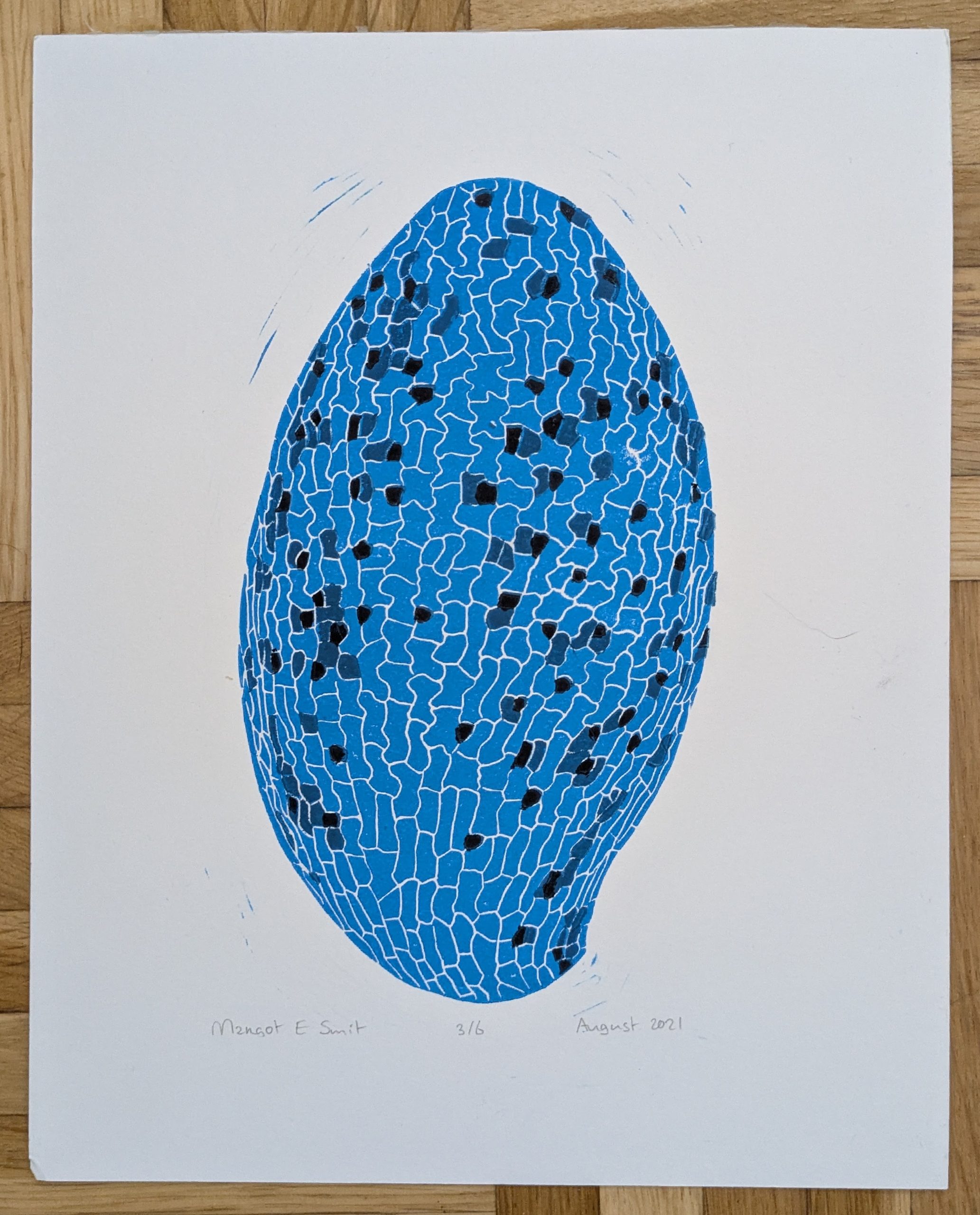
In this SciArt profile, we meet Margot Smit, a plant developmental biologist whose lab is in the ZMBP, University of Tübingen. Margot enjoys linocut printmaking of designs inspired by Arabidopsis images from the lab and scenes from nature.
Arabidopsis genetics. 6 stages of Arabidopsis rosettes arranged into 2 helices. Hand printed Spring 2024.
Can you tell us about your background and what you work on now?
I’m a Plant Developmental Biologist working on the temporal regulation of cell identity. In my lab we are using the Arabidopsis embryo and its stomatal lineage to study examples of temporal cell identity control. I studied Biotechnology in Wageningen (Netherlands) and quickly became interested plant developmental biology and during my MSc and PhD I studied the regulation of vascular identity specification during Arabidopsis embryogenesis. After years studying vascular development, I moved to Stanford University to study stomata. Right now, in Tübingen, we use embryonic stomatal development to study the temporal regulation of developmental decisions.
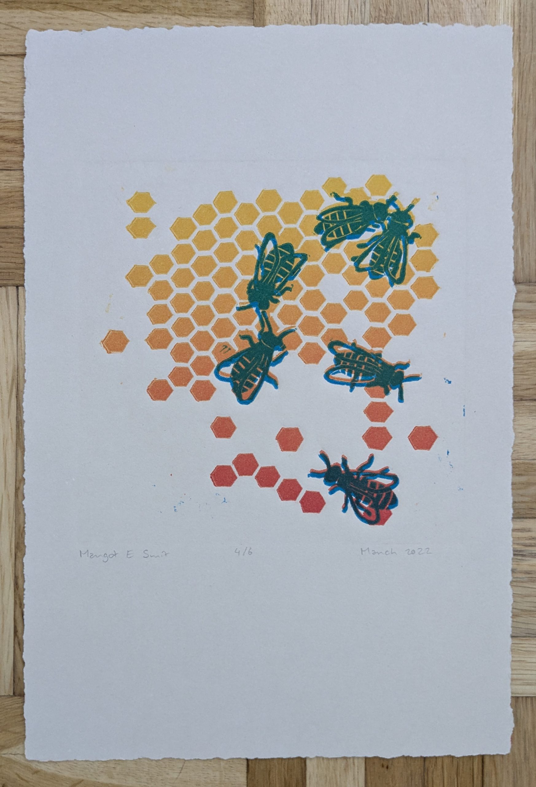
Bees and hexagons. Two-layer print of blue bees on a gradient of hexagons. Press printed March 2022.
Were you always going to be a scientist?
No, I didn’t really realize that scientist was a real job until I was doing my BSc thesis. I thought it was like becoming an astronaut; some people get to do it but it’s very out of reach for me. During my BSc thesis project, I fell in love with the scientific method of identifying and trying to answer basic questions in biology. I’ve always loved puzzles and all of it felt and still feels like a lovely, complex puzzle. I didn’t know whether I would be able to continue in academia, but I’ve just been taking it one step at a time and enjoying the journey.
Mushrooms. 3 of 5 total mushrooms. Press printed March 2023.
And what about art – have you always enjoyed it?
I’ve always enjoyed art and got a lot of opportunities to play around with it growing up but was never super confident. Several of my family members are very artistic and have attended and/or taught at art schools which got me exposed at a young age. However, I am not incredibly talented and do not have the patience or practice with drawing or painting: I make a lot of mistakes and easily give up on it. Instead, during my PhD I discovered a love for using Illustrator to create fun compositions and designs.
Young leaf. Print based on a microscopy image of a 3-day old Arabidopsis leaf. Press printed August 2022.
What or who are your most important artistic influences?
My mother, grandma and aunt were my first artistic influences growing up, there were always plenty of art supplies and examples around. While traveling in Eureka few years ago, I saw prints from Lynn M Jones and got inspired to give it a try. I then took Katie Gilmartin’s awesome printmaking course in San Francisco and she’s been a great inspiration. In recent years I’ve also been really inspired by online communities sharing their printmaking. For me, most prints are based on images from the lab or from nature.
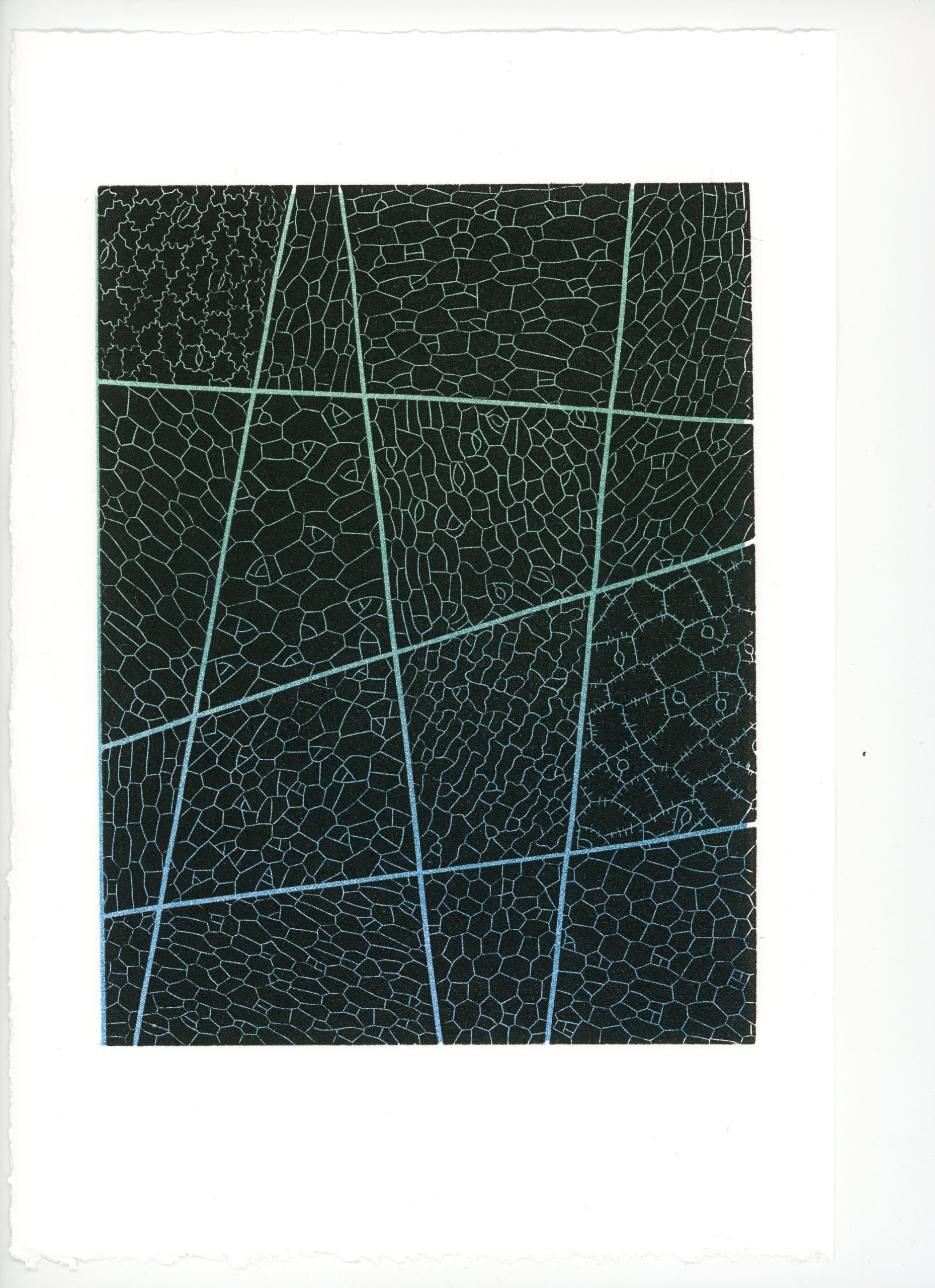
Embryogenic epidermises. Two-layer print of leaf surface patterns on a gradient. Each of the 16 sections is based on a microscopy image of the surface of an embryonic leaf surface. Press printed December 2021.
How do you make your art?
Each print starts with a series of images or ideas. The challenge for me is to figure out how to work with the restrictions that are part of my linocut printmaking process. I usually only carve one or two blocks per print and a design needs to be high contrast, relatively simplistic, and mostly two-tone with limited shading options. First, I try lots of different designs using Illustrator and my drawing tablet. Once I am satisfied with the design, I transfer it to the linoleum and start carving. When I think I am done I will do some test prints and maybe adjust the carving before moving to the real printing. To print I use oil-based inks that I mix to the desired color and apply using a roller. Then I place a sheet of damp paper on top and start to transfer the ink: for small blocks I can use my press, for larger blocks I have to hand print using a baren. Depending on the depth of the lines I will print each block five to twenty times.
Time-lapse video of the carving processTime-lapse video of the printing process
Does your science influence your art at all, and vice versa, or are they separate worlds?
For me they are very connected. A lot of my prints are based on images from the lab or based on scientific interests. That’s one of the cool aspects of working with plants, patterns and embryos.
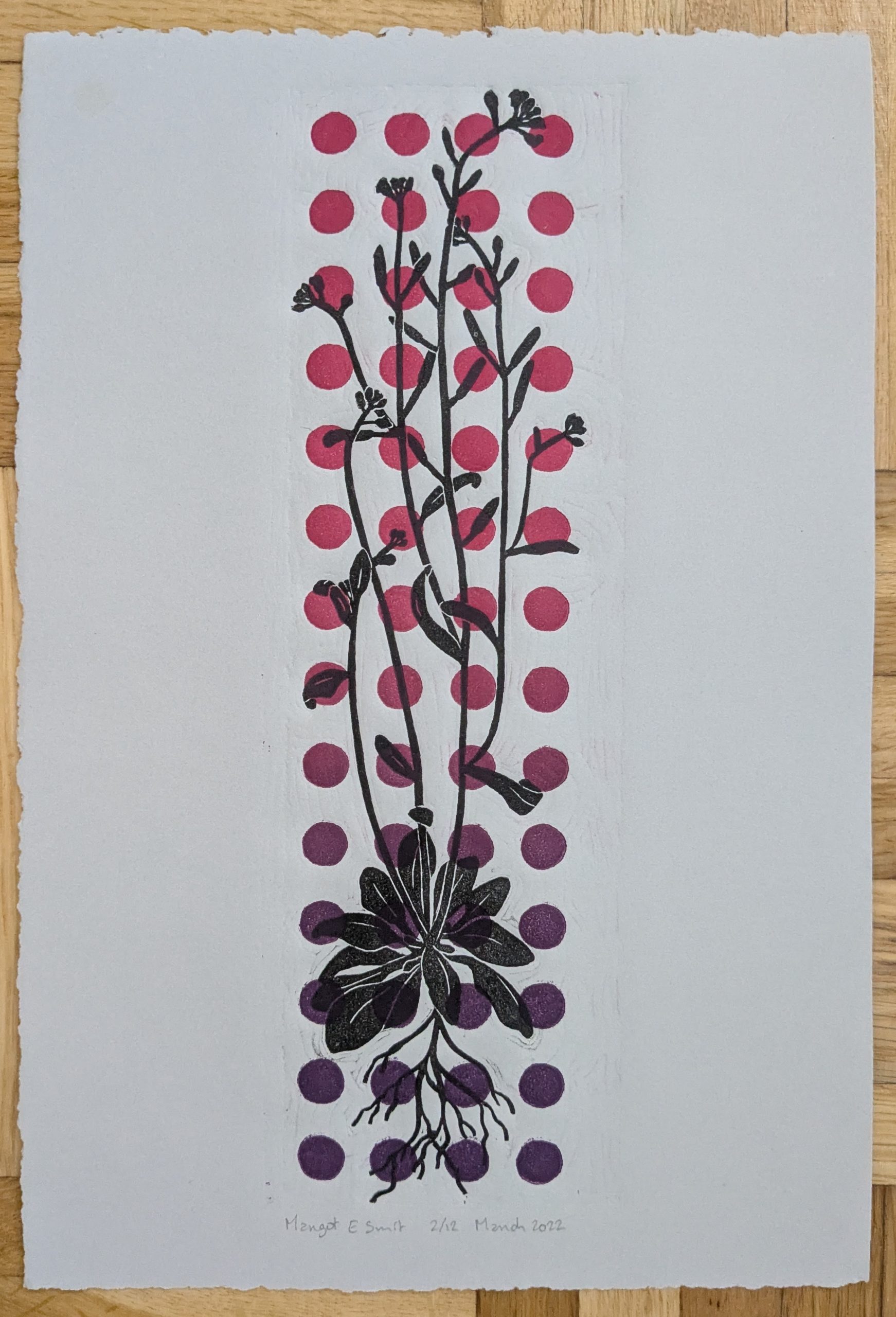
Artsy Arabidopsis. Two-layer print of adult Arabidopsis on a background of colorful circles. Press printed March 2022.
What are you thinking of working on next?
I’ve started a new project with a bunch of scientific model organisms. But things have been busy at work recently and while I am done with the design, I haven’t progressed far into the carving yet. I think it’ll be a challenge to print but that’s part of the fun.
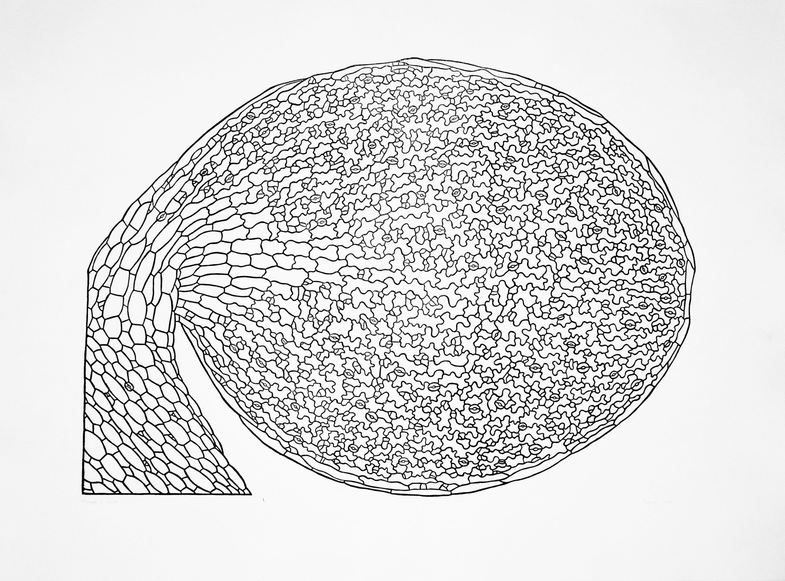
Embryonic overview. Three-layer reduction print of an Arabidopsis embryonic leaf. Hand printed August 2021.
How can people find more about you and your artwork?
I have a website where I have an (old) overview of my prints. When I moved back to Europe, I also sold some from there with proceeds going to charity, something that I am planning to do again though I’m not sure when. Otherwise, they just take up space in my drawers… I also sometimes post them on my BlueSky though I haven’t created a lot recently.
Reproduction is a metabolically costly process. Therefore, the proper regulation of physiological changes in the mother during pregnancy and lactation is crucial for fetal development and neonatal growth. In many mammals, including humans, pregnancy induces systemic changes in hormonal, metabolic, and immune functions.1 Reproductive-associated changes are also known to occur at the organ level, in particular, an increase in intestinal size during pregnancy and lactation has been observed in several mammals such as mice, rats, sheep, and pigs.2,3 However, despite the first report of this phenomenon, documented in 1939,4 the molecular mechanisms underlying maternal intestinal remodelling during reproduction, as well as their physiological significance, have only recently begun to be investigated.5
Studies using fruit flies have shed light on the molecular mechanisms regulating reproductive plasticity and sex differences in adult digestive tract. 6–13 Instead of studying the fly, we decided to focus on the maternal intestinal growth in mice.
Characterising maternal gut growth in epithelium
We first examined the organ size of the small intestine during reproduction and observed that the maternal small intestine is significantly longer and heavier than that of non-pregnant female mice, consistent with previous studies.14,15 Notably, the small intestine remains elongated even one month after weaning, suggesting that gut elongation is an irreversible (or only partially reversible) process.
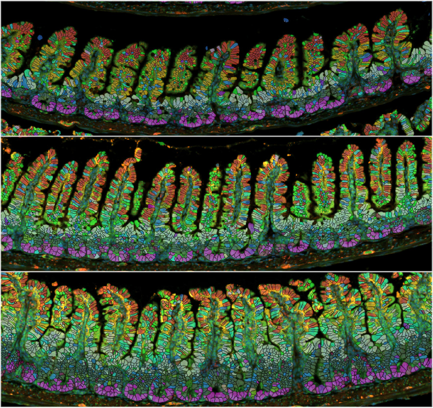
Next, we investigated the mucosal morphology of the small intestine, which consists of absorptive villi (finger-like projections into the lumen) and crypts (which house progenitor cells). We observed that both villi and crypts increased in size during pregnancy and lactation (Figure 1). Since the size of the crypt-villus structure can be determined by the number and size of epithelial cells—or both—we focused on epithelial proliferation. The intestinal epithelium has one of the fastest turnover rates among all tissues, typically renewing every 3–5 days.
Figure 1. Remodelling of the maternal gut epithelium. The image shows that the inside of the small intestine—composed of villi (protrusions that absorb nutrients from the lumen) and crypts (where progenitor cells produce the villi)—grows in non-pregnant (top), pregnant (middle), and lactating (bottom) mice.
We found that this turnover rate was accelerated during reproduction, with lactating mice exhibiting the highest turnover rate—renewing in just 1.3 days. In contrast to the irreversible elongation of the gut tube, the growth of the gut epithelium is reversible: both the crypt-villus structure and epithelial turnover returned to pre-pregnancy levels within one week after weaning. These results suggest that gut tube elongation and epithelial growth are regulated by different mechanisms.
Remodelling of epithelial cell types in the gut
We have two hypotheses of how the crypt-villus structure enlarges:
Do epithelial cells increase proportionally across different cell types at a similar rate?
Or do epithelial cells increase preferentially in specific cell types?
To address this question, we first analysed our single-cell RNA sequencing data from FACS-sorted intestinal epithelial cells, where we did not observe significant differences in cell composition between reproductive and non-reproductive mice. However, since it was unclear whether this lack of difference was due to technical—such as the dissociation of epithelial cells and the FACS-sorting process—or biological reasons, we decided to perform a spatial imaging analysis using the Xenium in situ platform. We profiled subtypes of intestinal epithelial cells—including absorptive enterocytes, secretory cells, and progenitor cells—to compare cell composition between intestines from reproductive and non-reproductive mice.
We actually found that lactating mice had significantly more progenitor cells, specifically isthmus progenitors in the upper crypts, along with their neighbouring enterocytes located at the base of the villi. This indicates that epithelial cell expansion during reproduction occurs preferentially in specific cell types.
While the reason for this preferential increase is still unclear, we speculate that the expansion of the progenitor population supports the accelerated epithelial turnover observed during lactation.
What is the molecular mechanism behind this?
To uncover potential drivers of maternal gut growth, we focused on a transporter gene called SGLT3a, whose expression was upregulated in our transcriptomics datasets—including whole small intestine tissue, FACS-sorted intestinal epithelial cells, and spatial imaging analysis—during reproduction. SGLT stands for sodium-glucose cotransporter, but previous studies have shown that, unlike SGLT1 (which transports glucose from the lumen into epithelial cells), SGLT3a does not function as a sugar transporter, at least at physiological pH.16
To investigate its function in vivo, we performed a series of phenotypic analyses using SGLT3a knockout mice. The knockout mice appeared physiologically normal under standard conditions; their food intake, body weight, and glucose tolerance were comparable to those of control littermates.
Importantly, however, maternal gut epithelial growth—both in terms of crypt-villus morphology and epithelial turnover—was significantly reduced in SGLT3a knockout mice during lactation. This effect appears to be specific to the reproductive state, as we did not observe similar phenotypes in non-pregnant female knockout mice. We also observed that progenitor proliferation was dampened in intestinal organoids—mini-guts grown in vitro—derived from SGLT3a knockout mice, suggesting a gut-specific role of SGLT3a in regulating maternal gut epithelial growth.
What are the downstream signals?
How does SGLT3a control villus growth during reproduction?We found that the population of progenitor cells expressing Fgfbp1—a known marker for isthmus progenitors17,18—was reduced in SGLT3a knockout mice. Since SGLT3a is not expressed in progenitor cells themselves, we hypothesise that SGLT3a expressed in enterocytes regulates progenitor proliferation extrinsically through Fgfbp1 expression.
Our electrophysiological experiments demonstrated that SGLT3a responds to protons and sodium, but not to sugars. Sodium is an especially intriguing candidate, as we observed that dietary sodium can mimic the effects of reproduction on the gut epithelium. Therefore, it would be interesting to further explore whether dietary sodium is essential for maternal organ remodelling, including that of the small intestine.
What are the upstream signals?
Could the upregulation of SGLT3a be driven by increased food intake during reproduction? This seems unlikely, as SGLT3a expression is already elevated by early pregnancy (gestational day 7), which precedes the marked increase in maternal food intake observed in mid-pregnancy. This suggests that other signals may be responsible for inducing SGLT3a expression in the intestinal epithelium.
One possible group of candidates is reproductive hormones. In our single-cell transcriptomic analysis of intestinal epithelial cells, we observed broad expression of the prolactin receptor, whereas estrogen and progesterone receptors were either undetectable or expressed at very low levels. Supporting this, we found that treating intestinal organoids with prolactin led to upregulation of SGLT3a expression.
These findings suggest that reproductive hormones—particularly prolactin—may play a role in this process by driving pregnancy-specific SGLT3a induction. Further research is needed to investigate how hormonal signals, or signals originating outside the intestinal epithelium,5 contribute to SGLT3a induction and maternal gut growth during reproduction.
What is the physiological relevance?
Despite no significant difference in litter size at birth, we observed a lower survival rate among pups born to SGLT3a knockout mothers compared to those born to wild-type mothers. Although we cannot exclude the possibility that this phenotype is influenced by SGLT3a function outside the gut—given that whole-body knockout mice were used—our findings suggest that SGLT3a contributes to reproductive success by sustaining maternal gut growth during pregnancy. It would be intriguing to investigate the long-term effects on both mothers and offspring, as excessive weight gain during pregnancy and after childbirth in humans is known to be associated with an increased risk of chronic diseases in mothers and future obesity in their children.
Access the article: Ameku T, Laddach A, Beckwith H, Milona A, Rogers LS, Schwayer C, Nye E, Tough IR, Thoumas JL, Gautam UK, Wang YF, Jha S, Castano-Medina A, Amourda C, Vaelli PM, Gevers S, Irvine EE, Meyer L, Andrew I, Choi KL, Patel B, Francis AJ, Studd C, Game L, Young G, Murphy KG, Owen B, Withers DJ, Rodriguez-Colman M, Cox HM, Liberali P, Schwarzer M, Leulier F, Pachnis V, Bellono NW, Miguel-Aliaga I. Growth of the maternal intestine during reproduction. Cell. 2025 Mar 19:S0092-8674(25)00200-4. doi: 10.1016/j.cell.2025.02.015. Epub ahead of print. PMID: 40112802.
References
Napso T, Yong HEJ, Lopez-Tello J, Sferruzzi-Perri AN. The Role of Placental Hormones in Mediating Maternal Adaptations to Support Pregnancy and Lactation. Front Physiol. 2018 Aug 17;9:1091. doi: 10.3389/fphys.2018.01091. PMID: 30174608.
Hammond KA. Adaptation of the maternal intestine during lactation. J Mammary Gland Biol Neoplasia. 1997 Jul;2(3):243-52. doi: 10.1023/a:1026332304435. PMID: 10882308.
Speakman JR. The physiological costs of reproduction in small mammals. Philos Trans R Soc Lond B Biol Sci. 2008 Jan 27;363(1490):375-98. doi: 10.1098/rstb.2007.2145. PMID: 17686735.
Poo LJ, Lew W, Addis T. Protein anabolism of organs and tissues during pregnancy and lactation. J. Biol. Chem. 1939 Volume 128, Issue 1, Pages 69-77, ISSN 0021-9258.
Onji M, Sigl V, Lendl T, Novatchkova M, Ullate-Agote A, Andersson-Rolf A, Kozieradzki I, Koglgruber R, Pai TP, Lichtscheidl D, Nayak K, Zilbauer M, Carranza García NA, Sievers LK, Falk-Paulsen M, Cronin SJF, Hagelkruys A, Sawa S, Osborne LC, Rosenstiel P, Pasparakis M, Ruland J, Takayanagi H, Clevers H, Koo BK, Penninger JM. RANK drives structured intestinal epithelial expansion during pregnancy. Nature. 2025 Jan;637(8044):156-166. doi: 10.1038/s41586-024-08284-1. Epub 2024 Dec 4. PMID: 39633049.
Blackie L, Gaspar P, Mosleh S, Lushchak O, Kong L, Jin Y, Zielinska AP, Cao B, Mineo A, Silva B, Ameku T, Lim SE, Mao Y, Prieto-Godino L, Schoborg T, Varela M, Mahadevan L, Miguel-Aliaga I. The sex of organ geometry. Nature. 2024 Jun;630(8016):392-400. doi: 10.1038/s41586-024-07463-4. Epub 2024 May 29. PMID: 38811741.
White MA, Bonfini A, Wolfner MF, Buchon N. Drosophila melanogaster sex peptide regulates mated female midgut morphology and physiology. Proc Natl Acad Sci U S A. 2021 Jan 5;118(1):e2018112118. doi: 10.1073/pnas.2018112118. PMID: 33443193.
Hadjieconomou D, King G, Gaspar P, Mineo A, Blackie L, Ameku T, Studd C, de Mendoza A, Diao F, White BH, Brown AEX, Plaçais PY, Préat T, Miguel-Aliaga I. Enteric neurons increase maternal food intake during reproduction. Nature. 2020 Nov;587(7834):455-459. doi: 10.1038/s41586-020-2866-8. Epub 2020 Oct 28. Erratum in: Nature. 2020 Dec;588(7839):E36. doi: 10.1038/s41586-020-3013-2. PMID: 33116314.
Zipper L, Jassmann D, Burgmer S, Görlich B, Reiff T. Ecdysone steroid hormone remote controls intestinal stem cell fate decisions via the PPARγ-homolog Eip75B in Drosophila. Elife. 2020 Aug 10;9:e55795. doi: 10.7554/eLife.55795. PMID: 32773037.
Ahmed SMH, Maldera JA, Krunic D, Paiva-Silva GO, Pénalva C, Teleman AA, Edgar BA. Fitness trade-offs incurred by ovary-to-gut steroid signalling in Drosophila. Nature. 2020 Aug;584(7821):415-419. doi: 10.1038/s41586-020-2462-y. Epub 2020 Jul 8. PMID: 32641829.
Hudry B, de Goeij E, Mineo A, Gaspar P, Hadjieconomou D, Studd C, Mokochinski JB, Kramer HB, Plaçais PY, Preat T, Miguel-Aliaga I. Sex Differences in Intestinal Carbohydrate Metabolism Promote Food Intake and Sperm Maturation. Cell. 2019 Aug 8;178(4):901-918.e16. doi: 10.1016/j.cell.2019.07.029. PMID: 31398343.
Hudry B, Khadayate S, Miguel-Aliaga I. The sexual identity of adult intestinal stem cells controls organ size and plasticity. Nature. 2016 Feb 18;530(7590):344-8. doi: 10.1038/nature16953. PMID: 26887495.
Reiff T, Jacobson J, Cognigni P, Antonello Z, Ballesta E, Tan KJ, Yew JY, Dominguez M, Miguel-Aliaga I. Endocrine remodelling of the adult intestine sustains reproduction in Drosophila. Elife. 2015 Jul 28;4:e06930. doi: 10.7554/eLife.06930. PMID: 26216039.
CAMPBELL RM, FELL BF. GASTRO-INTESTINAL HYPERTROPHY IN THE LACTATING RAT AND ITS RELATION TO FOOD INTAKE. J Physiol. 1964 May;171(1):90-7. doi: 10.1113/jphysiol.1964.sp007363. PMID.
Casirola DM, Ferraris RP. Role of the small intestine in postpartum weight retention in mice. Am J Clin Nutr. 2003 Dec;78(6):1178-87. doi: 10.1093/ajcn/78.6.1178. PMID: 14668281.
Barcelona S, Menegaz D, Díez-Sampedro A. Mouse SGLT3a generates proton-activated currents but does not transport sugar. Am J Physiol Cell Physiol. 2012 Apr 15;302(8):C1073-82. doi: 10.1152/ajpcell.00436.2011. Epub 2012 Feb 1. PMID: 22301059.
Capdevila C, Miller J, Cheng L, Kornberg A, George JJ, Lee H, Botella T, Moon CS, Murray JW, Lam S, Calderon RI, Malagola E, Whelan G, Lin CS, Han A, Wang TC, Sims PA, Yan KS. Time-resolved fate mapping identifies the intestinal upper crypt zone as an origin of Lgr5+ crypt base columnar cells. Cell. 2024 Jun 6;187(12):3039-3055.e14. doi: 10.1016/j.cell.2024.05.001. PMID: 38848677.
Malagola E, Vasciaveo A, Ochiai Y, Kim W, Zheng B, Zanella L, Wang ALE, Middelhoff M, Nienhüser H, Deng L, Wu F, Waterbury QT, Belin B, LaBella J, Zamechek LB, Wong MH, Li L, Guha C, Cheng CW, Yan KS, Califano A, Wang TC. Isthmus progenitor cells contribute to homeostatic cellular turnover and support regeneration following intestinal injury. Cell. 2024 Jun 6;187(12):3056-3071.e17. doi: 10.1016/j.cell.2024.05.004. PMID: 38848678.
A workshop to explore how synthetic biology can help us understand how embryos build themselves
20 – 21 October 2025, Brighton, UK. A Royal Society Theo Murphy meeting organised by Jake Cornwall-Scoones, Dirk Benzinger, and Sally Lowell.
Image credit: Shannon Taylor
As systems-level measurement of embryogenesis reaches maturity, developmental biologists are returning to foundational questions of how embryos build themselves. Synthetic biology has demonstrated how bottom-up explanations reveal design principles of biological transitions, and is increasingly looking towards embryos for inspiration, holistic contextualisation, and evolutionary interpretation. This meeting brings together these two disciplines towards a science of generative biology.
Speakers:
Philip Ball : David Brückner : Francesca Ceroni : Jamie Davies : Pulin Li : Mattias Malaguti : Yolanda Schaerli : Ricard Solé : Berna Sozen : Ben Steventon : Jared Toettcher : Vikas Trivedi : Berta Verd : Sara Wickström
At the end of each month, I pick the same month from a random year from the past 15 years of the Node, and take a look at what people were talking about back then.
Previously, I travelled back to February 2011 and March 2013 to have a look around the Node. This time, let’s fasten our seat belts and turn the dial to April 2014…
Our publisher, The Company of Biologists, offers Travelling Fellowships for a collaborative visit to another lab. Many of the recipients have written about their wonderful experiences on the Node. Read about their experiences, and find out more about the fellowships.
This week we will get to know insights from Dr. Eudald Pascual-Carreras, who is a postdoctoral researcher in the Multicellgenome lab at IBE Barcelona where he’s studying how metabolism regulates the cell cycle at the origin of animal multicellularity. Before joining IBE, he conducted postdoctoral work in the Steinmetz Group at the Michael Sars Centre, University of Bergen, Norway. Eudald has long been fascinated by how nutrient-dependent signaling influences stem cell proliferation and growth, approaching these questions using unique model systems like the planarian flatworms and the sea anemones. Keep reading to learn about his journey through the world of metabolism—and why curiosity driven basic science remains at the heart of it all. Along the way, he’s embraced the value of mentorship, stayed motivated through scientific challenges, and remained rooted in a deep curiosity for basic biology. Discover his journey through metabolism and learn about the mindset that keeps him going. Give him a follow over twitter or bluesky and check out his work here .
What was your first introduction to the field of metabolism? What inspired you to specialize in metabolic studies using two incredibly unique systems – the planaria flatworms and the sea anemones ?
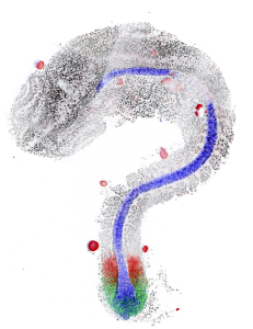
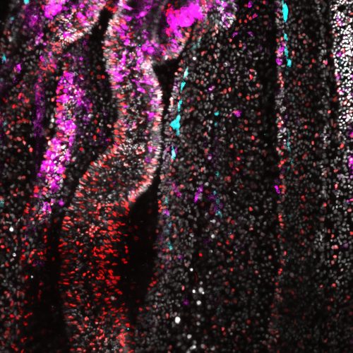
A confocal image of Nematosella mesenteries with the nuclei shown in gray, stem/progenitor cells in cyan, cycling cells with EdU in red and Tor signalling with pRPS6 in magenta. Image source : EPC
I can clearly remember my first introduction to metabolism during high school biology class. My teacher explained how glucose is broken down, the Krebs cycle, how ATP is generated. I was fascinated by the intricated biochemical pathways that sustain life. Later, in university, I took an animal physiology course that provided a broader biochemical perspective. For my PhD, I decided to study planarians due to their remarkable body plasticity. These animals can regenerate and modulate their body size depending on the nutrient availability. Initially, my research focused on regeneration, and metabolism wasn’t my main area of interest. However, over time, my focus shifted toward understanding the regulation of animal growth. This eventually led me to pursue a postdoctoral position in a Nematostella lab, where I kept exploring how nutritional changes influence organismal growth. Classic research organisms such as Drosophila, C. elegans and mice have a predetermined, fixed body size. In contrast, the unique organisms I study can grow and shrink throughout their lifetime, a trait common among many non-bilaterian animals, including sponges, corals, sea anemones and ctenophores. This suggests that body plasticity is likely ancestral to all animals. Studying these organisms has allowed me to explore fundamental questions about the evolution of animal growth, the mechanisms that regulate it and the intricate interplay between metabolism and genetics.
What sparked your interest in exploring nutritional and metabolic aspects of animal development through the lens of cell cycle and cell fate? Your work intersects metabolism, development and evolution. How do you integrate these disciplines in your research, and what unique insights have emerged from this approach?
Planarians and Nematostella can rapidly adjust their body size in response to nutrient availability. In both cases, cell number drives organismal growth, making the regulation of cell proliferation a crucial factor. This sparked my interest in understanding the cellular and molecular mechanisms that enable these animals to adapt. Therefore, I began studying the cell cycle, which I consider a fundamental cornerstone of this process. I found it fascinating that a such a highly conserved process as cell cycle can exhibit remarkable plasticity – pausing or adjusting its duration to accommodate different nutrient conditions. Trained as a developmental biologist, I became increasingly interested in evolutionary questions during my PhD. Deciphering how developmental processes have evolved has always been on my interest. Integrating a metabolic perspective into this field adds a new layer of complexity that has been overlooked in evolutionary developmental biology. I believe this perspective has the potential to reshape fundamental concepts in cell biology, physiology and developmental biology.
Can you briefly summarize how you’re using Nematostella to uncover unique mechanisms by which body plasticity responds to environmental inputs, particularly how stem or progenitor cell populations driving this plasticity adapt to nutritional cues?
Nematostella vectensis has emerged as a powerful research organism for studying body plasticity in response to environmental changes, including nutrient availability and temperature. Nematostella polyps exhibit a remarkable resilience during starvation conditions, capable of surviving over 200 days without food. In Steinmetz lab, we observed that after prolonged starvation, these animals can be refed and return to their original body size within two weeks. Our research demonstrated that changes in cell number and cell proliferation directly correlated with organismal growth (doi: 10.1242/dev.202926). This led us to ask a fundamental question: which cells contribute to this remarkable organismal growth? At the same time, the lab was also investigating the identification and characterization of a multipotent stem/progenitor population that contributes to both germline and somatic tissues (doi: 10.1038/s41467-024-52806-4). My project naturally evolved from these findings – I studied how this multipotent stem/progenitor population behaves under starvation and refeeding conditions. Essentially, my goal was to move from the organismal level to the cellular and molecular level, dissecting how this specific population adapts to the extreme nutritional shifts (doi: 10.1101/2025.02.27.640509).
Tell us about your work on nutritional control of cellular quiescence and how Nematostella is a unique model to answer questions about nutrient dependent quiescence. Summarize your key findings on cell cycle dynamics, the signaling pathways involved, and how these insights differ from known mechanisms in yeast and mammals.
When we began studying how starvation affects this stem/progenitor population, we considered different hypotheses based on observation in planarians and Hydra. In these organisms, stem cell populations (neoblast for planarians and i-cells for Hydra) continue dividing even under starvation. However, in Nematostella, we observed that the stem/progenitor population exhibited a low division rate, suggesting that these cells might enter a state of cellular quiescence. We then found differences in cell cycle phase distribution depending on the duration of the starvation. The longer the starvation period, the deeper the quiescent state these cells entered! This progressive deepening of quiescence following nutrient withdrawal had only been observed in yeast and cell culture models, never in an organismal level! What is fascinating is that after refeeding, these cells are primed to re-enter the cell cycle in short-term condition while the re-entry was delayed following prolonged starvation. Our findings position Nematostella as a unique in vivo model to study nutrient-dependent quiescence, and all it requires is subjecting animals to different starvation durations. Surprisingly simple yet incredibly powerful! Specifically, we have found that starvation induces a G1/G0 quiescence state. During short-term starvation, some cells remain cycling, and after refeeding, quiescent cells rapidly re-enter the cell cycle. However, after long term starvation, the majority of the cells have entered a deep quiescent state, and their cell cycle re-entry is delayed upon refeeding! While we are still investigating the molecular mechanisms underlying quiescence acquisition, we have identified that TOR signalling is essential for feeding-dependent cell cycle re-entry!
Tell us about the experimental challenges you encountered during this project?
This project was a major challenge from the start. I spent my first year establishing quantitative flow cytometry analysis in Nematostella. Since the stem/progenitor population is located deep with the tissue and cannot not be imaged directly, I quickly realized that understanding cell cycle dynamics would require a tightly controlled time course experiments. This meant meticulous planning, and of course, having the support of colleagues and undergrads was essential. To optimize time course experiments, I decided to try something slightly different. From T0 to 12 hours post feeding (hpf), I could sample continuously, which meant spending at least 14 hours in the lab. However, for the later time points (15, 18 and 21 hpf), instead of staying up all night, I strategically delayed the last feeding 3, 6 or 9 hours, allowing me to collect the samples the following morning. Naturally, I ensured that this adjustment had no circadian effects. This approach not only made the experiment more manageable but also allowed me to generate high-resolution temporal data without requiring overnight shifts. Though long hours were certainly still part of the process!
Before this, you worked on planaria and identified a novel gene family – blitzschnell, which coordinates cell proliferation and differentiation in response to nutritional availability. Please tell us about your exciting findings.
This was a truly unique project. Characterizing the function of a novel gene family came with significant challenges, as we had no reference to guide our understanding of the phenotype. It took time to piece everything together, but our findings were exciting! One of our key discoveries was that the transcription of this gene family, blitzschnell, is directly regulated by nutrient intake! Moreover, its function is critical for controlling the cell number by balancing cell proliferation and cell death. In planarians, this regulation may be linked to the requirement for continuous and rapid modulation of cell numbers in response to nutrient availability (doi: 10.1242/dev.184044)
How do you think the fields of studying evolution and metabolism overlap? How do the two model systems you worked with differ in terms of nutrient-dependent regulation ?
Evolution and metabolism are deeply intertwined! Nutritional regulation is likely one of the most ancient evolutionary mechanisms. In unicellular eukaryotes, one of the earliest forms of cellular regulation is nutrient-dependent division: cells divide when food is available, and halt division when it is scarce. In multicellular organisms, this regulation has become more complex due to cell type and tissue specialization, certain tissues sense the nutrients and signal other cells to divide. While nutrient-dependent growth regulation has been well studied in some animal tissues and cell cultures, we still lack a broader understanding from an organismal perspective. Using planarians and Nematostella as models, we can explore how stem cell populations that drive organismal growth respond to nutritional cues. One of the key differences we have observed is that in Nematostella, stem cells enter a quiescent state during starvation, whereas in planarians, this is less clear. Neoblasts continue proliferating even in the absence of food. The cell cycle dynamics of neoblasts during starvation remain poorly understood, with some results suggesting that starvation prolongs the G2 phase, allowing some neoblast to re-enter the cycle upon refeeding. However, this has not been definitively proven. To gain a more comprehensive understanding of how nutrient-dependent regulation evolved, more organisms with body plasticity, such as ctenophores, Ciona, and sponges, should be studied. These models could provide crucial insights into the cellular mechanisms underlying metabolic control of animal growth.
How have different animals evolved to respond to nutritional and metabolic stresses? TOR signaling is conserved from yeast to humans, but are there organism specific differences in which the pathway is utilised ?
Yes, the signalling pathways are highly conserved across species. TOR signalling is required for organismal growth and cell proliferation in both Nematostella and planarians. What is particularly interesting is how these different animals utilize the same conserved pathway in distinct ways. While both rely on TOR signalling, they employ different strategies to cope with nutrient availability. As I mentioned earlier, Nematostella stem cells enter a quiescent state during starvation, while planarian neoblasts continue proliferating, even under nutrient-deprived conditions. Despite these differences, both organisms use the same fundamental molecular toolkit, illustrating the remarkable versatility of conserved signalling mechanisms across evolution.
What role does curiosity play in your life, both within and outside of science? How important it is for you to answer basic science questions on metabolic signaling and how do you see its impact on human health/relevance ?
Curiosity is at the core of my scientific journey. I am deeply interested in basic science, particularly in understanding how developmental processes are regulated, how cells integrate surrounding signals, and how the metabolome interacts with the transcriptome and signalling pathways. These fundamental questions drive my research, as uncovering mechanisms not only expands our knowledge of biology but also lays the groundwork for better understanding of human biology and health. Although my research is not explicitly focused on human biology, I believe that the questions I explore have significant implications for human health. Fundamental discoveries in model organisms often provide insights into conserved biological processes, ultimately influencing biomedical research and our understanding of disease mechanisms.
What are your upcoming plans? What metabolic pathways or signals you aim to investigate further to understand their role in stem cell regulation?
The Steinmetz group at the Michael Sars Center (UiB, Bergen, Norway), where I conducted my postdoc, remains deeply interested in studying the metabolic regulation of this stem/progenitor cell population. Our ongoing work aims to uncover the transcriptomic and epigenetic changes these cells can undergo in response to nutritional shifts. Additionally, the group is exploring metabolic changes at the organismal level. Personally, I am about to start a new postdoctoral position, where I will investigate the metabolic regulation of the cell cycle in the context of the transition of animal multicellularity. As mentioned, nutritional regulation is likely one of the most ancient evolutionary mechanisms. I plan to leverage a facultative multicellular organism, whose life cycle includes distinct unicellular and multicellular stages. I am particularly curious to understand how metabolism influences multicellularity transition, whether nutritional gradients are generate within cell aggregates and whether shifts in metabolic state serve as prerequisites for multicellularity.
What changes have you seen in the scientific community in regard to studying these unique aspects of metabolic signaling in unconventional systems? How do you think scientific paradigms around gaining new insights from non-model organisms will evolve in the coming decades? Are we moving toward a more nuanced understanding, or do you see potential pitfalls?
The scientific community is increasingly recognizing that non-classic research organisms can provide valuable insights into the more fundamental questions. As a developmental biologist, I am well aware of the critical role that non-classic research organisms have played in advancing our understanding of core processes. For example, the discovery of cyclins in sea urchins. Similarly, I believe that studying unconventional model can unveil new and extraordinary metabolic processes that may have previously been thought to exist only in unicellular eukaryotes. Moreover, one aspect that has often been overlooked in recent years is the metabolic state of organisms when designing experiments. As we gather more data, it will become increasingly important for each scientific community to establish standardized protocols to improve reproducibility and ensure more meaningful interpretations of results. By integrating a more nuanced understanding of metabolic context, we can refine experimental approaches and uncover deeper insights into the fundamental principles of biology.
How do you see the future of metabolism evolve with the new upcoming techniques – what are you most excited about ?
I would love to establish metabolic sensing lines, as my work over the past few years primarily relied on fixed tissue, making it challenging to assess the dynamic nature of metabolic processes. Having live metabolic sensor lines would be a game-changer, allowing us to directly observe and analyze the metabolic state of a cell under the microscope in real time! Additionally, I believe it is crucial to move beyond relying solely on metabolomics. While metabolomic profiling provides valuable insights, integrating real-time metabolic imaging with other approaches will offer a more comprehensive understanding of cellular metabolism and its regulation. These advancements will open new avenues for studying metabolism in a more dynamic and physiologically relevant context.
Were there any pivotal moments that shaped your career path? What’s an unexpected place you’ve found inspiration for your work?
I don’t think I’ve had a single pivotal moment that shaped my career. Instead, I see my scientific journey as continuous progression. However, one thing I am certain of is that having good professors and mentors was essential in building my scientific confidence, which, in itself, is crucial for a successful career. Equally important is making time to disconnect and relax. Some of my ideas have come not while working in the lab, but while running, hiking, spending time with my family, or even having a beer with friends, often in moments when I wasn’t thinking about science at all. Stepping away from research can provide the mental space needed for creative problem-solving.
What advice would you offer to students and early career scientists interested in exploring the intersections of nutrition/metabolism and cell fate decisions ?
For early-career scientists interested in exploring the intersections of nutrition, metabolism, and cell fate, my advice would be to choose an organism with a significant body or developmental plasticity. These are the most fascinating systems, and there is still so much to learn from them!
How do you maintain a balance between your rigorous research activities and personal life? Are there hobbies or practices you find particularly rejuvenating?
Finding balance isn’t easy, and I’ve learned mostly through trial and error. I try to be as focused and productive as possible while I’m in the lab, but once I leave, I make a conscious effort to disconnect. That doesn’t mean I never check an email or skim through a paper in the evenings, but I don’t make it a habit. Setting boundaries has helped me maintain a healthier work-life balance. I also make time to run at least once a week. It’s a great way to clear my mind, organize my thoughts, and stay active. At the end of the day, having a fulfilling life outside of academia is essential for me. It keeps me motivated and ultimately makes me a better scientist.
If you hadn’t embarked on a career in biological research, what other profession might you have pursued, and why?
I would be a high school science teacher. Over the past few years, I’ve had the opportunity to teach both high school and undergraduate students, and I’ve genuinely enjoyed the experience. Teaching allows me to share my passion for science while inspiring the next generation of students. I also had an incredible high school biology teacher who played a significant role in shaping my path. His enthusiasm and teaching style sparked my interest in biology, and I wouldn’t be where I am today without that influence.
All the world’s a metabolic dance and early career scientists are leading the way. Check out all the MetabolismMondays posts !
From the flicker of a dividing cell to the sweeping arc of evolutionary adaptation, metabolism drives cell behavior and shapes organismal function — choreographing life at every scale. Metabolism is no longer confined to textbook pathways of energy production, today it takes a center stage in driving decisions of cell fate, growth, development, immunity, adaptation and evolution. It is an exciting time to study metabolism, as the field is wide open — metabolic signals are now recognized for their capacity to drive gene regulatory networks, enabling organisms to adapt and survive across different environments. This dynamic interplay offers new opportunities to explore the mechanisms that sustain life in ways previously unimagined.
A field is shaped as much by its discoveries, as by the early career scientists who ask new questions and chart innovative directions. In this new #MetabolismMondays series, we highlight early career scientists including graduate students, postdocs, and new faculty — who are pioneering metabolism research. Through a series of interviews, they share what drew them to metabolism, they reflect not only on their work, but also on the personal and professional moments that shaped their paths, and what continues to excite them into future. Their insights not only illuminate the vibrant energy of this scientific community but also reflect how metabolism, in all its complexity, continues to surprise and inspire.
In #MetabolismMondays, alongside their research, we aim to personify their scientific journey — capturing their perspectives on curiosity, unexpected sources of inspiration, work-life integration, and moments of growth that shaped their path as scientists. They also shed light on the importance of basic science in uncovering metabolism’s role in both animal and human health. The early career scientists featured in this series work across a remarkable range of model systems — including fruit flies, zebrafish, planarians, sea anemones, butterflies, mosquitoes, cavefish, mice, mammals, human organoids, and cell culture. Each model offers distinct advantages for probing key questions in metabolism. In this series, we will explore topics ranging from cell fate, growth, and patterning, to toxicology, seasonal rhythms, host-pathogen interactions, and the evolutionary impact of habitat change. Their work highlights how studying metabolism in diverse organisms and systems can reveal fresh insights not only into fundamental biological and evolutionary processes, but also in disease occurrence and progression.
As an emerging field, metabolism research is being shaped in real time — by new tools, interdisciplinary approaches, and bold ideas. From studying unconventional model systems to challenging long-standing scientific paradigms, these early career voices offer a fresh lens on where metabolism is headed — and why it resonates across different disciplines within biology.
Get ready to learn — and to become part of these unique journeys, where discovery and purpose unfold together, shaping new directions in the field of metabolism. Tune in every Monday morning to begin your week with #MetabolismMondays.
Week 1: If you are interested in how nutrient dependent signaling shapes cell fate decisions and developmental plasticity — or fascinated by aquatic organisms like sea anemones and planarians — this article is for you! Of Tor and Tide (Eudald Pascual-Carreras)
Week 2: If you are keen about how males and females are different metabolically – from the perspective of rewiring lipid storage and utilization, this article is for you – The Fat of the Matter (Lianna Wat)
Week 3: To know about the impact of environmental toxins on animal development and their resilient coping mechanisms from “genotypic” to “phenotypic” perspective, check out the article – From shifting Skies to Toxic Tides (Lautaro Gandara)
Week 4: If you’re keen about knowing how basic science research using Drosophila genetics is transforming lives via precision medicine for treating rare metabolic disorders, check out the article – Finding Fruit in Flies (Holly Thorpe)
Week 5: If you’re keen about knowing how viruses rewire and utilize host lipid metabolism using mosquitoes as a host model system with Wolbachia and dengue as viral players, check out the article – Lipids and Labyrinths (Cassandra Koh). Cassandra is a new PI, studying metabolic interactions of symbiosis and virus-virus host interactions. She is seeking motivated students and collaborators.
Week 6: To learn about how cancer cells rewire their metabolism to alter their cell fate and proliferate, check out – Switching Gears – Metabolic rewiring in cancer (Luis Cedeno-Rosario).
Week 7: To learn about the role of metabolism in seasonal adaptations and phenotypic plasticity using two uniques insect models – butterflies and budworms. Check out – The season’s script: Tales of Metabolic adaptation (Karin Van Der Burg). Karin is a new PI at Clemson University and is seeking motivated students and postdocs, please reach out to her if you’re interested !
Week 8 : To learn about how metabolism controls embryogenesis and development from a cell fate and patterning perspective, check out – Metabolic Origins: Steering of early developmental fate (Kristina Stapornwongkul). Kristina will soon be starting her new lab at IMBA, Vienna and is seeking motivated students and postdocs, please reach out to her if you’re interested !
Week 9 and 10 : To learn about how metabolism shapes controls cell fate from both developmental and evolutionary perspective, check out – Currents of Change: Metabolism shaping cell fate and evolution (Anna-Lenna Vigil). In part 2 of her interview – Between Molecules and Milestones Anna discusses about her transition from working in US to moving to Germany for grad school, why metabolism continues to excite her, and why curiosity is an extremely essential part for growing both as a scientist and as a person.
After two fulfilling years working at the Node and Development, I have decided to move on and embark on a new adventure. That means, my job is up for grabs! I’ll probably write a more personal reflection closer to my leaving day in mid-July, but for now, I’d like to briefly talk about what this role entails and hopefully persuade some of you to apply!
If you’re interested in science communication and would like to help contribute to the developmental and stem cell biology community in a unique way, this role might be a good fit for you! You’ll still be very much in touch with the scientific community, even if you’ll no longer be running experiments. If you know anyone who might be interested, do share this opportunity with them.
Below are my main tasks:
Running the Node
Commission content: behind the paper stories, interviews, meeting reports, discussions, perspectives… anything you think the community will be interested in reading about
Provide editorial feedback on drafts from researchers
Write and produce your own content
Manage the Node’s social media channels: Bluesky, X, LinkedIn, Facebook
Maintain and develop the site: make sure the site is up-to-date, provide user support, and steer the development of the site (we are turning 15 this year and as the online environment continues to evolve, there are always opportunities to try out new things)
Working as part of the ‘Community Managers’ team
Collaborate with the two other community managers of preLights and FocalPlane: we share tips on running our sites, bounce ideas and co-organise webinars and workshops
Provide cover and support
Working as part of the Development team
Write Research Highlights: usually once a week
Conduct interviews: ‘people behind the papers interviews’, longer single-person features of scientists from a variety of career stages
Manage the journal’s social media channels: Bluesky, X, Facebook, Instagram
Help out with the Development presents… webinars: promotion, video editing of the recordings
Write press releases
Attending conferences
Go to meetings and conferences, both within the UK and abroad, mostly society/community-based meetings: represent the Node/Development, promote our work, network with people, learn about the latest science
Jamie: A bit of a mad mixture. The lab has always worked on organogenesis and self-organization in development, at the bench and at the computer. For around 20 years, we have combined natural development with tissue engineering, with a special interest in 3Rs applications as well as eventual clinical use. We were among the founders of the now burgeoning field of synthetic morphogenesis (our first publication, 2008). Many in the lab want to use it to make useful living materials; the PI has an interest in using synthetic biology ‘re-creating’ developmental mechanisms that we think we understand, in a very simple form, to check that we really do understand them (a bit like testing understanding of aerodynamics by making a paper ‘plane’). We began this by making synthetic patterning systems, and then added creation of shape. The next step is engineering agency and purpose. In addition to this, and almost by accident, we have a strong interest in pharmacoinformatics and run the global drug database for IUPHAR, the International Union of Basic and Clinical Pharmacology, and the antibiotic database for the Global Antibiotic Research and Development Partnership. Sadly, the IUPHAR meetings, which I have to attend, almost always clash with the BSDB spring conference.
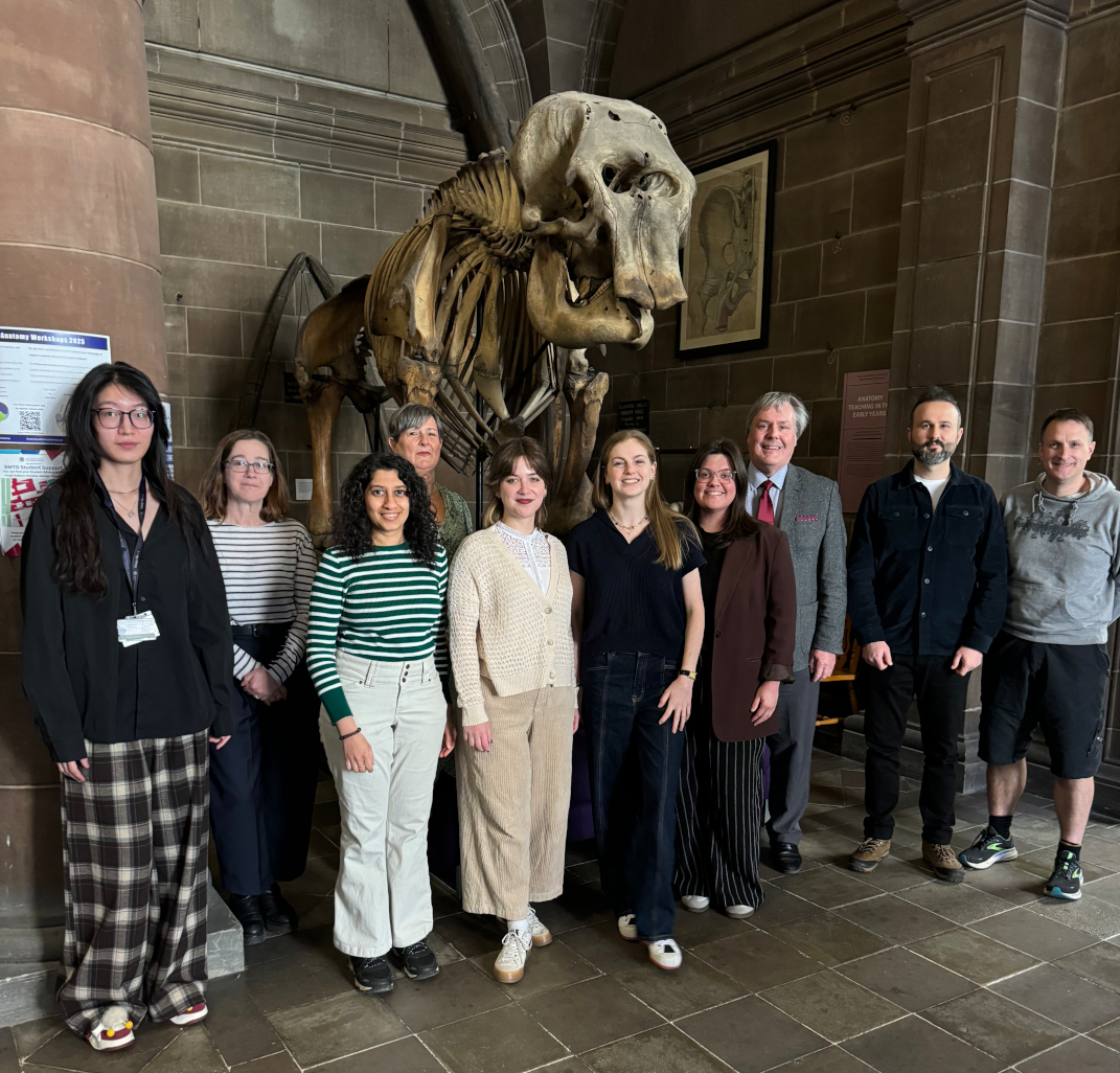
From left to right: Wentong, Jane, Sneha, Elena, Natalia, Louise, Rhiannon, Jamie, Hüseyin, Simon.
Roll call (in order of length of time in the lab):
The lab gremlin, in the lab from time immemorial; spends its time corroding electrical connections, moving critical antibodies to the wrong fridge, and emptying CO2 cylinders overnight. Like magnetism, it is invisible but we know it to be present by its observable effects.
Jamie Davies, Professor and anyone’s-technician-in-a-crisis (the jobs go together); now grey-haired enough to have made most of the mistakes people make in research, so is able to help young scientists avoid them and instead discover new and exciting mistakes of their own.
Jane Armstrong, Senior Curator, who joined as a wet-lab post-doc over 25 years ago and later discovered the joy of informatics.
Simon Harding, Senior Developer, a molecular biology post-doc turned software wrangler, with the group for over a decade now.
Elena Faccenda, Curator, a pharmacology post-doc turned informatician, also in the group for more than a decade. As well as managing the database, Elena, Simon and Jane write high-profile papers together, and have each acquired h-indices higher than many full professors.
Rhiannon Beadman, a post-doc working on homeorhesis and error-correction in organ development, uses a mix of classic 1920s-style cut-and-paste embryology and up-to-date molecular techniques.
Hüseyin Gül, a PhD student of this lab turned post-doc; works on a new drug target he identified for polycystic kidney disease.
Natalia Penar, a PhD student; combines optogenetics and morphogenetic control to test hypotheses about symmetry-breaking in branching morphogenesis.
Sneha Ravi, a PhD student; uses our new patented biofabrication technique to engineer ureters and similar tubular tissues.
Louise Goossens, a PhD student who will use Sneha’s ureters to model bacteria-tissue interactions, and how they affect antibiotic resistance, in urinary tract infections.
Nick Younger, a post-doc just joining us to work on optogenetics and axioloids.
Wentong Fu, an MSc project student working on dynamics of pattern coarsening following wounding of a synthetic patterning system.
Favourite technique
Jamie: Reading developmental literature from a century or more ago; researchers then did not have our fancy tools, but they had observation, insight and imagination and their work is an unfailing source of inspiration.
Outside your own work, what are you most excited about in developmental or stem cell biology?
Jamie: Evo-devo generally. I have never worked in this area, but seeing links develop between understanding of developmental opportunities and constraints, and the areas of morphospace visited and avoided over phylogenetic evolution, is amazing and is a ‘new’ new synthesis.
How do you manage your group and your various tasks?
Jamie: I ‘manage’ with as light a touch as possible. If you give bright young people the right encouragement, they will ask interesting questions and do interesting things, many of which I would never have imagined. In recruitment, I value diversity, not just in terms of ethnicity, gender and religion, but also in terms of academic culture; my lab has included people with first degrees in philosophy, anthropology, mathematics, and engineering as well as in biological subjects, and has benefited from the different ways of thinking they bring. As for managing my own tasks, the golden rule is not to procrastinate, and to reserve time just to think; I mean actually reserve it in a calendar, with the same non-negotiability as one marks out a timetabled lecture or conference. Thinking is part of a scientist’s job, and it is entirely reasonable to demand time in which it can be done.
What’s the best thing about where you work?
Answers given by members of the lab in general include: “I like how everyone in the building helps each other…. I love my daily commute to work through the Meadows.” “It’s a space where complex problems turn into exciting challenges that motivate me to learn, experiment and grow. It’s a mix of curiosity, teamwork, and the occasional lab triumph—it’s never dull!” “The people! As a biocurator and office-based scientist, it is great to still be part of an academic research laboratory, especially one that is so supportive. Being based on a campus right in the heart of Edinburgh is pretty special as well.” “The lab is friendly and supportive — and seems to benefit from lots of different experiences.” “I enjoy the intellectual stimulation that researching for the Guide to Pharmacology brings. A huge bonus is working with competent and friendly colleagues who help you through the days when things aren’t quite going to plan.” “The best part about working here is the friendly and supportive lab environment, where you can always count on others and never feel like you are asking too much.” “The best thing is friendly atmosphere and cooperation in our lab. It makes dealing with setbacks easier, celebrating successes even more pleasant, and keeps us motivated.” “The people—everyone is approachable, motivated, and genuinely passionate about their work”.
What’s there to do outside the lab?
Answers given by members of the lab in general include: “Dancing! Edinburgh has a vibrant community for various dancing styles (my pick is Latin dances). It’s amazing for meeting new friends and staying healthy.” “I like to go for walks in the Pentland Hills followed by a well-deserved beer in one of the many pubs.” “The Meadows is a great spot to unwind and soak in some greenery or a hike up Arthur’s seat offers stunning views of the city. Plus, the area is filled with great eateries, making it easy to grab a bite between experiments” “The Scottish Highlands and Islands are all within a half-day of travel. Fantastic if you enjoy epic scenery and being outdoors.” “Being based in the centre of Edinburgh is great – there’s usually lots going on, especially during the festival. Lots of nice green spaces close by is another positive.” “There is no shortage of historical, cultural, sporting, entertainment and foody places and events to explore in the heart of Scotland’s capital city. What makes it really special is that beaches, hills and the great outdoors are within very easy reach, whether you’re a thrill seeker or just want to chill.” “I love exploring Scotland, with its endless hikes and breathtaking scenery, though it is most enjoyable on the rare occasion when the weather is not cold and wet!” “The best place to go if you find yourself outside the lab is back inside the lab again!”

 (No Ratings Yet)
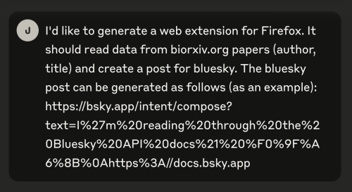

 (3 votes)