Glaucoma is a devastating retinal degenerative disease without effective treatments, and its causes remain elusive. Our laboratory uses mice as a model to study glaucoma pathogenesis at the molecular level. A NIH-funded postdoctoral position is available immediately to investigate glaucoma pathogenesis in mice. In addition, our laboratory also studies the molecular mechanisms underlying adult stem cell self-renewal and differentiation. The candidate also has a unique opportunity to investigate the potential of using stem cells to treat retinal degenerative diseases. Our research has been published in high-profile journals, including Science, Nature, Cell Stem Cell and PNAS. Preferably, the candidate has a Ph.D. degree in molecular biology, developmental biology and/or genetics (research experience with mouse research and vision research is a plus). We offer highly competitive salary plus medical benefits. Our lab is accessible to state-of-art expert-run facilities, including high throughput sequencing, microarrays, proteomics, histology, FACS and imaging. For more information about the research in Dr. Xie’s laboratory, please visit the website: http://www.stowers.org/faculty/xie-lab. If interested and any questions, please send an email to Dr. Ting Xie attgx@stowers.org.
Zika infection in humans is associated with birth defects including microcephaly. Zika has two major lineages – the Asian lineage, which has been associated with birth defects, and the African lineage, which has not – but the relative effects of each strain on brain development, and the effects of the related dengue virus that co-circulates with Zika, are not understood. This week we feature a paper published in the latest issue of Development that uses a mouse model to compare the individual effects of these viruses. Co-first authors, Qiang Shao and Stephanie Herrlinger, and PI Jian-Fu (Jeff) Chen told us more.
Qiang Shao, Stephanie Herrlinger and Jeff Chen
Jeff, can you give us your scientific biography and the main questions your lab is trying to answer?
JFC My lab is interested in mechanisms underlying microcephaly and neurodegeneration. We use mouse and human pluripotent stem cell-derived neural cells to model neurological disorders followed by mechanistic studies at molecular, cellular, and circuit levels.
Qiang and Stephanie, how did you come to join the Chen lab?
SH I have always been very passionate about studying the brain and I was looking for a mentor who was as enthusiastic as I was. Jeff’s previous work studying brain development and neural tube defects gave me the opportunity to explore brain development and embryology, and his enthusiasm for understanding biology is infectious. After the first week of my rotation with his lab, it was obvious that I was going to do my PhD with Jeff.
QS I did my PhD in China, focusing on the discovery of antiviral genes and drugs. I am always passionate about brain development and neuro-infectious disease. Before my graduation, one of my committee members, Dr. He, recommended me to Jeff for a postdoc position, knowing that he is an expert in neurodevelopmental disorders. After a few rounds of interviews, Jeff offered me this position. And here I am studying Zika virus and microcephaly.
What was known about the differences between the two lineages, both in the lab and in the human population, before your work?
JFC We knew that Asia Zika virus (ZIKV) is sufficient to cause microcephaly and additional brain abnormalities from both animal models and the human population. On the other hand, Africa ZIKV had not yet been associated with any birth or brain defects. Before our work, we also knew there was about a 110 amino acid difference between the Africa ZIKV isolate and Asia isolate in our lab, but we had no idea about their comparative toxicity and their impact to the developing brain.
Can you give is the key results of the paper in a paragraph?
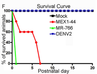
JFC The key findings are that Africa Zika virus (ZIKV) grows faster, causes more cell death in neural progenitor cells (NPCs) and neurons, and leads to more severe brain damage and postnatal death than Asia ZIKV. Both viruses similarly infect NPCs and trigger microglial activation and astrogliosis. Meanwhile, Dengue virus also infects NPCs and grows robustly in the developing brain, but fails to cause brain damage and postnatal death. Therefore, Africa ZIKV and Asia ZIKV have some intrinsic differences that account for their differential impacts on the developing brain.
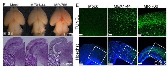
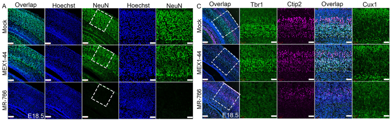
Africa Zika causes a smaller cortex and increased progenitor and neuronal death than Asian Zika. From Figures 4 and 7, Shao et al, 2017.
Why did you test this hypothesis in a mouse model as opposed to using human cerebral organoids?
JFCWe previously have established the first postnatal microcephaly mouse model associated with ZIKV infection (Shao et al., Development, 2016). It is handy for us to use this system. It is absolutely important to further test this hypothesis using human cerebral organoids, which have the advantage of modelling early human brain development.
SH Cerebral organoids are a great system to study NPCs and NPC behaviours at early stages; however, the developing brain includes a diverse set of neural progenitor, immune, vascular, and glial cell types. With our mouse model, we are able to specifically ask how the heterogeneous cell populations of the brain responds and contributes to ZIKV infection pathology and disease progression.
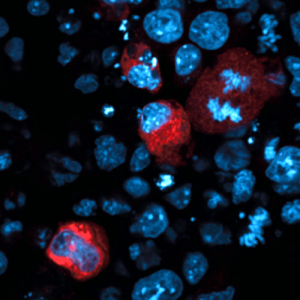
Africa Zika causes more neuronal loss than Asia Zika. From Figure 5, Shao et al, 2017.
You mention cases of co-infection between Zika and dengue, do you have plans to model this in mice and what effect do you hypothesise it will have on neural development?
JFC We would like to model it and we expect that co-infection between Zika and dengue will yield an intermediate brain defect.
What do you hypothesise the genetic/molecular basis might be for the different in virulence between the strains?
JFC We expect that certain mutations in one or more structural/non-structural viral proteins accounts for the difference. We are actively pursuing this direction now.
When doing the research, did you have any particular result or eureka moment that has stuck with you?
SH When we first saw the much more dramatic pathology of the African ZIKV strain on the developing brain, we were very surprised! The fact that this virus was not previously associated with birth defects was a big part of why we hypothesized that the Asian-ZIKV would be more deleterious than the African-ZIKV strain.
QS It was how the ZIKV African strain caused embryonic lethality that struck me the most. Before this study, there was no evidence suggesting an association between ZIKV African strain and microcephaly. I was expecting to see a mild phenotype in ZIKV African strain infected mouse embryos compared to Asian strain. It turned out that none of the ZIKV African strain infected embryo survived to postnatal stage. I was shocked by the phenotype of embryonic lethality. After this project, I realised unexpected results often lead to interesting and important discovery.
Postnatal survival of mice infected with the different viruses. From Figure 4, Shao et al, 2017.
And what about the flipside: any moments of frustration or despair?
SH It was frustrating at first to find that the African-ZIKV infected embryos did not survive gestation as the Asian-ZIKV infected embryos did. We were initially concerned that it was due to some complication of the surgeries as they can be arduous and the embryos are sensitive.
QS ZIKV African and Asian strain side by side comparison surgery is the most difficult part of this study. Statistical analysis requires three biological replicates. So I had to inject three embryos with mock, ZIKV-African, and ZIKV-Asian, respectively, which means nine embryos in one litter. This presents with many technical and experimental challenges including the potential of early embryo loss. It took me a whole month to optimize surgical conditions such as polishing glass needles, reducing viral titer and adjusting injection sites. Fortunately, through a long process of trial and error, I was able to obtain enough samples to work with.
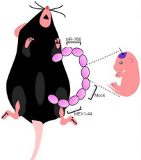
Infection of embryonic mice. From Figure 4, Shao et al, 2017.
What are your career plans following this work?
SH I successfully received a predoctoral to postdoctoral transition award from the NIH (D-SPAN F99/K00) to study the impact that ZIKV has on post-transcriptional regulation in infected neural progenitor cells. In the coming year I plan to graduate and move on to my postdoctoral career.
QS I am currently writing a research proposal to further study the mechanism underlying ZIKV caused neurovascular defect. Specifically, I am interested in understanding the role of individual ZIKV protein in neurovascular development disruption. This work will provide important molecular mechanism of ZIKV pathogenesis.
And what next for the Chen lab?
JFC We are trying to figure out the molecular mechanisms underlying Zika virus-induced microcephaly and associated neurological disorders.
Finally, what do you three like to do when you are not in the lab?
JFC I always like to spend more time with my family.
SHI like to spend time going to see live music, my family, hiking, and travelling.
QS I enjoy spending time with my partner and friends. I love cooking at home. It’s kind of like doing experiments in the kitchen with different ingredients. I find those moments very relaxing and full of delight.
The Mokalled lab in the Department of Developmental Biology at Washington University School of Medicine is hiring at all levels (http://www.mokalledlab.com/). Our lab uses zebrafish and mouse model systems to study neural regeneration after spinal cord injury or disease. Candidates with enthusiasm for neuroscience, regenerative biology, and zebrafish research are encouraged to forward a cover letter, CV, and list of 3 or more references to mmokalled@wustl.edu.
We are recruiting 1-2 PhD students (to begin Fall 2018) and a postdoctoral fellow (position immediately available) to study cell shape changes and rearrangement and underlying cytoskeletal regulation during organ formation in Dr. SeYeon Chung’s newly established lab at Louisiana State University.
We use the genetically tractable Drosophila salivary gland as a model system to understand how a flat sheet of epithelial cells becomes a three-dimensional tubular structure during development. The project will employ a combination of fly genetics, advanced imaging and image analysis, and molecular and biochemical approaches.
Related publication: Chung, S., et al., (2017) Uncoupling apical constriction from tissue invagination. eLife 6:e22235.
Grad students: Highly motivated candidates with a strong undergraduate degree in any area related to the biological sciences are encouraged to apply.
Postdocs: Highly motivated candidates who recently obtained or are about to obtain a PhD in a field of cell biology, developmental biology or biochemistry are encouraged to apply.
If interested, please send your CV, a brief description of your research interests, and contact information of three references to Dr. SeYeon Chung (seyeonchung@lsu.edu).
Here are the highlights form the current issue of Development:
Cross-border control of stem cell behaviour
Cell identity and proliferation differs between organs, raising the question of how cells at interorgan boundaries are regulated to maintain organ integrity. On p. 4091, Don Fox and colleagues identify a specialised transition zone at the midgut/hindgut boundary in the Drosophila intestine. This ‘hybrid zone’, which shows gene expression profiles from both organs, changes in size during development but is maintained into adult life, and cells within it contribute to both midgut and hindgut tissue. The authors describe a new population of stem cells – the organ-boundary intestinal stem cells (OB-ISCs) – that reside in the midgut immediately adjacent to the hybrid zone and show slower division rates. Injury to the hybrid zone increases proliferation of these OB-ISCs, and if the injury is severe enough, hyperplastic OB-ISCs can cross the boundary and invade the hindgut. The authors find that OB-ISC proliferation is induced by release of the JAK-STAT ligand Unpaired-3 from the hindgut and the hybrid zone following injury. The hybrid zone therefore serves as a focal point of interorgan interaction to influence the behaviour of OB-ISCs and preserve the midgut/hindgut distinction, raising the possibility that interorgan regulation of stem cell behaviour may be a common mechanism to maintain organ identity.
Migrating interneurons get active
During the development of the vertebrate central nervous system, interneuron precursors migrate extensively before they reach their final destination in the brain, where they then differentiate and become integrated into functional circuits. It is known that developing interneurons are sensitive to neurotransmitters but now, on p. 4125, Ronald Jabs and co-workers show that interneuron precursors require synaptic input for correct migration. Focussing on a population of cerebellar molecular layer interneurons in mice, the authors use time-lapse imaging and patch-clamp recordings to determine the morphological and electrophysiological characteristics of migrating interneuron precursors. Their analyses reveal that interneuron precursors exhibit spontaneous postsynaptic currents and receive both glutamatergic and GABAergic input as they migrate. Ultrastructural studies further demonstrate the presence of synaptic structures on these cells, and the authors also show that the density of synaptic elements increases along the migratory route of these interneuron precursors. Finally, the researchers report that blocking synaptic transmission, using tetanus toxin and Co2+ to abrogate presynaptic release, perturbs migration; both the speed and directionality of migration are reduced. Together, these findings reveal that the direct synaptic innervation of migrating interneuron precursors regulates their migratory behaviour and highlights an important and unprecedented role for synapses during neuronal pathfinding.
Zika: strain-specific impacts on brain development
Infection with Zika virus during pregnancy can lead to severe birth defects in humans, including microcephaly. Zika has two major lineages – the Asian lineage, which has been associated with birth defects, and the African lineage, which has not – but the relative effects of each strain on brain development, and the effects of the related dengue virus that co-circulates with Zika, have not been addressed. On p. 4114, Jian-Fu Chen and colleagues address this problem by performing intracerebral inoculation with Zika and dengue virus on embryonic mouse brains and comparing their effects on neural development. They show that both dengue and Zika viruses cause microcephaly through impaired neural progenitor proliferation and increased neuronal apoptosis, though the effect is much greater for Zika than dengue. Surprisingly, given the apparent absence of virus-related pathology in affected human populations, the African strain grows faster and causes greater progenitor and neuronal cell death, and higher postnatal mortality, than the Asian lineage. This study generates insights into the neurodevelopmental phenotypes generated by these viruses, and provides a foundation for future investigations into the molecular and genetic causes of Zika pathogenesis.
PLUS:
Fibroblast growth factors: key players in regeneration and tissue repair
Both regeneration and repair are orchestrated by a highly coordinated interplay of different growth factors and cytokines. Among the key players are the fibroblast growth factors (FGFs), which control the migration, proliferation, differentiation and survival of different cell types. In addition, FGFs influence the expression of other factors involved in the regenerative response. In their Review, Sabine Werner and colleagues summarize current knowledge on the roles of endogenous FGFs in regeneration and repair in different organisms and in different tissues and organs.
The evolution of cortical development: the synapsid-diapsid divergence
During evolution, the cortex appeared in stem amniotes and evolved divergently in two main branches of the phylogenetic tree: the synapsids (which led to present day mammals) and the diapsids (reptiles and birds). Comparative studies in organisms that belong to those two branches have identified some common principles of cortical development and organization that are possibly inherited from stem amniotes and regulated by similar molecular mechanisms. These comparisons have also highlighted certain essential features of mammalian cortices that are absent or different in diapsids and that probably evolved after the synapsid-diapsid divergence. In his Review, Andre Goffinet discusses this data and provides an evolutionary perspective on cortical neurogenesis, neuronal migration and cortical layer formation and folding.
Developing a sense of touch
The sensation of touch is mediated by mechanosensory neurons that are embedded in skin and relay signals from the periphery to the central nervous system. During embryogenesis, axons elongate from these neurons to make contact with the developing skin. Concurrently, the epithelium of skin transforms from a homogeneous tissue into a heterogeneous organ that is made up of distinct layers and microdomains. Throughout this process, each neuronal terminal must form connections with an appropriate skin region to serve its function. In their Review, Blair Jenkins and Ellen Lumpkin present current knowledge of the development of the sensory microdomains in mammalian skin and the mechanosensory neurons that innervate them.
Development is pleased to welcome submissions for an upcoming special issue on human development. This issue will focus on advances in our understanding of how human organs and tissues are formed, and how the processes and mechanisms involved compare to those in other species.
This special issue will be published in early autumn 2018, to coincide with our upcoming meeting ‘From stem cells to human development‘, and continues Development’s efforts to support and promote the growing community of researchers interested in the development of our own species. Until recently, our understanding of human embryogenesis has been hampered by the inaccessibility of the system. Recent advances in the stem cell field – the generation of human pluripotent stem cells and the development of organoid cultures systems – now allow us to investigate developing human tissues in vitro, complementing analyses of primary human cells and tissues. Together, these resources and technologies are enabling us to gain significant insights into human cell fate specification and tissue organisation, and informing our efforts to treat developmental disorders and develop regenerative therapies.
As the leading journal focussed on advances in developmental biology and stem cells, Development is the natural home for papers on human development. We therefore invite you to submit your breakthrough research for consideration for this special issue. The issue will be widely marketed and distributed at relevant conferences worldwide, providing prominent exposure for your work. We encourage submissions of Research Articles and Reports, and Techniques & Resources papers, that use in vitro stem cell and organoid systems as well as human tissue samples.
Articles should be submitted by 1st March 2018 for consideration for the special issue
We also welcome proposals for Review Articles for this special issue. Please send us a short synopsis detailing the scope and structure of the proposed article, and including key references. The deadline for submission of proposals is 15th January 2018, and articles should be submitted by 1st March 2018.
Please refer to our author guidelines for information on preparing your manuscript for Development, and submit via our online submission system. Please highlight that your submission is to be considered for the special issue in your cover letter. For any queries about the special issue, or for any presubmission enquiries, please get in touch by email.
This will be our second special issue on human development; we invite you to browse our earlier issue, published in September 2015.
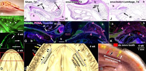
A position at the Postdoctoral level is available December 1, 2017 to investigate the evolution and development of teeth in reptiles. Our lab has been studying the processes of tooth development and tooth replacement in snake and lizard embryos (see review Richman and Handrigan, Genesis, 2011; Handrigan et al., 2010, Dev Biol, vol. 348, 130-141). In addition we have identified populations of putative stem cells in the dental epithelium using the gecko model (Handrigan et al., Development, 37: 137 3545-3549, 2010). This NIH-funded project will focus on the signalling pathways that control tooth replacement as well as the contribution of putative stem cells to this process. Approaches used will include primarily in vivo manipulations on adult geckos followed by single-cell RNAseq on the responding dental epithelium and mesenchyme. Applicants should have recently completed a PhD (in the last 2 years or less) and have related research experience in bioinformatics, RNAseq, developmental biology and/or evolution. Salary support is available from research grants but applicants will be encouraged to apply for independent support. Please email a CV, statement of research interests and contact information for at least three referees to:
The composition of extracellular microenvironment is dynamically regulated in time and space during embryonic development, and our lab discovered that cell-type specific expression of the extracellular matrix protein fibronectin is essential for mammalian embryogenesis. Furthermore, we found that fibronectin regulates distinct morphogenetic processes in cell type-specific manner, and functions both in cell-autonomous and non cell-autonomous manner. We are searching for a motivated postdoctoral researcher to uncover differences in the mechanisms by which cell-autonomous and non cell-autonomous fibronectin signals to cells and regulates cell fate decisions. The successful applicant will apply state-of-the art confocal and super-resolution microscopy techniques, utilize mouse genetics, CRISPR, and global profiling of gene expression and signaling pathways to uncover the mechanisms, by which extracellular microenvironment guides morphogenetic programs. Our lab is located in the heart of Philadelphia, USA. For further information about our lab and publications, please visit our lab’s website: http://www.jefferson.edu/university/research/researcher/researcher-faculty/astrof-laboratory.html
To apply, please send a letter of interest detailing your expertise, CV and names and contact information of three references to sophie.astrof@gmail.com
As an employer, Jefferson maintains a commitment to provide equal access to employment. Jefferson values diversity and encourages applications from women, members of minority groups, LGBTQ individuals, disabled individuals, and veterans.
Addgene is a global, nonprofit repository that was created to help scientists share plasmids. Before we go over the developmental biology resources available at Addgene, here’s a little background on our organization. Our mission is to accelerate research and discovery by improving access to useful research materials and information. Labs deposit plasmids with Addgene at no cost, and we handle storage, distribution, and record keeping. Researchers can request plasmids and, in some cases, ready-made lentivirus and AAV, from our collection, and we coordinate Material Transfer Agreements to facilitate easy sharing.
We also provide numerous educational resources on our blog and website. Our Plasmids 101 and CRISPR 101 blog series were designed to help scientists of all levels learn more about molecular biology, cloning, genome engineering, and other popular technologies. To make it easy to find plasmids for a given purpose, we provide curated pages on our website for various plasmid collections like CRISPR, stem cells, cancer, and fluorescent proteins. Addgene’s technical support team also provides scientists with real-time plasmid troubleshooting. By providing these services, Addgene’s goal is to create a lasting resource for research and discovery around the world.
Developmental biologists have deposited many of Addgene’s most popular plasmids. I’ll cover the general collections most relevant to developmental biology and also highlight a few standouts from our collection.
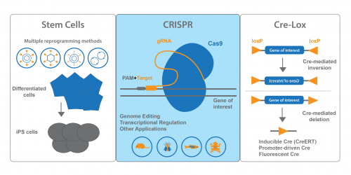
Stem Cell Tools
Our stem cell collection is a true Addgene strength. We have many plasmids that cover methods for iPS cell generation – retroviral, lentiviral, adenoviral, episomal and non-viral methods. We also distribute plasmids for direct differentiation and transdifferentiation. These plasmids are conveniently organized on our Stem Cell Collection page. Keep an eye out for the blue flame plasmids – these each have been requested over 100 times and have proven useful for many researchers!
Lentiviral and retroviral reprogramming methods carry the caveat of insertional mutagenesis, while plasmid and adenoviral reprogramming methods suffer from lower efficiency. Woltjen et al. created a Tet-inducible reprogramming system using the Piggybac transposase. This system allows you to easily reprogram fibroblasts into iPS cells using only plasmids – there’s no need to prepare or deliver virus to your cells. Instead, you co-express PB-TET-MKOS containing the four Yamanaka factors, PB-CA-rtTA Adv containing the rtTA Tet transactivator, and a plasmid containing the Piggybac transposase (available from Transposagen and others.) Doxycycline treatment then turns on MKOS expression, promoting reprogramming. Once reprogrammed lines have been isolated, transient expression of Piggybac transposase will remove the MKOS transgene from the genome.
CRISPR Plasmids for Use With Developmental Models
Addgene’s CRISPR collection also has resources for scientists interested in developmental biology. On our CRISPR landing page, you can find plasmids sorted by model organism or system. In addition to our many mammalian expression plasmids, we have plasmids available for common developmental models like Drosophila, C. elegans, Xenopus and zebrafish. Addgene also maintains a list of Pre-designed gRNAs that have been used in previous publications, and you can easily search the table for your favorite gene. We also encourage you to deposit your own gRNA plasmids.
Zebrafish have proven very amenable to CRISPR genome engineering, and the Zon lab Gateway constructs can help you to knock out your gene of interest in a tissue-specific manner. Their Tol2 based system enables you to examine mosaic disruption of your gene in F0 embryos or to generate stable tissue-specific knockout lines. All four plasmids from this publication have earned blue flames for 100+ requests! To use this system, you’ll combine Gateway destination vector pDestTol2pA2-U6:gRNA or pDestTol2CG2-U6:gRNA with a middle entry vector containing Cas9 (e.g. pME-Cas9). You’ll also need a 5’ entry vector containing your tissue-specific promoter and a 3’ entry vector with a polyA sequence, both of which can be obtained from the Tol2kit. A Gateway reaction will bring the pieces together, allowing you to examine the effects of gene knockout in a specific tissue. A detailed protocol is available from the Zon lab on the plasmid pages of each Addgene plasmid.
Cre-Lox Systems for Spatio-Temporal Control of Gene Expression
If you’re using Cre-lox recombination to turn gene expression ON or OFF, chances are Addgene has plasmids you can use. In addition to standard Cre recombinase, we also provide inducible, promoter-regulated, and optimized Cres, to name a few. Cre-dependent vectors expressing fluorescent proteins or luciferase are also available. FLEx (FLip-Excision) vectors, which conditionally turn off one gene and activate another, are available from many Addgene depositors. PhiC31 recombinase and other non-Cre recombinases are available for use in Drosophila.
This plasmid expresses CreERT, or tamoxifen-inducible Cre, in a mammalian expression system. In cell lines transfected with this plasmid, tamoxifen will activate Cre, allowing removal or inversion of a floxed gene of interest. This plasmid has been requested over 1100 times, showing its versatility and applicability to many kinds of studies.
Lineage tracing plasmids make up another exciting mini-collection at Addgene. Brainbow is a combinatorial Cre-lox based system that colors single cells with as many as 90-160 different colors. Many of Addgene’s Brainbow plasmids have blue flames. Although originally developed for neuronal mapping, these plasmids have been adapted for lineage tracing. Multibow plasmids adapted from the Brainbow system for use in zebrafish create unique “barcodes” allowing researchers to track individual cells and their clonal progeny.
It’s impossible to cover all of the resources available at Addgene in one blog post, so we encourage you to take a look at our website. If you’re looking for plasmids from a specific publication, start by searching for the paper’s corresponding author to see if she or he has deposited with us. You can also sign up for Addgene Alerts for your favorite PIs and you’ll get emails whenever new plasmids are available from their labs. If you have trouble finding what you need, or you’d like us to reach out to a potential depositor, feel free to email us at help@addgene.org. We also encourage you to deposit your own plasmids thereby making them easily accessible to other members of the research community.
Mary Gearing is a Scientist at Addgene. She enjoys developing educational content to help scientists learn more about molecular biology. Follow her on Twitter @megearing.
piggyBac transposition reprograms fibroblasts to induced pluripotent stem cells. Woltjen K, Michael IP, Mohseni P, Desai R, Mileikovsky M, Hamalainen R, Cowling R, Wang W, Liu P, Gertsenstein M, Kaji K, Sung HK, Nagy A. Nature. 2009 Apr 9;458(7239):766-70. doi: 10.1038/nature07863. PMID: 19252478
A CRISPR/Cas9 Vector System for Tissue-Specific Gene Disruption in Zebrafish. Ablain J, Durand EM, Yang S, Zhou Y, Zon LI. Dev Cell. 2015 Mar 4. pii: S1534-5807(15)00075-1. doi: 10.1016/j.devcel.2015.01.032. PMID: 25752963
High-Efficiency FLP and PhiC31 Site-Specific Recombination in Mammalian Cells. Raymond CS, Soriano P. PLoS ONE. 2007 Jan 17;2(1):e162. PMID: 17225864
Multiple new site-specific recombinases for use in manipulating animal genomes. Nern A, Pfeiffer BD, Svoboda K, Rubin GM. Proc Natl Acad Sci USA. 2011 Aug 23;108(34):14198-203. doi: 10.1073/pnas.1111704108. PMID: 21831835
Controlled expression of transgenes introduced by in vivo electroporation. Matsuda T, Cepko CL. Proc Natl Acad Sci U S A. 2007 Jan 16;104(3):1027-32. PMID: 17209010
Improved tools for the Brainbow toolbox. Cai D, Cohen KB, Luo T, Lichtman JW, Sanes JR. Nat Methods. 2013 May 5;10(6):540-7. doi: 10.1038/nmeth.2450. PMID: 23817127
Multibow: digital spectral barcodes for cell tracing. Xiong F, Obholzer ND, Noche RR, Megason SG. PLoS One. 2015 May 26;10(5):e0127822. doi: 10.1371/journal.pone.0127822. eCollection 2015. PMID: 26010570
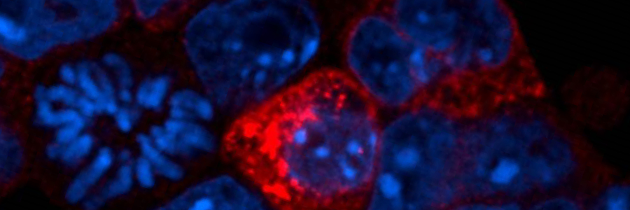
Production of proteins from transposons (red). DNA shown in blue.
Molecules called endosiRNAs help us avoid genetic chaos, according to a new study from a team at the Babraham Institute. Much of the human genome contains pieces of DNA called transposons, a form of genetic parasite. When active, transposons can damage genes so it is important to keep them inactive. At a certain point early in the human life cycle controlling transposons is particularly difficult. This latest research, published in Cell Stem Cell reveals how endosiRNAs keep our genes safe during this vulnerable stage.
Transposons, also called transposable elements, are ancient viruses that have become a permanent part of our genes. Around half of the human genome is made of transposons, many are damaged, but some can become active. Active transposons can be harmful because they move about the genome. When transposons move they can damage genes, leading to genetic illnesses and playing a part in some cancers.
Chemical markers in DNA called methylations can keep transposons inactive. Cells often use methylations to inactivate pieces of DNA, whether they are genes or transposons. Yet, in each new generation most methylations are temporarily erased and renewed by a process called epigenetic reprogramming. This means that, during sperm and egg production there is a short time when methylations do not control transposon activity, leaving them free to damage genes and shuffle DNA.
The new findings show that transposons become active when cells erase DNA methylation and they are shut down by the endosiRNA system. Just like active genes, active transposons produce messages in the form of RNA molecules, which have many similarities to DNA. The study reveals that cells can detect these transposon RNA messages and use them to create specific endogenous small interfering RNAs (endosiRNAs). The endosiRNAs then act like a trap allowing a protein called Argonaute2 (Ago2) to seek and destroy transposon messages before they cause any harm.
Speaking about the research, lead author on the paper, Dr Rebecca Berrens, said: “Epigenetic reprogramming plays a vital role in wiping the genome clean at the start of development, but it leaves our genes vulnerable. Understanding the arms race between our genes and transposon activity has been a long-running question in molecular biology. This is the first evidence that endosiRNAs moderate transposon activity during DNA demethylation. EndosiRNAs provide a first line of defence against transposons during epigenetic reprogramming.”
The effects of active transposons vary, often they have no effect, only occasionally will they alter an important gene. Yet, transposons can affect almost any gene, potentially leading to different kinds of genetic disease. Studying the control of transposons, adds to our understanding of the many ways that they can impact on human health.
This work highlights that a transposons often sit within genes and are read in the opposite direction to the surrounding gene. It is this arrangement that allows cells to identify RNA messages from transposons. RNA messages read from the same piece of DNA in opposite directions are complementary, meaning they can join to form a structure called double-stranded RNA (dsRNA), which initiates the creation of endosiRNAs.
Senior scientist on the paper, Professor Wolf Reik, Head of the Epigenetics Laboratory at the Babraham Institute, said: “Transposons make up a large part of our genome and keeping them under control is vital for survival. If left unchecked their ability to move around the genome could cause extensive genetic damage. Understanding transposons helps us to make sense of what happens when they become active and whether there is anything we can do to prevent it.”
Much of this research was carried out using embryonic stem cells grown in the lab, which had been genetically modified to lack DNA methylations. Natural epigenetic reprogramming happens in primordial germ cells, the cells that make sperm and eggs, but these are harder to study. The researchers used primordial germ cells to verify the key results from their study of stem cells.
Notes:
Publication Reference
Berrens, RV., Andrews, S., Spensberger, D., Santos, F., Dean, W., Gould, P., Sharif, J., Olova, N., Chandra, T., Koseki, H., von Meyenn, F., Reik, W.. An endosiRNA-based repression mechanism counteracts transposon activation during global DNA demethylation in embryonic stem cells. Cell Stem Cell
DOI: http://dx.doi.org/10.1016/j.stem.2017.10.004
Research Funding
This work and the researchers that contributed to it were generously supported by SNSF, Gates Cambridge Trust, BBSRC (Epigenetics Institute Strategic Programme Grant), Wellcome Trust, EU BLUEPRINT and EpiGeneSys
Image Credit
Credit: R. Berrens
Header – Image of fluorescently labelled embryonic stem cells. These cells have been modified to mimic the effects of epigenetic reprogramming. DNA in the cells is marked in blue. Red indicates cells that contain active transposons.
Embedded – Graphical abstract of the key findings from the paper. The opposing directions of a transposon and a gene result in double stranded RNA that can be used to produce endosiRNA to prevent transposon activation. Credit: Veronique Juvin
Animal Statement:
As a publicly funded research institute, the Babraham Institute is committed to engagement and transparency in all aspects of its research. Animals are only used in Babraham Institute research when their use is essential to address a specific scientific goal, which cannot be studied through other means. The main species used are laboratory strains of rodents, with limited numbers of other species. We do not house cats, dogs, horses or primates at the Babraham Research Campus for research purposes.
Samples of primordial germ cells were collected from C57Bl/6J transgenic mice at embryonic day 13.5 and 14.5. The use of animals in this study was performed in accordance with the Animal (Scientific Procedures) Act 1986, and regulated by the Babraham Institute Animal Welfare and Ethical Review Body (AWERB). Experiments were planned and designed in accordance with the 3Rs.
About the Babraham Institute:
The Babraham Institute receives strategic funding from the Biotechnology and Biological Sciences Research Council (BBSRC) through an Institute Core Capability Grant to undertake world-class life sciences research. Its goal is to generate new knowledge of biological mechanisms underpinning ageing, development and the maintenance of health. Research focuses on signalling, gene regulation and the impact of epigenetic regulation at different stages of life. By determining how the body reacts to dietary and environmental stimuli and manages microbial and viral interactions, we aim to improve wellbeing and support healthier ageing.
 (No Ratings Yet)
 (1 votes)
 Cell identity and proliferation differs between organs, raising the question of how cells at interorgan boundaries are regulated to maintain organ integrity. On p.
  During the development of the vertebrate central nervous system, interneuron precursors migrate extensively before they reach their final destination in the brain, where they then differentiate and become integrated into functional circuits. It is known that developing interneurons are sensitive to neurotransmitters but now, on p.
  Infection with Zika virus during pregnancy can lead to severe birth defects in humans, including microcephaly. Zika has two major lineages – the Asian lineage, which has been associated with birth defects, and the African lineage, which has not – but the relative effects of each strain on brain development, and the effects of the related dengue virus that co-circulates with Zika, have not been addressed. On p.
  Both regeneration and repair are orchestrated by a highly coordinated interplay of different growth factors and cytokines. Among the key players are the fibroblast growth factors (FGFs), which control the migration, proliferation, differentiation and survival of different cell types. In addition, FGFs influence the expression of other factors involved in the regenerative response. In their
  During evolution, the cortex appeared in stem amniotes and evolved divergently in two main branches of the phylogenetic tree: the synapsids (which led to present day mammals) and the diapsids (reptiles and birds). Comparative studies in organisms that belong to those two branches have identified some common principles of cortical development and organization that are possibly inherited from stem amniotes and regulated by similar molecular mechanisms. These comparisons have also highlighted certain essential features of mammalian cortices that are absent or different in diapsids and that probably evolved after the synapsid-diapsid divergence. In his
  The sensation of touch is mediated by mechanosensory neurons that are embedded in skin and relay signals from the periphery to the central nervous system. During embryogenesis, axons elongate from these neurons to make contact with the developing skin. Concurrently, the epithelium of skin transforms from a homogeneous tissue into a heterogeneous organ that is made up of distinct layers and microdomains. Throughout this process, each neuronal terminal must form connections with an appropriate skin region to serve its function. In their
 Plasmid spotlight: