This interview was first published in Development.
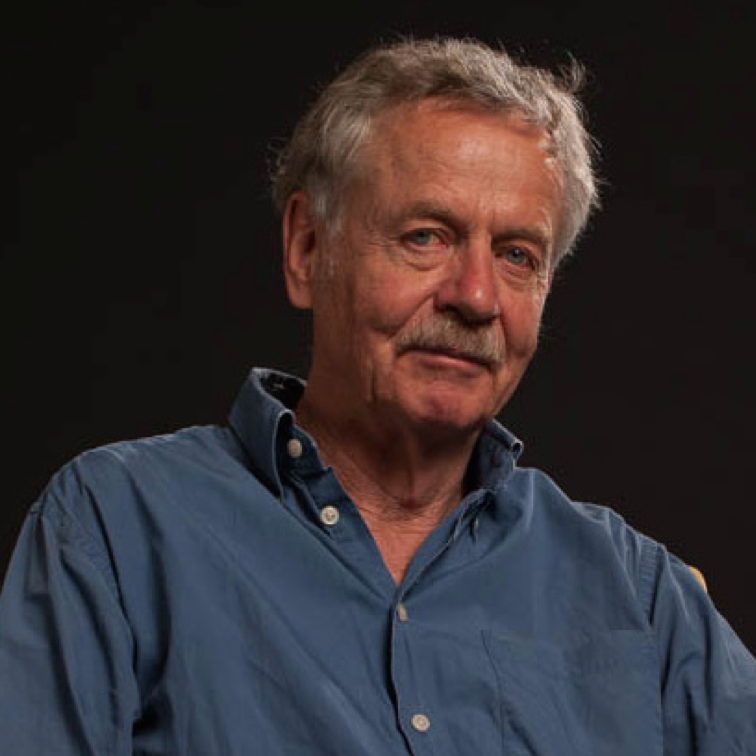
Rudolf Jaenisch is a Professor of Biology at Massachusetts Institute of Technology, a founding member of the Whitehead Institute for Biomedical Research and the current president of the International Society for Stem Cell Research (ISSCR). His contributions to the stem cell field span from making the first transgenic mouse to seminal advances in the reprogramming field, and much more. In recognition of his pioneering research leading to induced pluripotency, he recently received the 2015 March of Dimes Prize in Developmental Biology. At the recent Keystone Meeting on ‘Transcriptional and Epigenetic Influences on Stem Cell States’ in Colorado, we had the opportunity to talk to him about his life and work.
 You initially studied medicine and then transitioned to basic research. What motivated or inspired you to make the change?
My parents and grandparents were medical doctors so I was interested in studying medicine, although my father suggested I shouldn’t. I actually liked going to medical school, especially the pre-clinical work: physiology, chemistry, anatomy and pathology. Then, in the 1960s, at the beginnings of molecular biology, I did my thesis on phages in Peter-Hans Hofschneider’s lab at the Max Planck Institute in Munich. I was totally fascinated by experimental biology: doing your own experiments, asking questions and solving them. So I decided that medicine was probably not the right thing for me to do. I really wanted to go into science.
Is there someone who has been a particular influence on or inspiration for your career?
Beatrice Mintz had a major influence on my scientific carrier. She really is an amazing developmental geneticist. With her, I learned how to look at mice, how to use mouse coat colour genetics to ask questions and how to inject DNA into mouse embryos.
Arnie Levinewas a fantastic mentor and I have the highest respect for him. When I started in his lab in Princeton, I was his first postdoc and three weeks after I started there he told me: “By the way, I am going on sabbatical to Europe, you run the lab”. That was before there was email, and calling was expensive and not a good option! I was a bit shocked. But I managed it and the time I spent there was really key.
Paul Berg was also incredibly generous. When he and Peter Rigby had just invented nick translation, a technique to make hot DNA probes, they allowed me to use this unpublished technique for a paper with Beatrice (Jaenisch and Mintz, 1974) and he wasn’t even an author! I think that this would be unlikely to happen today.
The time I spent at the Salk Institute with young colleagues like Tony Hunter, who showed me how to make labelled triphosphates for the nick translation, was also crucial. There, Inder Verma and Hung Fan introduced me to the Moloney murine leukaemia virus (MMLV) system, which I later used to generate transgenic mice.
David Baltimore made a very significant contribution to my career too. My first contact with him was when he visited the Salk Institute. He communicated our first articles describing the generation of transgenic mice to PNAS, and when he established the Whitehead Institute he offered me a position; I was really excited because I think he’s one of the most impressive scientists I know.
You started your scientific career by working on SV40 during your postdoc, trying to understand how virus tropism works. How did that lead to your lab being one of the first to report the reprogramming of a somatic cell to a pluripotent state?
In Arnie’s lab we conducted some really interesting experiments on the replication of SV40, which was the model system to learn about eukaryotic DNA replication. SV40 is a tumour virus and that got me thinking: why does it only induce sarcomas and not other types of tumour when you inject it into a mouse? And then I read a paper on striped mice by Beatrice Mintz (Mintz, 1967), which was really one of the most influential papers I have read in my entire career. I was fascinated by the fact that she was able to culture mouse embryos in a dish and derive adult mice. I thought that if I could inject SV40 DNA into these early embryos, all the tissues would carry the viral DNA and I could answer my question about virus tropism. I told Beatrice my idea, but nobody had injected DNA into embryos before so she was very sceptical. Eventually, she agreed to let me do the experiment in her lab. I injected the SV40 DNA into mouse embryos and got mice. I’d hoped there would be tumours growing all over, but the resulting mice were totally normal because, as I would learn much later, the viral DNA gets silenced in early embryos and in embryonic stem cells (ESCs). This looked like a dead-end project. Only after getting my first job at the Salk Institute did I choose to use MMLV, which is highly expressed in mouse, to revisit this problem. I exposed mouse embryos to MMLV and was able to show that it got inserted into the genome and into the germline and was transmitted to the next generation (Jaenisch, 1976). The integration of a virus into the mouse genome can cause mutations in genes by ‘insertional mutagenesis’ and, if the mice are homozygous for the insertion, this can result in lethality. Using insertional mutagenesis we found that in one transgenic strain the virus had mutated the collagen 1 gene, which was not a very exciting developmental gene at first but later taught us a lot about how a virus silences a gene – how it inserts and spreads DNA methylation (Schnieke et al., 1983). We then realised that if a virus integrates when it infects an embryo or ESC, it gets silenced. However, if it infects a fibroblast it is highly expressed. This process was clearly developmentally regulated (Jähner et al., 1982).
Then, when I was at the Whitehead Institute, homologous recombination became possible in ESCs and we immediately adapted this technique. I was really interested in DNMT1, the only methyltransferase known at the time. Until then, DNA methylation and gene expression had only been correlated: there was no direct evidence that methylation could silence gene expression. The generation of a Dnmt1 knockout mouse allowed us to address this question. The phenotype of the Dnmt1 mutant was very informative because the mice died at gastrulation – with their genome hypomethylated, so methylation was required for normal development (Li et al., 1992). We then used this mutation to analyse the role of DNA methylation in imprinting, X-inactivation and to define a causal role of methylation in cancer. We also worked on how viruses get methylated and silenced. So we came full circle and understood why the mice I derived from SV40 DNA-injected embryos during my postdoc did not develop tumours: the virus was epigenetically silenced in all tissues.
And then came Dolly the sheep, the first animal to be cloned by nuclear transfer. I thought that working on cloning would be the most unbiased way to study epigenetics and reprogramming. When the Yanagimachi lab published the cloned mouse Cumulina (Wakayama et al., 1998), I immediately arranged a collaboration and shifted a major part of my lab to work on this topic. I had really good students in the lab like Kevin Eggan and Konrad Hochedlinger who adapted this technology very quickly and we learned a lot about reprogramming. The key question was how to reprogram a somatic cell without the egg cytoplasm and we had similar ideas to Shinya Yamanaka, except that he beat us to the first publication (Takahashi and Yamanaka, 2006)! A year later three labs – Konrad’s, Shinya’s and mine – showed that induced pluripotent cells (iPSCs) were indistinguishable from ESCs and were able to contribute to chimaeras and to the germline (Takahashi et al., 2007; Maherali et al., 2007; Wernig et al., 2007). Many people doubted the original Yamanaka paper (though I did not!), but with three independent labs confirming the quality of iPSCs there was no
doubt that differentiated cells can be reprogrammed to a pluripotent state that was indistinguishable from ESCs. The key issue is now to understand the molecular mechanisms of reprogramming.
Which big questions still pique your curiosity? Where do you think the stem cell field is going?
One big question that intrigues me and on which I am actively working is understanding diseases by using iPSCs. Most people concentrate on monogenic diseases, such as Parkinson’s disease, where certain gene mutations have 100% penetrance. But monogenic diseases are rare and most diseases are more complex, involving multiple genes with mutations in each gene slightly increasing the risk of developing the disease. When you start using patient-derived iPSCs you soon realise that comparing different cell lines is basically like comparing apples with oranges. Cells from patients and control individuals have different genetic backgrounds and this profoundly influences the phenotype of differentiated cells in an unpredictable way. Thus, if you find something different between the control and the patient-derived iPSCs you have to worry whether the difference is due to the pathology of the disease or to the variability between cell lines. For this reason, years ago, we started generating isogenic cell lines that differed from one another exclusively by a disease-causing mutation in order to generate a molecularly defined system in which we could study genetic diseases (Soldner et al., 2011). However, an important part of medically relevant diseases is that most are sporadic. Genome-wide association studies (GWAS) identify genomic loci that are associated with an increased risk of developing a certain disease by a few percent. But what really is a GWAS hit? Most of them are in regulatory regions, such as enhancers, but the target genes of such regions are often unknown. In on-going studies we are trying to answer these questions by applying our techniques to Parkinson’s disease. Using isogenic cell lines we have significantly improved the sensitivity of GWAS and I think that we can provide one of the first mechanistic insights into the nature of a GWAS hit.
Concerning where the field is going, a potential problem when studying a complex pathology such as Parkinson’s or Alzheimer’s disease is being limited to a 2D dish. These are long-latency diseases. Can you study them in the short time that you have in a dish? We need to find a more complex model. I think 3D systems like organoids are going to transform the field. For example, Juergen Knoblich’s ‘mini-brains’ (Lancaster et al., 2013) are very promising. They are far too complex for screening, so you would screen for molecules or genes involved in a pathology using a highthroughput system like neurons in a culture dish. But once you get there, you can try to understand the pathological mechanism in the organoid system where you have cell-cell interactions in a 3D context. Another issue to resolve is how to study late-onset pathologies like Parkinson’s disease. Can you get these organoids to mature in vitro? I think there are still many unresolved issues.
Induced pluripotency has anumber of potential applications. What do you think is its realistic impact on regenerative medicine? And from a more basic perspective, how do you think it impacts our understanding of developmental processes?
iPSCs are already being used and evaluated clinically, for example for sickle cell anaemia. The next step is to find candidate genes and molecules for pathologies with this culture system. How this can be adapted to cell therapy is further away, but surely it will work at some point. It is a very promising and exciting field.
From a more basic point of view, ESCs have already opened the door to studying some aspects of human development, but iPSCderived organoids like the ‘mini-brains’ are going to be crucial to find out more about human brain development and developmental diseases of the central nervous system.
Last year you succeeded Janet Rossant as President of the ISSCR. What do you think is the role of this organisation and what are the aims of your presidency?
The ISSCR is an institution that can assess and advise to improve and maintain the quality of research; it is a trusted voice in the public debate on the therapeutic use of stem cells. There are many issues around stem cells and most of them are of public interest: thousands of rogue clinics offer unproven therapies, the possibility of human germline modification, and so on. During my time at the ISSCR, I want to make sure that we properly inform the public and clearly explain some of the complicated issues around stem cell research and its application for therapy. Hopefully, people will consult the ISSCR book on stem cell therapy (Patient Handbook on Stem Cell Therapies, Appendix I of the Guidelines for the Clinical Translation of Stem Cells, December 3, 2008, ISSCR) to learn about what you should look for if you sign up for stem cell therapy, the problems associated with these therapies and the standards you should expect. These are important issues and I think the ISSCR is the right voice to give advice on this topic. The ISSCR also has very clear guidelines designed to guide research centres in using stem cells in a clinical context. Some of the most prominent people in the stem cell field are involved in the ISSCR, so that makes it a trusted, believable and authoritative voice in the stem cell community.
Another very important role of the ISSCR that we take very seriously is the education of young scientists. That is why we have training courses as part of the annual meeting, or as extra meetings. And, of course, our annual meeting is a real success story and always has an exciting programme – organised this year by Leonard Zon, the founding president of the ISSCR. It is the most important stem cell meeting of the year, attended by almost 4000 people.
In recent years, several high-profile ethics issues have brought the stem cell field under scrutiny, including discussion in post-publication peer review forums. Do you think that post-publication discussions are useful for the scientific community?
In my opinion, post-publication peer review forums may have a role in clarifying issues. However, a problem is that people can comment and attack anonymously. Such discussions are often not constructive – you talk to a wall if you answer. If someone writes to me and questions our data without stating their identity, I will not answer. However, if someone puts a name or a journal name behind their comment then, of course, I will address the issue. Each paper both solves and raises questions. For example, there is a lot of controversy, discussion and unresolved issues around the naïve pluripotency state. All of these are genuine disagreements over divergent datasets and they need to be discussed openly; ‘below-the-belt’ attacks are not necessarily useful.
What is your advice for young researchers?
This is a difficult question – so much has changed over the years! I was never driven by what I thought my job or my career should be; I was driven because I wanted to solve an interesting question, and sometimes I took some risks. For example, the experiment I undertook to generate the first transgenic mouse was a risk. If it hadn’t worked, I would be a doctor now, somewhere (although I swore I would never practice!). That experiment was funded by the first grant I got from the NIH, even though that project was very risky. These days, such a project would probably be rejected right away.
I really think that being driven and excited by what you are doing is very important. Nowadays, people often come to your lab and when you ask them what their goal is, they say: “Oh, having a Cell paper”. I don’t think this is the right motivation. What is important is the problem to be solved rather than having a Cell paper as a goal.
What would people be surprised to find out about you?
Rick Young showed me how to fly a plane, though I’m not sure he’d admit to this. He and I have a long history, and we’ve been on many hikes and treks in the Himalayas over the years.
References
Jaenisch, R. (1976). Germ line integration and Mendelian transmission of the exogenous Moloney leukemia virus. Proc. Natl. Acad. Sci. USA 73, 1260-1264.
Jaenisch, R. and Mintz, B. (1974). Simian virus 40 DNA sequences in DNA of healthy adult mice derived from preimplantation blastocysts injected with viral DNA. Proc. Natl. Acad. Sci. USA 71, 1250-1254.
Jähner, D., Stuhlmann, H., Stewart, C. L., Harbers, K., Lö hler, J., Simon, I. and Jaenisch, R. (1982). De novo methylation and expression of retroviral genomes during mouse embryogenesis. Nature 298, 623-628.
Lancaster, M. A., Renner, M., Martin, C.-A., Wenzel, D., Bicknell, L. S., Hurles, M. E., Homfray, T., Penninger, J. M., Jackson, A. P. and Knoblich, J. A. (2013). Cerebral organoids model human brain development and microcephaly. Nature 501, 373-379.
Li, E., Bestor, T. H. and Jaenisch, R. (1992). Targeted mutation of the DNA methyltransferase gene results in embryonic lethality. Cell 69, 915-926.
Maherali, N., Sridharan, R., Xie, W., Utikal, J., Eminli, S., Arnold, K., Stadtfeld, M., Yachechko, R., Tchieu, J., Jaenisch, R. et al. (2007). Directly reprogrammed fibroblasts show global epigenetic remodeling and widespread tissue contribution. Cell Stem Cell 1, 55-70.
Mintz, B. (1967). Gene control of mammalian pigmentary differentiation. I. Clonal origin of melanocytes. Proc. Natl. Acad. Sci. USA 58, 344-351.
Schnieke, A., Harbers, K. and Jaenisch, R. (1983). Embryonic lethal mutation in mice induced by retrovirus insertion into the alpha 1(I) collagen gene. Nature 304, 315-320.
Soldner, F., Laganière, J., Cheng, A. W., Hockemeyer, D., Gao, Q., Alagappan, R., Khurana, V., Golbe, L. I., Myers, R. H., Lindquist, S. et al. (2011). Generation of isogenic pluripotent stem cells differing exclusively at two early onset Parkinson point mutations. Cell 146, 318-331.
Takahashi, K. and Yamanaka, S. (2006). Induction of pluripotent stem cells from mouse embryonic and adult fibroblast cultures by defined factors. Cell 126, 663-676.
Takahashi, K., Tanabe, K., Ohnuki, M., Narita, M., Ichisaka, T., Tomoda, K. and Yamanaka, S. (2007). Induction of pluripotent stem cells from adult human fibroblasts by defined factors. Cell 131, 861-872.
Wakayama, T., Perry, A. C. F., Zuccotti, M., Johnson, K. R. and Yanagimachi, R. (1998). Full-term development of mice from enucleated oocytes injected with cumulus cell nuclei. Nature 394, 369-374.
Wernig, M., Meissner, A., Foreman, R., Brambrink, T., Ku, M., Hochedlinger, K., Bernstein, B. E. and Jaenisch, R. (2007). In vitro reprogramming of fibroblasts into a pluripotent ES-cell-like state. Nature 448, 318-324.
 (2 votes)
 Loading...
 (No Ratings Yet)