Department/Location: Wellcome Trust – Medical Research Council Cambridge Stem Cell Institute
Salary: £28,132-£36,661
Reference: PS03564
Closing date: 22 July 2014
Limited funding: The funds for this post are available until 30 June 2017 in the first instance.
The Stem Cell Institute is a world-leading centre of excellence in stem cell biology and regenerative medicine, supported by a strategic funding partnership between the Wellcome Trust and the Medical Research Council (www.stemcells.cam.ac.uk).
The Institute comprises over 300 researchers spanning fundamental science through to clinical applications. Our vision is of a deep understanding of stem cell biology for the prevention and treatment of human disease.
We are seeking a dynamic, innovative and self-motivated individual with the skills and experience to enhance the performance and profile of the Stem Cell Institute. The Coordinator will assist the Director and the Institute Steering Committee to optimise communication and connectivity within the Institute and to achieve greater identity and visibility.
The Coordination Officer will put in place a strategy to enhance the profile of the Institute with the research community, sponsors, policy makers and the media. They will work closely with the Institute’s Public Engagement Officer to ensure that there is effective and professional communication with both our internal and external stakeholders.
The role holder will be responsible for organising and facilitating communication, networking and information exchange within the Stem Cell Institute, which is currently spread across several sites in Cambridge. Therefore the role holder must demonstrate a proven track record in relationship building, report writing, event organisation and data management. The Coordination Officer will also assist in preparing the Institute’s annual reports and securing renewed funding for the Institute, and will be secretary to the Institute Steering Committee.
You will have outstanding organisational and administrative experience and be comfortable working to tight deadlines with minimal supervision. You should have demonstrable experience in web-based/social media communication and you should have excellent written and verbal communication and negotiation skills. You will be IT literate and able to work both on your own and as part of a team.
The post will report to the Institute Director. You will have a degree (or equivalent). Experience of working in the HE or research sector in a senior administrative role would be an advantage.
Once an offer of employment has been accepted, the successful candidate will be required to undergo a health assessment.
To apply online for this vacancy and to view further information about the role, please visit: http://www.jobs.cam.ac.uk/job/4160. This will take you to the role on the University’s Job Opportunities pages. There you will need to click on the ‘Apply online’ button and register an account with the University’s Web Recruitment System (if you have not already) and log in before completing the online application form.
Please upload your current CV and cover letter with your application by Tuesday 22nd July 2014.
Interviews will be held at the beginning of August 2014. If you have not been invited for interview by 31st July 2014, you have not been successful on this occasion.
Please quote reference PS03564 on your application and in any correspondence about this vacancy.
The University values diversity and is committed to equality of opportunity.
The University has a responsibility to ensure that all employees are eligible to live and work in the UK.
Here are the highlights from the current issue of Development:
Insights into familial dysautonomia
Familial dysautonomia (FD) is a germline autosomal recessive disease that is characterized by impaired peripheral sensory and sympathetic neuron development. The disease is known to be caused by mutations in the gene encoding Elp1 (also known as IKBKAP), but how Elp1 functions in neurons is unclear. Now, Warren Tourtellotte and colleagues investigate the role of Elp1 in mice (p. 2452). The researchers first generate conditional knockout mice in which Elp1 is ablated from the neural crest progenitors that give rise to sympathetic and sensory neurons. They demonstrate that the loss of Elp1 in these progenitors has no effect on their migration, proliferation, cell fate specification or survival. By contrast, target tissue innervation was perturbed following Elp1 ablation in neural crest progenitors, leading to increased apoptosis of post-migratory sympathetic and sensory neurons. Furthermore, they report that the ablation of Elp1 in post-migratory sympathetic neurons disrupts tissue innervation, and this is associated with attenuated axon branching. In line with this, the authors demonstrate that Elp1-depleted sympathetic and sensory neurons exhibit impaired neurite outgrowth and altered tubulin dynamics, suggesting a role for Elp1 in cytoskeletal regulation. These and future studies of this new mouse model for FD offer promising insights into the role of Elp1 in neural development and disease.
New atlas-builder software and the eNeuro atlas
The recent advent of tools for manipulating and monitoring gene expression calls for efficient ways to document, access and analyse these gene expression patterns. Although a number of databases and gene expression atlases have been compiled in recent years, many of them are limited with regards to their content and utility. Here, Chris Doe and colleagues develop new software that overcomes these limitations (p. 2524). This new ‘atlas-builder’ software can be used to create an atlas of gene expression in any tissue in any organism with stereotyped cell positions. Importantly, they report, the atlases generated by this software are three-dimensional, allow for the registration of an infinite number of markers, are searchable and are open-ended; additional markers can be added by users. To validate the software and to help demonstrate its advantages, the authors generated an ‘eNeuro’ atlas of the Drosophilaembryonic CNS. The authors initially populated the atlas with eight transcription factors that mark the major CNS cell types. The atlas was subsequently expanded to include data from 75 Gal4 lines expressed in sparse patterns, thereby allowing the identification of molecularly distinct subsets of interneurons and revealing unexpected diversity among motor neurons. The ‘atlas-builder’ software and the eNeuro atlas, both of which have been made publicly available, promise to be valuable resources for the developmental biology community.
Sp(1)ecifying haematopoietic cells
Haematopoiesis – the formation of blood cells – is regulated by a number of ubiquitous and tissue-specific transcription factors, but the extent of interplay between these factors is unclear. Sp1 is a transcription factor that is ubiquitously expressed and regulates the expression of thousands of genes, and it has been shown that Sp1-deficient mouse embryos die during early development. Now, on p. 2391, Sjaak Philipsen, Constanze Bonifer and colleagues reveal a crucial role for Sp1 during the early stages of haematopoiesis. Using mouse embryonic stem cells (ESCs) that express a DNA binding-deficient variant of Sp1, the researchers first show that Sp1 activity is required for the differentiation of ESCs to hematopoietic lineages; the cells can progress through most steps of blood cell development but are unable to complete terminal differentiation. Furthermore, they demonstrate that gene expression in Sp1-deficient ESCs becomes progressively deregulated as they differentiate. In particular, they report, some Cdx and Hox family genes that are direct targets of Sp1 are downregulated at an early stage of differentiation, and this is followed by the progressive deregulation of other genes that are implicated in haematopoiesis, suggesting that the effects of Sp1 deficiency are cumulative. Together, these findings identify a crucial role for Sp1 during haematopoiesis and provide detailed insight into the hierarchy of the transcriptional network that drives blood cell formation.
Getting to the root of nodule development
Root nodulation in plants is a form of de novoorganogenesis and involves the dedifferentiation of root cortical cells in response to rhizobia-derived factors. However, due to the complexity of this event, our understanding of the factors and mechanisms that initiate nodule formation is limited. Now, Takuya Suzaki and co-workers (p. 2441) reveal a role for endoreduplication during the onset of nodule development in Lotus japonicus. The researchers identify novel nodulation-deficient mutants that harbour mutations in VAG1, which encodes a protein that is orthologous to a component of the Arabidopsistopoisomerase VI complex that has been implicated as a regulator of endoreduplication. In line with this, the authors report that the number of endoreduplicated cells in vag1 mutant nodules is reduced compared with that of controls. Importantly, the analysis of nuclear size suggests that VAG1-mediated endoreduplication is crucial for the initiation of nodule formation. Finally, the researchers demonstrate that infection threads, the specialized structures used by rhizobia to invade host cortical cells, elongate towards endoreduplicated cells, and this directional elongation is perturbed in vag1 mutants. These findings highlight an essential role for endoreduplication during root nodule development and suggest that VAG1-mediated endoreduplication is required for the efficient guidance of symbiotic bacteria to host cells.
PLUS…
An interview with Phil Ingham
Philip Ingham is a geneticist and developmental biologist, based at the Imperial College, London – Nanyang Technological University, Lee Kong Chian School of Medicine in Singapore. Phil has made significant contributions to the developmental biology field over three decades and, in recognition of these achievements, was awarded the Waddington Medal at the 2014 BSDB Spring meeting, where we had the opportunity to interview him. See the Spotlight article on p. 2363
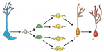
How to make a hippocampal dentate gyrus granule neuron
Granule neurons in the hippocampal dentate gyrus (DG) are known to be continuously generated throughout adult life, and the ongoing integration of newborn neurons into the existing hippocampal neural circuitry provides enhanced neuroplasticity, which plays a crucial role in learning and memory. In their Primer article, Gage and colleagues summarize the developmental principles that regulate the process of DG neurogenesis and discuss recent advances in harnessing these developmental cues to generate DG granule neurons from human pluripotent stem cells. See the Primer on p. 2366
Chromatin features and the epigenetic regulation of pluripotency states in ESCs
In pluripotent stem cells, the interplay between signaling cues, epigenetic regulators and transcription factors orchestrates developmental potency. Here, Maria-Elena Torres-Padilla and Ian Chambers review what is known about transcriptional heterogeneity in pluripotent stem cells, focusing on the underlying causes of heterogeneity and how transcriptional heterogeneity can be to the benefit of the whole stem cell population. See the Review on p. 2376
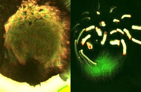
Many congratulations to Nathan Kenny (University of Oxford), Kathryn McClelland (Institute for Molecular Bioscience, University of Queensland), and Sophie Miller (University of Cambridge), who took this image at last year’s Woods Hole Embryology course. The image shows a squid (Loligo pealeii) embryo stained with anti-acetylated tubulin (red), anti-sorotonin (green), and DAPI (blue,nuclei).
The runners-up to this competition were the annelid larvae by Poulomi Ray (Clemson University), the live squid embryo by Brijesh Kumar (Indian Institute of Technology, Kanpur) and the squid eye by Ezgi Kunttas (Carnegie Mellon University).
The winning squid embryo image will feature in the cover of a coming issue of Development. This was only the first round though, so expect another set of beautiful images from the Woods Hole embryology course here on the Node very soon!
June 1st, 2014: Exactly one year after my departure flight from Bologna to Boston to attend the 2013 MBL Embryology course held at the Marine Biological Laboratory in Woods Hole, MA (https://thenode.biologists.com/six-weeks-in-woods-hole/events/), I was again in the Bologna airport, only that this time I was landing from the States.
I was returning to my home institution, the University of Modena and Reggio Emilia, Italy where I am a last year graduate student, from the Stowers Institute for Medical Research in Kansas City (MO, USA), where I spent the last 3 months working in the laborartory of Prof. Alejandro Sánchez Alvarado.
Thanks to the 2013 MBL Embryology course, where I met Prof. Sánchez Alvarado (Director of the course together with Prof. Richard Behringer), and thanks to the Development Travelling Fellowship, I could collaborate with a great research group in one of the best places anywhere in the world that a biologist can work at: the Stowers Insitute (http://www.stowers.org/stowers-report/fall-2013/best-places-to-work).
At the time of the 2013 MBL Embryology course, I was working mainly on the immune functions of the freshwater gastropod, Pomacea canaliculata, a mollusk which is considered a dangerous pest already invading North America and South-East Asia from the South America. Pomacea is also the intermediate host of the nematode Angiostrongylus cantonensis, which causes eosinophilic meningitis in humans. This snail is aggressively invading new territories because of its adaptability to different kinds of environmental conditions and resistance to multiple stress conditions. As such, the characterization of the immune system of this snail is important for controlling its diffusion by allowing to uncover features that may be vulnerable to intervetions for eradication.
However, after the 2013 MBL Embryology course in Woods Hole, I became highly interested in regeneration and the incredible mechanisms able to activate either stem or differentiated cells in order to increase the proliferative rate and build either amputated parts or whole organisms altogether. More than two hundred years ago, Lazzaro Spallanzani’s experiments (in part performed in Modena, by the way) showed that terrestrial snails are able to regenerate their heads after decapitation. In Woods Hole, I was really impressed by the regenerative capacity of planarians, which can be included with mollusks and other groups into the lophotrocozoan taxon. Because of this, when I returned to Modena in 2013, I tried and cut the sensory organs of P. canaliculata in order to verify if it is able to regenerate.
I was very excited when I observed that after 2, 3 and 4 weeks, respectively, the cephalic and oral tentacles, as well as the eyes were completely regenerated. This observation formed the basis for my period in Kansas City as a visiting student.
Before I left for Kansas City, I collected the samples of the regenerating cephalic tentacles, oral tentacles and eyes of Pomacea canaliculata at different time points. Once at the Stowers Institute, the RNA purification and sequencing of the samples and the bioinformatic analysis of the data allowed for the construction of a transcriptome database. A high-resolution time-course was produced for each organ and the analysis of the gene expression profiles uncovered a significant number of genes differentially expressed during the regeneration processes surveyed. The availability of the transciptome of these complex organs will allow for detailed molecular analyses of the pathways involved in regeneration, providing a solid foundation for my future studies.
These three months at the Stowers Institute, which directly stemmed from the 2013 MBL Embryology course, gave me the chance to write a personal three-month project, learn new techniques, work with a planarian model, perform experiments in the field of regeneration, and collect data that will be surely useful in my future studies. Last but not least, at the Stowers Institute I met a lot of great scientists, and attended many interesting lectures. Equally enjoyable were my interactions with post-docs from all over the world from whom I benefited extensively through their continuous and kind assistance in my lab activities.
I am confident that this experience will reveal of fundamental importance for me and my scientific career.
Cells move in (still) mysterious ways to achieve morphogenesis.
Prominently, cells of an early vertebrate embryo (blastula, a mass of undifferentiated cells) move extensively during gastrulation to generate the three basic layers of the organism: ectoderm at the surface, endoderm presaging the digestive tube, and the mesoderm in between. At the end of the process, the body plan is clearly visible, its main axes established.
This is perhaps most dramatically illustrated in the extremely large embryos of most amniotes (such as in birds and most mammalians, including the human): they consist of thousands or tens of thousands of cells, arranged in an epithelial disc (epiblast). Gastrulation is achieved through a seemingly fixed structure at the midline, the primitive streak (Figure 1).
Figure 1: Chick embryo at gastrulation. The primitive streak is the darker stricture along the midline.
Here, cells engage in epithelial-to-mesenchymal transitions (EMTs, similar to carcinoma progression [1]) to leave the epiblast and contribute to the inner layers. In the chick embryo – a good representative of most amniotes -, virtually all cells in the embryonic disc have long been observed to be engaged in large displacements, directed mainly toward the primitive streak ([2, 3], Figure 2).
Figure 2. Massive movements across the embryonic epiblast. Left, using transmitted light to observe development prior to streak formation. Right, fluorescent marking of the domain where the primitive streak will form (green) and tracks of other groups of cells (yellow; red indicates the direction of movement).
But how is the primitive streak maintained as a fixed structure in the middle of a field of constantly moving cells? How does it form in the first place? To add to the puzzle, massive cell movements in the epiblast actually start well before gastrulation in the form of counter-rotating whorls, the meeting point of which always correspond to where the primitive streak later forms. Is there a causal relationship?
We have now shown [4] that the primitive streak arises by a chain reaction of EMT events. This is primed by rare EMT events, which occur in the epiblast well before gastrulation. EMTs become highly cooperative through a community effect requiring TGF-b signalling. The gene coding for Nodal, a member of this superfamily, is transcribed early in a small domain on the edge of the blastula, but the activity of the protein is antagonised by signals emanating from an amniote-specific extra-embryonic layer, the hypoblast, which initially lines the epiblast. It is its displacement (and the disinhibition of Nodal activity) that triggers the chain reaction of EMTs and, therefore, the initiation and later the maintenance of the primitive streak.
The Nodal expression domain corresponds to a region in which we previously found cell intercalation to occur, in the plane of the epiblast (green in figure 2, right panel) – this is again regulated by the hypoblast, but positively and via different signals (FGFs, [5]). The intercalation region is initially arranged along the edge of the epiblast and intercalation displaces its cells along one of its radii, before gastrulation. So, one key invention of amniotes is a mechanism to regulate Nodal activity and displace it along the future anterior-posterior axis of the embryo, before it can trigger gastrulation. Interestingly, both intercalation and delaying Nodal-dependent chain reaction of EMTs are regulated by the hypoblast, the characteristic evolutionary acquisition of amniotes.
Two key, local cell behaviours therefore drive primitive streak formation. What about the movements in the rest of the epiblast? We turned to computer simulations and we show that these two components can indeed entrain cells in the entire epithelial sheet to move in the correct pattern, and that the model can also represent the experimental conditions of abrogating or ectopically creating one or two of these components. Local actions can trigger global movements.
A few directions now open, which we now pursue in my new lab in Cambridge. There is now a framework for interpreting experiments at molecular or cellular level and linking them quantitatively to large-scale morphogenesis, and refine both experiments and models in step with each other. Such models hold the key not only for understanding large, regulative embryos (including the human), but also for adequately discerning the common developmental patterns and the true acquisitions / losses during evolution, and the constraints that brought them about. Gastrulation also sets the stage for the development of the nervous system and the elongation of the main body axis; we now hope that we have better tools to continue exploring these key issues.
References
[1] Nieto, M. (2011). The Ins and Outs of the Epithelial to Mesenchymal Transition in Health and Disease Annual Review of Cell and Developmental Biology, 27 (1), 347-376 DOI: 10.1146/annurev-cellbio-092910-154036
[2] Gräper, L. (1929). Die Primitiventwicklung des Hühnchens nach stereokinematographischen Untersuchungen, kontrolliert durch vitale Farbmarkierung und verglichen mit der Entwicklung anderer Wirbeltiere Wilhelm Roux’ Archiv für Entwicklungsmechanik der Organismen, 116 (1), 382-429 DOI: 10.1007/BF02145235
[3] Wetzel, R. (1929). Untersuchungen am Hühnchen. Die Entwicklung des Keims während der ersten beiden Bruttage Wilhelm Roux’ Archiv für Entwicklungsmechanik der Organismen, 119 (1), 188-321 DOI: 10.1007/BF02111186
[4] Voiculescu, O., Bodenstein, L., Lau, I., & Stern, C. (2014). Local cell interactions and self-amplifying individual cell ingression drive amniote gastrulation eLife, 3 DOI: 10.7554/eLife.01817
[5] Voiculescu, O., Bertocchini, F., Wolpert, L., Keller, R., & Stern, C. (2007). The amniote primitive streak is defined by epithelial cell intercalation before gastrulation Nature, 449 (7165), 1049-1052 DOI: 10.1038/nature06211
If you are doing a PhD (or involved in research in any way), you probably take a lot of humour, procrastination and comfort from PHD comics. The comic strip ‘Piled Higher and Deeper’ has been portraying “life (or the lack thereof) in academia” since 1997, with a cast of characters ranging from a nameless graduate student struggling with his PhD to a postdoc with a knack for stealing free food from seminars.
But in the last few years PHD has become more than just the comics. The creator Jorge Cham, has travelled around the world giving talks about the power of procrastination, and PHD TV is a fantastic collection of interesting videos about different science topics and researchers, mixing real life footage with Jorge’s illustrations. Another exciting project spinning off from PHD comics was the PHD movie. Filmed on location at the California Institute of Technology, and starring real life graduate students and researchers as many of the main characters, this feature length movie brought many of the jokes that we have come to love to the big screen. The project was a huge success and the movie was screened at over 500 universities and research institutes across the world. Eva Amsen (the previous Node community manager) wrote for the Node about it when she attended one of these screenings in London (she also interviewed Jorge for the Node before).
Following the great success of the first movie, the PHD team is now keen on creating a sequel, and they need your help! A Kickstarter campaign has been launched in an attempted to raise the $100K required, and you can show your support by making a donation which will not only make the project possible but also give you access to exclusive rewards!
If you haven’t watched the first instalment and want to know what the movie is all about, you can watch the PHD movie for freefor this month only. To give you a taster, here is the trailer:
Postdoctoral positions are available in the laboratory of Amanda Dickinson to study craniofacial and skin development in Xenopus.
I am looking for highly motivated and tenacious applicants with a passion for developmental biology. Qualifications include a PhD in cellular/molecular or developmental biology and experience with Xenopus or zebrafish is preferred.
There are several projects in the lab which include:
-Vitamin deficiencies and orofacial development
-Epigenetic regulation of palate formation
-The biomechanics of forming a face
-Signaling and cell adhesion during skin development
The laboratory is part of a growing group of developmental biologists in the department of biology at Virginia Commonwealth University, Richmond Virginia. Check out these links to learn more about some of the great things about living in Richmond, VA.
Please send a letter of application (including a brief description of previous research experience), CV, and the name and contact information of 3 references to Amanda Dickinson at ajdickinson@vcu.edu.
– How good is a ‘use it or loose it’ model at explaining neuronal connections in the vertebrate retina? The students of the developmental neurobiology seminar at Reed College posted their last journal club discussion.
– And we reposted an article from the F1000Research blog about a recent study where Kenneth Lee and colleagues attempted to reproduce the STAP study.
Outreach:
– Interested in making your own outreach videos? Lilian came across an easy-to-use app that can help you make simple and effective videos using images and sound.
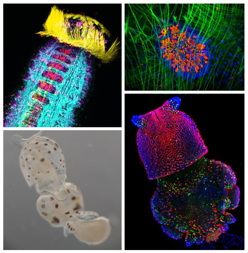
– Our popular Woods Hole cover competition is back! Which image from last year’s course would you like to see in the cover of Development?
– Switzerland is not just about cheese and chocolate- Monika visited the Mosimann lab in Zurich to learn how to use the zebrafish tamoxifen-controlled Cre/lox system.
A postdoctoral fellowship for 3 years is available from October 2014 in Delphine DUPREZ’s team, located in the Developmental Biology laboratory in Paris (bio–dev.snv.jussieu.fr/). The team focuses on tendon and muscle development using animal models. The objective of the project is to better understand the molecular pathways involved in tendon development. The project will involve the use of the chick embryos and mesenchymal stem cells in 2-dimensional and 3-dimensional culture systems.
DUPREZ – Related references:
Guerquin et al.,(2013). J Clin Invest123, 3564-3576.
Lejard et al., (2011) J. Biol. Chem286(7), 5855-5867.
Wang et al., (2010) Dev Cell, 18, 643-654.
Applicants may be of any nationality and should have obtained the equivalent of a PhD less than 2 years. A background in developmental biology or/and an expertise in the use of mesenchymal stem cells will be an advantage.
Applications should be sent to Delphine DUPREZ to Delphine.duprez@upmc.fr
Applications should comprise the following:
– CV
– Description of previous research experiences, publication list and a personal statement describing research interests and career goals
– List of 3 names of referees including email address and telephone number
Any mammal who celebrated Mother’s Day earlier this month realizes how important mothers are for us and the tight bond between them and their children. Forget clean shirts and packed lunch every day; for us developmental biologists, there is no better reflection of this bond than the extraembryonic membranes that support the growth of the fetus in the uterus. These tissues, among other roles, serve as circulatory, digestive and excretory systems until we develop our own. As a matter of fact, they are so important that they begin to form even before the fetus itself – during the preimplantation period, before the embryo attaches to the uterus (check out the cartoon below). Therefore, the very first decisions cells need to make during a mammal’s life are whether to become part of the extraembryonic lineages (called trophectoderm and primitive endoderm at this stage) or become the foundation of the fetus (the epiblast).
Historically, the first of these decisions (whether or not to become trophectoderm) has received more attention by researchers. However, the second one, whether to become primitive endoderm or the pluripotent, embryonic epiblast, has only been studied in more detail over the last decade or so. A number of molecular markers and the cellular behaviors involved in this process have been described, although no transcription factor has yet been shown to be essential for primitive endoderm specification. In the Hadjantonakis lab, we have recently looked at the role of the main suspect – the GATA family transcription factor GATA6 – and we are publishing the results in the current issue of Developmental Cell [1].
Cartoon and timeline for the main stages of mouse preimplantation development, from the zygote (left) to the blastocyst (right). Cell lineages and their contributions later in development (boxes) are color coded: trophectoderm (green), primitive endoderm (blue), epiblast (red). Inner cell mass (purple). Implantation takes place approximately at day 4.5 of development.
This study is important for two reasons: one biological and the other one technical. As I mentioned earlier, this is the first study to show a transcription factor that is absolutely required for primitive endoderm specification. Gata6 null embryos completely lack primitive endoderm because all cells of the inner cell mass default prematurely to epiblast fate (the future fetus). Although they show no other phenotypic defect, these embryos die upon implantation, presumably because there is no tissue to support the growth of the epiblast. We also show that Gata6 heterozygous null embryos have reduced levels of GATA6 and, as a consequence, show a delay in primitive endoderm specification. Finally, this study clarifies the love triangle formed by GATA6, the pluripotency champion NANOG and the fibroblast growth factor (FGF) signaling pathway. FGF4 (or basic FGF/FGF2) is the instructive signal for primitive endoderm specification among inner cells in the blastocyst; however, as we show for the first time in this paper, GATA6 is necessary for cells to respond to FGF4, consequently downregulate NANOG, and acquire primitive endoderm identity. Our paper not only provides a mechanism for the induction of primitive endoderm fate, but also suggests that the relative levels of GATA6 to NANOG in each cell could tip the balance towards either an extraembryonic or an embryonic fate. How these levels change and how cells can measure them remains to be seen…
Proposed interaction between GATA6, NANOG and the FGF signaling pathway. Dashed lines represent hypothesized regulation, grey lines represent weak effects due to low protein concentrations. From Schrode N, Saiz N, Di Talia S, Hadjantonakis A-K (2014) GATA6 Levels Modulate Primitive Endoderm Cell Fate Choice and Timing in the Mouse Blastocyst. Dev Cell 29: 454–467
Our study is also exciting because of the approach we used to analyze the Gata6 allelic series. For those less familiar with it, the mouse preimplantation embryo is as good a model for (live) imaging and single-cell analysis as it is unfit for biochemistry. It is very small, with few cells, and can develop in vitro, without maternal support, in standard culture conditions. This means we can image and analyze the behavior of every single cell within the embryo using a rather simple experimental setup, making it an ideal model to study mammalian cell differentiation in situ. Despite these features, only recently have there been attempts at quantifying gene expression and doing single cell analyses, partly fueled by the technology that has become available [see references 2-4, or, more recently, 5-7, for examples].
In this work, we have used MINS, a piece of software recently developed in our lab [8] that can segment nuclei in series of confocal images with higher accuracy and lower manual input than any other software we have tried so far – including popular (and powerful) solutions like ImageJ or Imaris. MINS collects spatial coordinates and fluorescence intensity data for every channel on each cell, gives them a unique ID, and generates a data matrix for each embryo. After some manual curation for under/oversegmentation and to remove the occasional outlier (generally dead cells), the data is ready for you to do maths and statistics to your heart’s content. Being biologists, large data matrices send shivers down our spines, so we teamed up with physicist-turned-biologist Stefano Di Talia and, lo and behold, the numbers began to make sense! Furthermore, because MINS also collects positional information, we were able to relate changes in protein levels and cell identity to position within the embryo. To a certain extent, this kind of analysis allows us to bypass Western Blots and study protein expression in individual cells, while preserving positional information, which is lost when cells are disaggregated for gene expression analyses. This study is the first of several from our lab where we will apply this pipeline to three- and four- (time lapse) dimensional datasets to understand better the earliest cell fate decisions in mammalian development. We hope the implementation of this analysis pipeline by other labs will improve it and help generate even higher quality data in future studies. The combination of these advanced algorithms for image analysis with single-cell expression profiling techniques will let researchers study with high resolution the molecular mechanisms controlling cellular processes in situ, in intact tissues or embryos. For further proof that this is becoming a thing, have a look at these recent comments: 9, 10.
If you want to discuss or comment, do it below or you can reach me on twitter
References:
1.Schrode, N., Saiz, N., Di Talia, S., & Hadjantonakis, A. (2014). GATA6 Levels Modulate Primitive Endoderm Cell Fate Choice and Timing in the Mouse Blastocyst Developmental Cell, 29 (4), 454-467 DOI: 10.1016/j.devcel.2014.04.011
2.Kurimoto, K., Yabuta, Y., Ohinata, Y., Ono, Y., Uno, KD., Yamada, R., Ueda, H., & Saitou, M. (2006). An improved single-cell cDNA amplification method for efficient high-density oligonucleotide microarray analysis Nucleic Acids Research, 34 (5) DOI: 10.1093/nar/gkl050
3.Plusa, B., Piliszek, A., Frankenberg, S., Artus, J., & Hadjantonakis, A. (2008). Distinct sequential cell behaviours direct primitive endoderm formation in the mouse blastocyst Development, 135 (18), 3081-3091 DOI: 10.1242/dev.021519
4.Guo, G., Huss, M., Tong, G., Wang, C., Li Sun, L., Clarke, N., & Robson, P. (2010). Resolution of Cell Fate Decisions Revealed by Single-Cell Gene Expression Analysis from Zygote to Blastocyst Developmental Cell, 18 (4), 675-685 DOI: 10.1016/j.devcel.2010.02.012
5.Frum, T., Halbisen, M., Wang, C., Amiri, H., Robson, P., & Ralston, A. (2013). Oct4 Cell-Autonomously Promotes Primitive Endoderm Development in the Mouse Blastocyst Developmental Cell, 25 (6), 610-622 DOI: 10.1016/j.devcel.2013.05.004
6.Le Bin, G., Munoz-Descalzo, S., Kurowski, A., Leitch, H., Lou, X., Mansfield, W., Etienne-Dumeau, C., Grabole, N., Mulas, C., Niwa, H., Hadjantonakis, A., & Nichols, J. (2014). Oct4 is required for lineage priming in the developing inner cell mass of the mouse blastocyst Development, 141 (5), 1001-1010 DOI: 10.1242/dev.096875
7.Ohnishi, Y., Huber, W., Tsumura, A., Kang, M., Xenopoulos, P., Kurimoto, K., Oleś, A., Araúzo-Bravo, M., Saitou, M., Hadjantonakis, A., & Hiiragi, T. (2013). Cell-to-cell expression variability followed by signal reinforcement progressively segregates early mouse lineages Nature Cell Biology, 16 (1), 27-37 DOI: 10.1038/ncb2881
8.Lou, X., Kang, M., Xenopoulos, P., Muñoz-Descalzo, S., & Hadjantonakis, A. (2014). A Rapid and Efficient 2D/3D Nuclear Segmentation Method for Analysis of Early Mouse Embryo and Stem Cell Image Data Stem Cell Reports, 2 (3), 382-397 DOI: 10.1016/j.stemcr.2014.01.010
9.Wen, L., & Tang, F. (2014). Reconstructing Complex Tissues from Single-Cell Analyses Cell, 157 (4), 771-773 DOI: 10.1016/j.cell.2014.04.024
10.Robson, P. (2014). Deciphering Developmental Processes from Single-Cell Transcriptomes Developmental Cell, 29 (3), 260-261 DOI: 10.1016/j.devcel.2014.04.032
 (No Ratings Yet)
 Familial dysautonomia (FD) is a germline autosomal recessive disease that is characterized by impaired peripheral sensory and sympathetic neuron development. The disease is known to be caused by mutations in the gene encoding Elp1 (also known as IKBKAP), but how Elp1 functions in neurons is unclear. Now, Warren Tourtellotte and colleagues investigate the role of Elp1 in mice (p.
  The recent advent of tools for manipulating and monitoring gene expression calls for efficient ways to document, access and analyse these gene expression patterns. Although a number of databases and gene expression atlases have been compiled in recent years, many of them are limited with regards to their content and utility. Here, Chris Doe and colleagues develop new software that overcomes these limitations (p.
  Haematopoiesis – the formation of blood cells – is regulated by a number of ubiquitous and tissue-specific transcription factors, but the extent of interplay between these factors is unclear. Sp1 is a transcription factor that is ubiquitously expressed and regulates the expression of thousands of genes, and it has been shown that Sp1-deficient mouse embryos die during early development. Now, on p.
  Root nodulation in plants is a form of de novoorganogenesis and involves the dedifferentiation of root cortical cells in response to rhizobia-derived factors. However, due to the complexity of this event, our understanding of the factors and mechanisms that initiate nodule formation is limited. Now, Takuya Suzaki and co-workers (p.
 Granule neurons in the hippocampal dentate gyrus (DG) are known to be continuously generated throughout adult life, and the ongoing integration of newborn neurons into the existing hippocampal neural circuitry provides enhanced neuroplasticity, which plays a crucial role in learning and memory. In their Primer article, Gage and colleagues summarize the developmental principles that regulate the process of DG neurogenesis and discuss recent advances in harnessing these developmental cues to generate DG granule neurons from human pluripotent stem cells. See the Primer on p.
 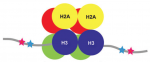 In pluripotent stem cells, the interplay between signaling cues, epigenetic regulators and transcription factors orchestrates developmental potency. Here, Maria-Elena Torres-Padilla and Ian Chambers review what is known about transcriptional heterogeneity in pluripotent stem cells, focusing on the underlying causes of heterogeneity and how transcriptional heterogeneity can be to the benefit of the whole stem cell population. See the Review on p.
  (1 votes)
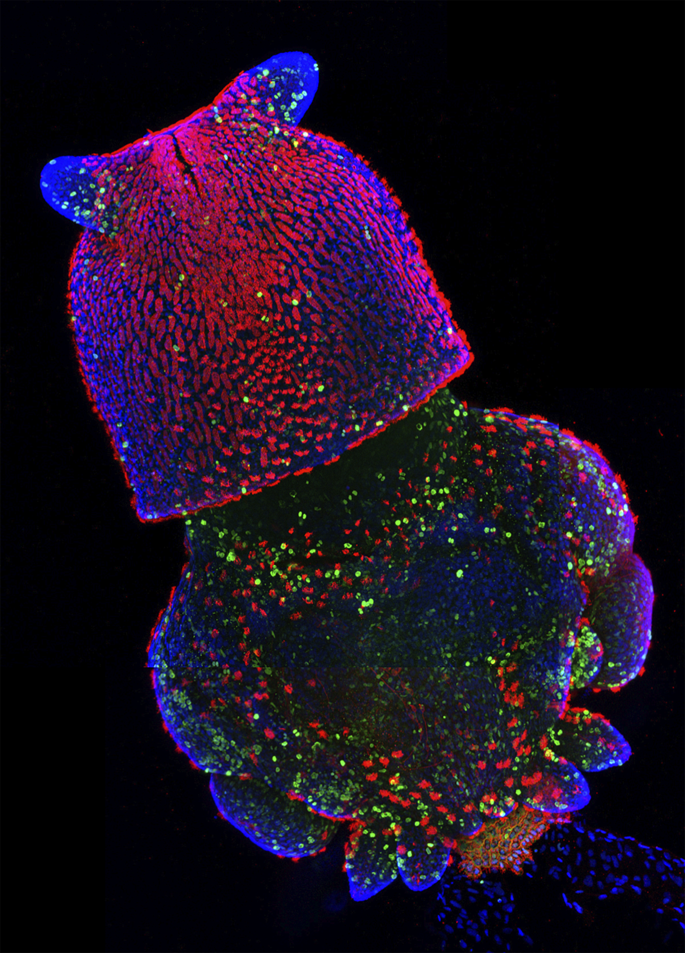


 (5 votes)