Researchers are being encouraged now more than ever to communicate their science to the public. First and foremost science communication is a way to share the enjoyment and excitement of science to others. But in a time of austerity, successfully communicating research is also important to justify to the public that funds it what scientists do.
But science communication can sometimes be daunting. There are so many different ways to do it- giving talks, having a stall at a science festival, collaborating with artists. Where to start? In this outreach series on the Node, we aim to showcase the variety of outreach activities out there, and provide ideas and resources to those interested in getting involved. We have invited various researchers and science communicators to write a series of posts on the different types of outreach that they do, and how they got first involved in science communication. In addition, we have asked them to suggest an easy outreach activity to get you started. From promoting science at music festivals to getting involved in a departmental open day, we hope that you will find something for you in this series, and that you will feel inspired to get involved and communicate your science to others!
Disease Models & Mechanisms (DMM) is pleased to welcome submissions for a Special Issue scheduled for publication in late 2014. This issue will focus on translational advances made using the zebrafish model, including insights into disease mechanisms and therapeutic targets, new resources and technologies, and drug discovery and development.
The issue will be guest edited by:
James Amatruda (UT Southwestern Medical Center, USA) Liz Patton (University of Edinburgh, UK) Lalita Ramakrishnan (University of Washington, USA)
Topics to be covered in invited review articles include advances in regenerative medicine and cancer, mechanisms underlying neurodevelopmental and neurodegenerative diseases, small-molecule screening using zebrafish and more.
We invite you to showcase your breakthrough zebrafish research in this Special Issue. Submissions should describe original research in the form of a Research Article, Resource Article or Research Report. Please read the author guidelines for information on preparing a manuscript for DMM, and submit your manuscript online. For rapid feedback on a paper, send us a presubmission enquiry.
Key benefits of publishing in DMM include:
– Open Access (CC-BY)
– High visibility and impact
– Rapid peer-review
– Author videos featured on Company of Biologists YouTube channel – Accepted manuscripts online within 1 week
– All articles included in Medline (PubMed), Scopus and ISI Web of Science
– PMC deposition
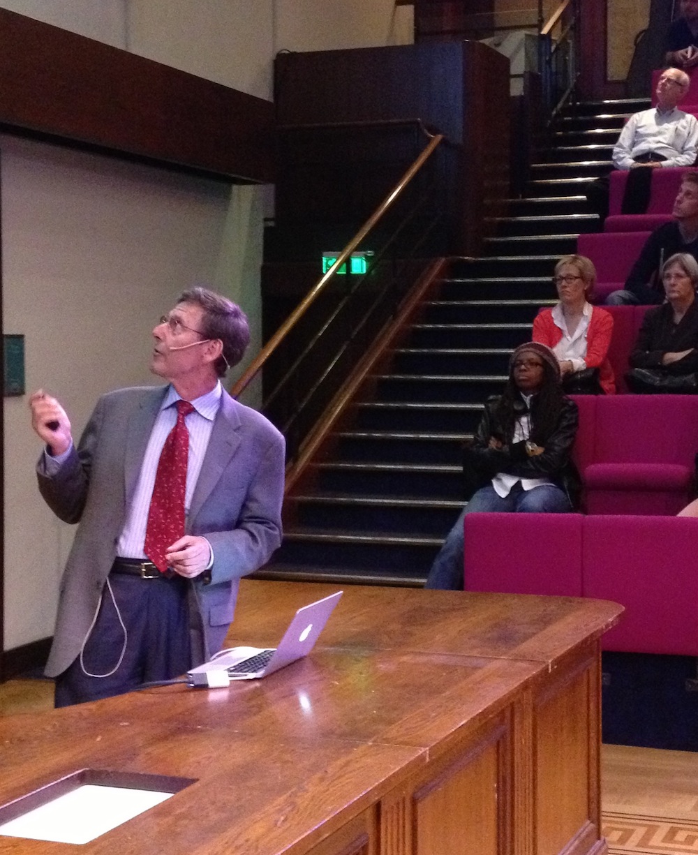
The 13th FASEB Plant Biology Conference was held from August 11- 16, 2013, in Saxtons River, Vermont, a modest but beautiful setting. This was a special meeting, since it marked 25 years since the first FASEB Plant Molecular Biology conference- the theme changed to Plant Development in the mid ‘90’s. Around 160 attendees spent 5 days hearing the latest “developments” in plant development, and enjoying casual discussions in the swimming pond and the bar.
The meeting kicked off with an inspiring plenary talk by Prof. Liam Dolan (Oxford University). Liam’s lab has been studying root hair patterning and other aspects of root development in Arabidopsis for many years, but in this talk he described new work that seeks to understand the evolutionary origin of root- like structures in plants. using a recently adopted liverwort model, Marchantia.
The following morning, the first session of the conference focused on the latest research on local signals. The speakers showed their work on plasmodesmata-dependent miRNA movement (Ykä Helariutta, University of Helsinki), endosome-dependent protein movement (Kimberly Gallagher, University of Pennsylvania) and recognition of Calcium signals in roots (Giles Oldroyd, John Innes Centre), sensing of CO2 concentration in leaves (Cawas Engineer, UCSD) and cytokinin levels in secondary meristems (Yuval Eshed, Weizmann Institute). Furthermore, recent findings on the control of Arabidopsis embryo maturation and maize meristem size homeostasis (Michael Pautler, CHSL) were shared with the community.
In the polarity session, several different examples of cell- and tissue-level polarity were discussed. Jeff Long (Salk Institute) presented new work on the genetic specification and maintenance of apical-basal polarity via TOPLESS-dependent and –independent pathwaysin the Arabidopsis embryo, while Laurie Smith (UCSD) presented insights into the regulation of asymmetric cell divisions during stomatal development in maize. Remko Offringa (Leiden University) described how regulation of the protein kinase PINOID (PID) can influence the pattern of PIN polarity – and hence auxin transport within a tissue. Finally, Xana Rebocho (John Innes Centre) described the importance of the regulation of tissue polarity, and growth rates, for the morphogenesis of the Antirrhinum flower and the insights gained from modeling studies.
Shoot architecture of higher plants is determined by developmental events occurring at the growing tips. Meristems are the site of organogenesis, giving rise to lateral organs, such as leaves and axillary branches, which themselves have meristematic properties. A meristem contributes immobile cells to the formation of lateral organ primordia, and it is self-renewing. A major outstanding question is precisely how cells dynamically accept cell fate at the meristem-incipient organ boundary during organ initiation. An enthralling session devoted to boundaries featured studies from Rüdiger Simon (Heinrich-Heine University), Marcus Heisler (EMBL) and Klaus Theres (Max Planck Institute for Plant Breeding Research) that honed in on the genetic and molecular dissection of boundary specification and formation.
The meeting then moved on to considering long range signaling in plants. Leslie Sieburth (University of Utah) presented new findings on the bypass signal that coordinates the development of shoots and roots. David Braun (University of Missouri) discussed the Tie-Dyed (TDY) genes of maize; mutations in which show defects in phloem differentiation and sucrose trafficking. Micro-RNAs can also be transported long distances in the plant, and Tzyy-Jen Chiou (Academia Sinica, Taiwan) presented her research on the regulation of phosphate uptake and translocation, which involves miR399 acting as a systemic signal. In the last talk of the session, Catherine Rameau (INRA Versailles) discussed how Strigolactones and other long-range signals regulate shoot branching in pea.
Compared to Arabidopsis, many crops exhibit unique inflorescence structures requiring more coordinated transition from stem cell maintenance to floral organ formation. Inflorescence architecture is largely determined by the fate of meristems, population of stem cells under specific genetic control. In the session on switches, novel molecular regulators of this inflorescence development were presented in three major crops: tomato (Zachary Lippman, CSHL), rice (Junko Kyozuka, University of Tokyo), and corn (Paula McSteen, University of Missouri).
The signal integration session focused on the integration of molecular signals that coordinate complex environmental inputs and govern developmental outputs. The session highlighted an exciting array of developmental responses to the environment, from the phototracking of sunflowers to root patterning in heterogeneous soils. Jennifer Nemhauser (University of Washington) started out the session by introducing the hypocotyl as a model system for measuring the role of complex environmental input on developmental output. In the next talk, José Dinneny (Carnegie Institution) elucidated the role of heterogeneous water availability on lateral root patterning, a phenomenon referred to as “hydropatterning”. Stacey Harmer (UC Davis) was the next speaker. She emphasized the impact of the circadian clock on transcriptomic patterns. The Harmer lab harvested tissue every 4 hours for 2 days and found that half of all genes are differentially expressed in a time dependent manner. Harmer also introduced sunflower solar tracking as a model system to study circadian clock entrainment. The final speaker for the session was José Alonso (North Carolina State University), who presented a very clever mutant screen that was performed to fish out genes coordinating the relationship between auxin signaling and ethylene response.
Talks then turned to regulation of morphogenesis and shape. Olivier Hamant (Lyon) presented research on mechanical signals in plant morphogenesis, and the proposal that coupling between mechanical cues, microtubule (MT) alignment and PIN1 orientations plays a key role in morphogenesis and the generation of phyllotactic patterns. Uptal Nath (Indian Institute of Science) focuses on the molecular basis of polar leaf growth. Leaf growth is often allometric, where leaves experience polar growth – more growth at the base and progressively less towards the tip. His work has shown that the polarity of leaf growth can be diverse, and that this may be related to the expression of miR396. Next, Elliot Meyerowitz (Caltech) presented work describing how physical and chemical signals control morphogenesis at the shoot apical meristem. He suggested an approach in which all developmental biologists should consider the physical stresses acting in tissues and study morphogenesis as a whole using all parameters. Adrienne Roeder (Cornell) discussed how coordination of cell division and cell type control the morphogenesis of the Arabidopsis sepal, while Siobhan Braybrook presented data on how cell wall mechanics and pressure affect the overall shape of hypocotyl extension.
In the last session, participants returned to the Vermont Academy auditorium after an afternoon of outdoor activities. Refreshed by a game of volleyball, a jog through the woods, or a swim at the local pond, attendants were pleased to hear three fantastic stories outlining recent discoveries in developmental phenomena related to species evolution. Vincent Colot (Institut de Biologie de l’Ecole Normale Supérieure) presented his group’s recent work exploring the link between genome sequence, DNA methylation status, and plant phenotype. Claudia Köhler (Linnean Center of Plant Biology, Uppsala, Sweden) presented her group’s recent breakthroughs in understanding the mechanisms behind the ‘triploid block’ that causes the post-zygotic isolation of newly formed polyploid progeny. George Coupland (Max-Planck-Institute for Plant Breeding Research) finished off the session by presenting his group’s recent findings on the molecular mechanisms behind vernalization (induction by cold treatment) and age requirements for flowering in the genus Arabis.
The conference ended with a thoughtful overview by Scott Poethig (U. Penn). He reminded us of how our field has come full circle, from a situation where everyone studied a different plant, through a bottleneck where almost everyone worked on Arabidopsis or one or two other model systems, to a new era where genomics allows us to again use diverse species to tackle important problems in plant development. He also reminded us how some concepts discussed at the meeting had been discussed much earlier, in particular reminding us of the seminal ideas of Paul Green in thinking about the role of mechanical forces in plant morphogenesis. Overall it was a truly memorable meeting, and we look forward to the next FASEB Mechanisms in Plant Development meeting in 2015, which will be organized by Dominique Bergmann and Rudiger Simon.
This report was written by several of the students who attended the meeting. If you’re interested in finding out more, you can read their full report here (pdf)
My name is Andrew Mathewson and I am a fourth year graduate student in the University of Washington’s Molecular and Cellular Biology program. Whereas most my friends from college are pursuing productive careers in medicine, social justice, or environmental preservation (or just trying to get by), I spend most my days deep in a basement encouraging small fish to mate so that I can steal their babies for science. I am currently pursuing a PhD in the lab of Dr. Cecilia Moens in the Basic Sciences division at the Fred Hutchinson Cancer Research Center (FHCRC) in Seattle, Washington. The Moens lab exclusively studies the zebrafish as a model for vertebrate development, focusing primarily on hindbrain patterning and cell migration. My project investigates the interplay between basic cell polarity signaling and the ability of neurons to move throughout the developing brain.
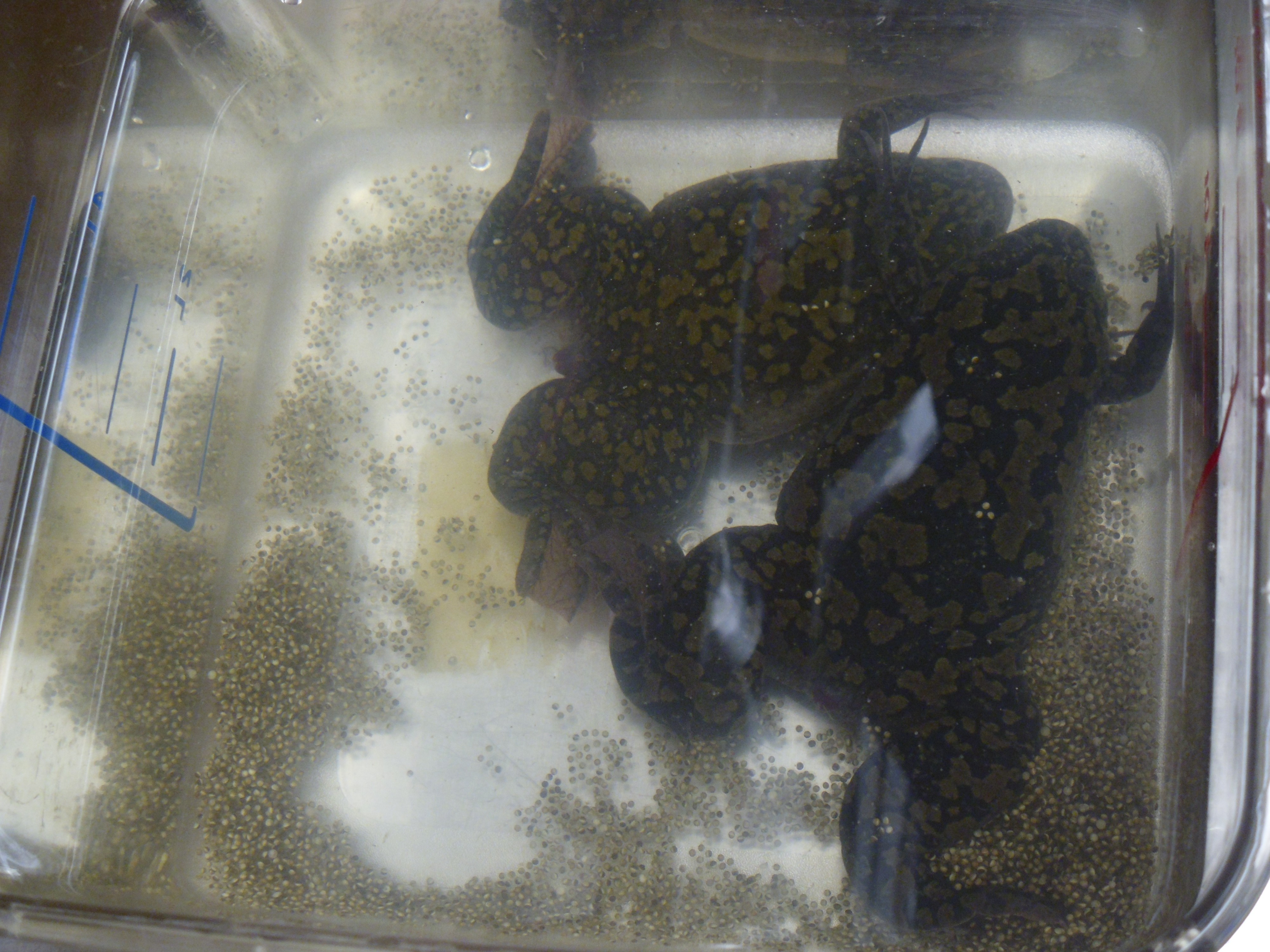
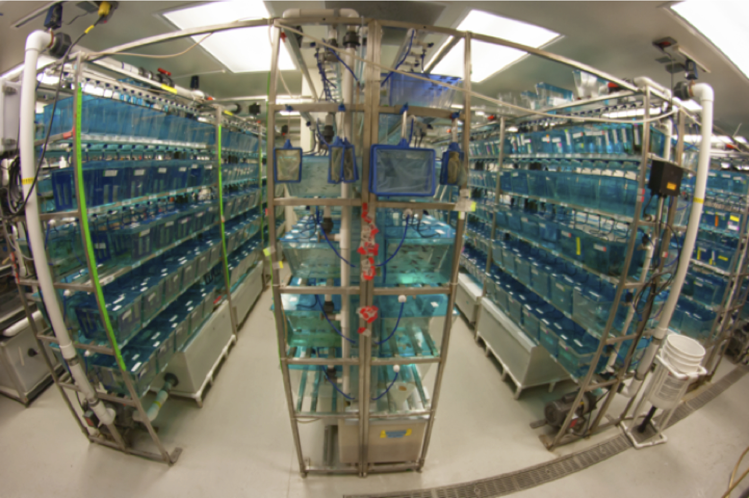
A tank of adult zebrafish expressing a red fluorescent protein in all muscle tissues bustles with excitement during their morning feeding.
Zebrafish are small tropical freshwater fish species native to the Indian subcontinent that were popularized as aquarium fish many years ago. They were pioneered as a model system for scientific research by George Streisinger and colleagues at the University of Oregon, where the Zebrafish International Resource Center (ZIRC) is now located. Many developmental biologists have adopted these robust little critters for their research in the last several decades due to the fact that their biology is very amenable to experimentation and observation. Zebrafish develop externally from a single cell to a rather complex larval fish in just a couple of days, meaning that in just a few hours, zebrafish speed through developmental processes that take weeks or months in other vertebrate models. Most incredibly – they remain entirely transparent while doing so. A single mating pair of zebrafish can produce upwards of a thousand embryos in a single morning, making zebrafish a powerful system for generating large experimental datasets. Zebrafish has triumphed as a system for forward genetic screens and researchers have developed innumerable transgenic lines for the scientific community in recent years. It is the unique combination of developmental speed, optical transparency, and the rapidly growing array of genetic tools that have made zebrafish a popular system for modern developmental research.
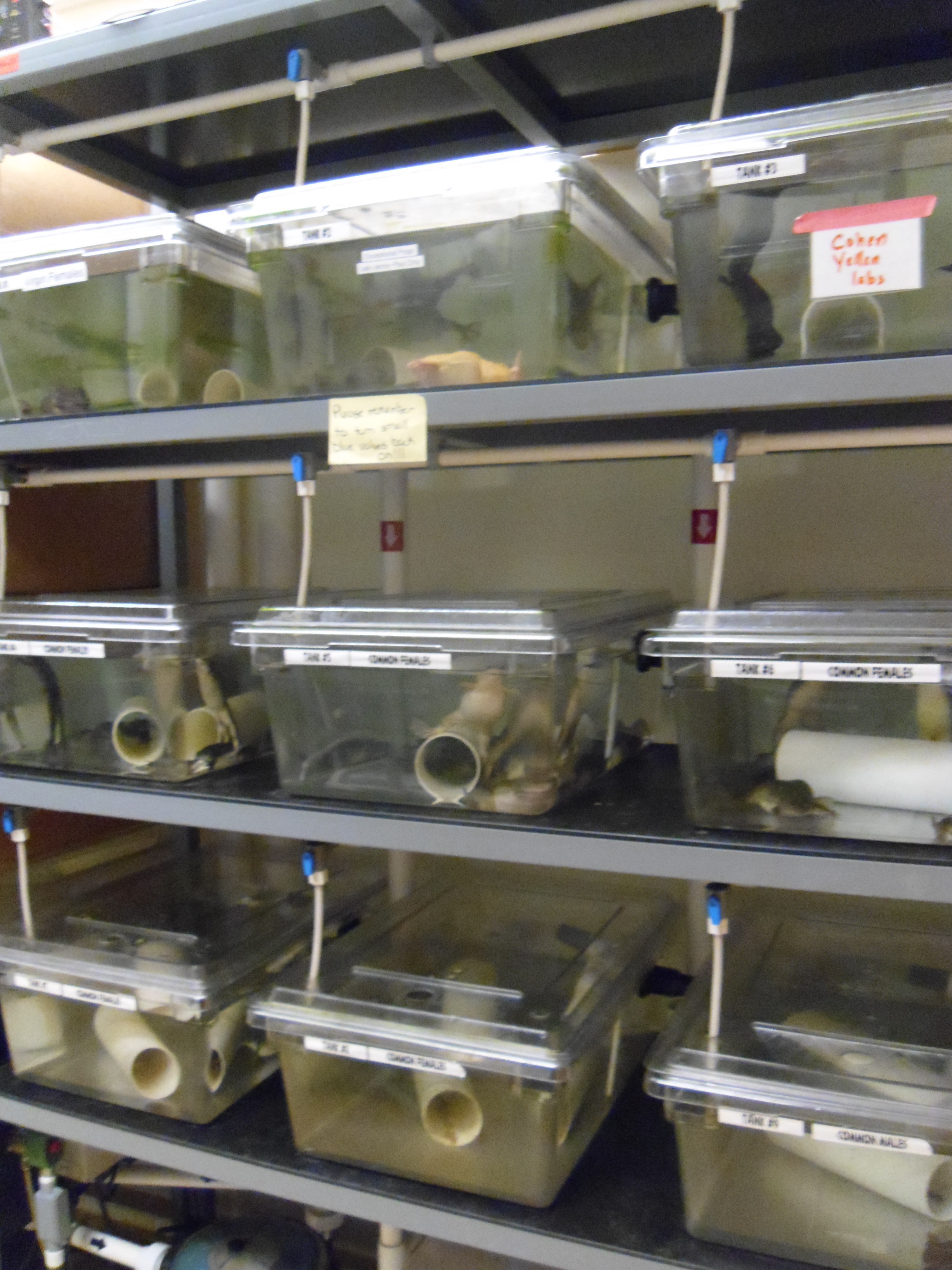
If you have never worked in a fish lab you might wonder what goes on behind the locked doors of our facility deep in the bowels of the FHCRC. In the evening before most of my experiments I set aside a lucky few male and female fish with the mutations and/or transgenes I am using and place them into crossing tanks that allow the fish to see and smell but not touch one another. At 9AM the next morning the facility lights turn on and the fish are frisky after a long night’s rest. One of my favorite parts of my job is when I get to remove the transparent dividers from crossing tanks, allowing the fish to finally interact after a long night of frustrated solitude. Healthy males will almost immediately begin aggressively courting any willing (or not) female in sight. This courting mostly looks like a game of tag, where the female is “it”. Usually in a matter of minutes the females will begin to release eggs that the males dutifully fertilize and I patiently collect for my morning experiments.
The Moens Lab main fish facility
While any research demands practice and patience, zebrafish work in particular demands exceedingly steady hands, sharp eyes, and an even sharper pair of forceps. A typical day can involve anything from molecular cloning and sequence analysis, confocal microscopy and imaging, visual or genetic screening of mutant embryos, all the way up to actual wrangling of adult fish. Most of my current work involves studying developmental processes that occur within the first two days of life. This means that I spend the first half of most work weeks crossing fish lines and manipulating embryos and the second half imaging and analyzing my results. One technique I’ve tried to master is cell transplantation between embryos of different genotypes in order to ask questions about cell-specific gene function. Zebrafish is a great system for this type of work since embryos develop externally, are produced in large numbers, and can tolerate a lot of manipulation. However, most of my current experiments revolve around cloning interesting bits of DNA together and injecting them into newly laid single-cell embryos with infinitesimally small glass needles. I can efficiently create mosaic embryos (where some cells express my DNA and others do not) by mixing my DNA with transposase to increase genomic integration. Most people can comfortably inject hundreds of embryos per hour making it possible to rapidly conduct multiple experiments in parallel. Once I’ve injected my embryos, I spend the rest of my days in dark rooms peering through dissection scopes trying to discern if my manipulations change how cells behave during development. Even though these cells tend to be labeled with at least one type of fluorescent protein, this process can be difficult because zebrafish embryos are quite small. Therefore, once I’ve identified embryos that express my injected DNA or contain successfully transplanted cells, I’ll typically anesthetize the embryos, mount them in agar, and image them on one of our confocal microscopes for closer analysis. The coolest part of this is that I do most of my imaging on living embryos, so I can see how cells behave in their natural environment as the fish continue to develop. This also allows me to use the fish in further experimentation or I can raise them to generate new transgenic lines. Depending on the type or combination of transgenes I want for my future experiments, it can sometimes take several generations and many months to actually generate a new stable transgenic line.
Not surprisingly, it can take a little bit of work keep my experimental stocks of hundreds (or thousands) of fish alive and happy throughout my experimental timeline which in some cases can extend for years. Fortunately the Moens lab is able to hire a full-time fish technician that cares for our facility and generally keeps things running smoothly. She maintains cultures of brine shrimp (aka sea monkeys, a zebrafish’s favorite snack) and rotifers (microscopic water creatures that baby fish love to munch on), feeds the entire facility both live and dry food several times a day, cleans used tanks and nets, helps keep track of our stocks as they mature and age, and euthanizes aging or diseased fish, to name just a few of the amazing things she does for us. Occasionally our technician takes a day off work (like any normal human being), leaving us researchers responsible for our own fish. It’s usually not a big deal—everyone in the lab is at least nominally trained in fish facility maintenance for our own work, and ever since the funding drop, everyone has gotten used to sacrificing the odd weekend to keep our stocks alive. Yet when things go wrong it forces one to recognize the sheer amount of effort required to generate the massive amounts of data produced by the zebrafish research community. Just the other day it was my turn to feed our baby tanks but it turned out that our rotifer cultures had crashed. Our babies went hungry that day—slowing their growth and decreasing their chances of survival, meaning that we can’t count on them for future experiments. Fortunately these small disasters are pretty rare because we have such an awesome fish technician and everyone in lab pays attention to the needs of the fish.
Disease and parasites are a major concern when running a large facility of animals. We try to minimize introducing unwanted pests into our facility by keeping an entirely separate “quarantine” facility for housing fish sent from other labs as well as regular sterilization of media and equipment. Even with these precautions it is impossible to prevent the occasional unwanted pest from getting in. It’s not uncommon to discover an odd arthropod swimming in one’s dishes while sorting embryos. Though tiny creatures like these are usually a normal part of a complex water environment in nature, they can devastate young (or even adult) zebrafish populations in lab. Instances like these remind us that the hard work we do to keep our facility clean and our fish happy isn’t just because the FHCRC requires us to—it’s so we all can continue to do our research.
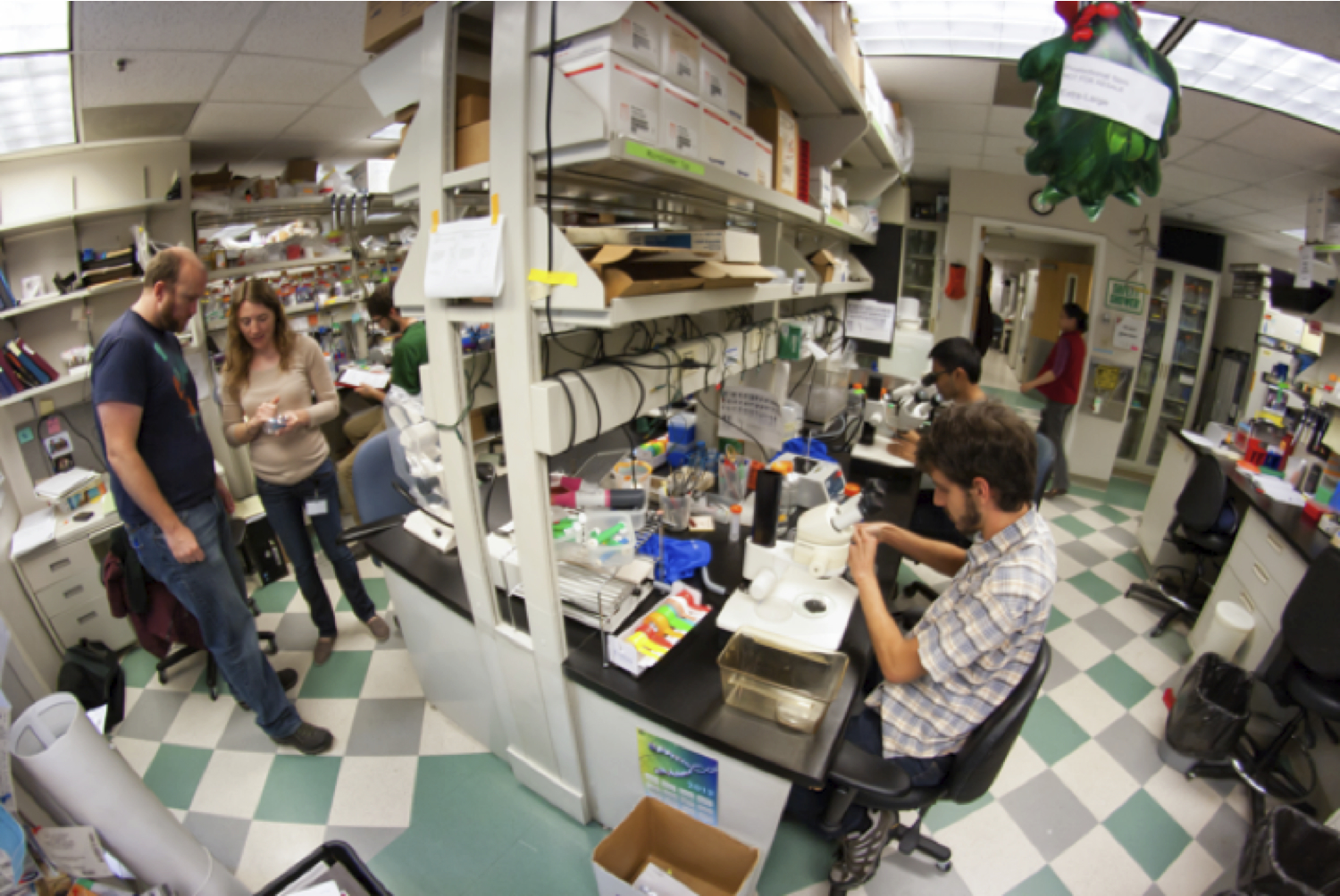
Lab mates cluster around our central work bench most afternoons. Adam Miller and Crystal Davey inspect injection needles (on the left) for cell transplantations while Daniel Berman and Arish Shah (on the right) sort zebrafish embryos.
I like working with zebrafish. While I find the pace of yeast and tissue culture almost frenetic, and the speed of Arabidopsis growthto be at times overly meditative, zebrafish grow and live at a rate that I can relate to. Most experiments fall into a natural daily rhythm in sync with our fish facility’s diurnal cycle. From my experience, zebrafish labs tend to foster a friendly and collaborative lab atmosphere due to the tiny bit of mutual dependence required for keeping stocks alive and experiments running (or maybe zebrafish work just attracts friendly people). Given that we usually want our fish to stay healthy, we have a pronounced lack of the toxic chemicals one usually associates with this type of research, which means the lab can be a comfortable space for everyone. One nice thing about zebrafish work in general is that the fish do most of the work. Once I’ve done my manipulations early in the week I place my embryos in a water filled dish in an incubator and don’t touch them until they are old enough to study. Each fish has its own yolk sac meaning that they don’t need food (or much of anything) for the first week of development. Though zebrafish is a great system for cell transplantation and other active manipulations, the fish really shine as a model organism once they are old enough to look at. Whether you are tagging an interesting protein with GFP, tracking transplanted cells with viable dyes, screening for mutant phenotypes, or just watching the precise choreography of development, zebrafish is the system to turn to. The fact that I can mount living transgenic embryos and image their (often glowing and multicolored) cells as they grow and interact with one another just blows my mind. This aspect is what caught my attention early in graduate school and is a big part what keeps me working towards my Ph.D. It’s hard to describe the beauty of a living, glowing brain as seen through a powerful confocal microscope, and even after several years of works it never fails to leave me in awe.
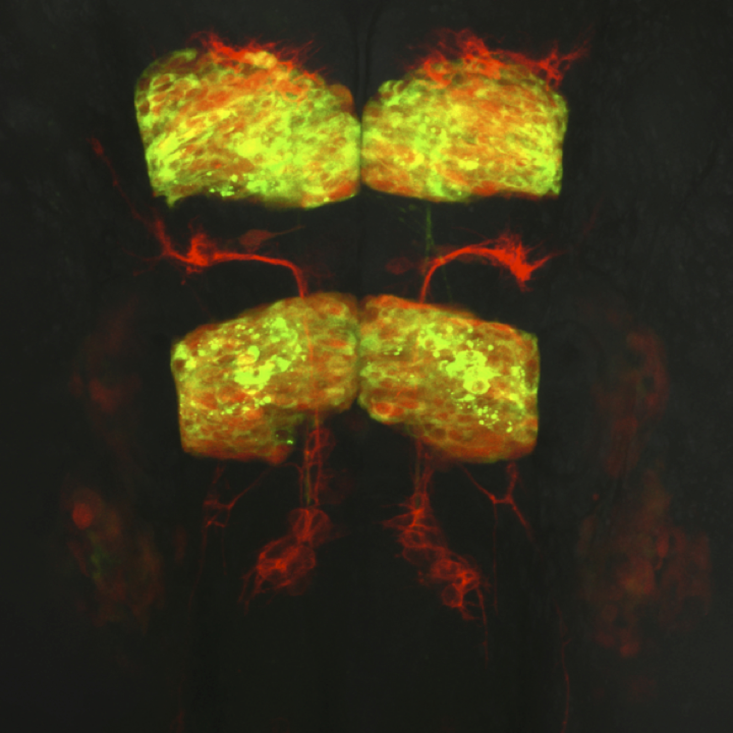
Dorsal view of a live transgenic zebrafish embryo’s hindbrain at 24 hours post fertilization. Facial branchiomotor neurons express membrane red fluorescent protein and migrate posteriorly through hindbrain segment rhombomere 5 while leaving behind trailing axons. Rhombomeres 3 and 5 expresses a red fluorescent protein marker as well as a cytoplasmic protein tagged with green fluorescent protein.
This post is part of a series on a day in the life of developmental biology labs working on different model organisms. You can read the introduction to the series here and read other posts in this series here.
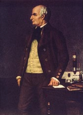
Jonathan Bard, long-time colleague and friend in Edinburgh, recalls the life and work of the mouse developmental anatomist Matt Kaufman, who died in August 2013.
Matt Kaufman was the leading mouse developmental anatomist of his generation and played key roles in three major aspects of mouse research. He was the person who initially dissected out the blastocysts from which he and Martin Evans went on to culture embryonic stem cells. He wrote The Atlas of Mouse Development that taught mouse developmental anatomy to a generation of molecular biologists and he subsequently worked with his colleagues in Edinburgh to produce the anatomical infrastructure of mouse informatics. In all, he wrote more than 200 papers and a dozen books on mouse development as well as on Scottish medical history.
Matt grew up in a very orthodox and poor Jewish family in London. His parents saw him as a future scribe, writing the five books of Moses on parchment. One result of this early training was his exquisite handwriting; another was some formal training in Jewish learning and a long-lasting love of scholarship. This plan fell foul of a grammar school education, medical training at the University of Edinburgh and a taste of research in reproductive physiology there with Professor Anne McLaren. He decided on an academic career and moved to the mouse lab of Professor C. R. (Bunny) Austin in Cambridge in 1970 to do a PhD on mouse parthenogenesis. After this, he spent two years at the Weizmann Institute in Israel, and returned to Cambridge as a demonstrator, then lecturer in anatomy. By 1980, he had published almost 30 mainly first-author papers – his capacity for hard work and rapid writing was extraordinary!
Around then, he started his collaboration with Martin Evans who had been working on embryonal carcinoma (EC) cells in Cambridge. Matt’s experience with isolating early mouse blastocysts and Evans’ knowledge of culture enabled them to obtain cell cultures from the inner cell masses: originally called Evans- Kaufman (EK) cells but now known to the world as embryonic stem cells (ESCs). The five papers that they published together in 1981-1984, together with those of Gail Martin in the USA at around the same time, provided the baseline knowledge for all subsequent work on the genetic manipulation of ESCs, their use in making transgenic mouse strains and their potential for regenerative medicine. It has to be said that Matt took no part in this later work because he was really interested in the anatomy of the developing mouse.
In 1985, Matt was awarded the chair of anatomy at the University of Edinburgh, a post first established in 1705, and remained there until he retired in 2007. The University did not come to view this as an entirely successful appointment for two main reasons. First, they had hopes that he could introduce molecular biology into the department, but he was not really equipped to do this. Second, they wished to reduce the amount of anatomical teaching in the curriculum to make room for genetics and social medicine; this shocked Matt because he believed that all of medicine stemmed from the cadaver and he fought the inevitable changes at every step. In hindsight, Matt lost more battles than he should have – probably because, for all his academic strengths, he was a dreadful university politician! Things were not helped by his ill health: in 1995 or so, he was diagnosed with polycythaemia (a blood malignancy) and spent the rest of his life on the maximum dose of methotrexate his body could tolerate – he lived in a state of continuous pain and discomfort.
Despite his political and medical problems, he continued research on the developmental anatomy of the mouse and on how it was affected by polyploidy and by external factors such as alcohol and anesthesia. These essentially minor papers were part of a much bigger project (on which he worked every evening): to put on paper everything that he knew about mouse development. This work all came together in 1992 when he published The Atlas of Mouse Development, a book that turned out to be, by the standards of mouse development, a major bestseller and is still in press (a supplement is being planned that will be part of his legacy). It contained around 1500 micrographs of sections from all 26 Theiler stages of mouse embryo development, each labeled to show the tissues, together with detailed explanations of what was happening at each stage. The book explained mouse development to a generation of molecular biologists making and analyzing transgenic mice, who had the shock of realizing that doing the molecular genetics was far easier than understanding the resulting phenotypic changes!
About then, mouse informatics was getting off the ground both at the Jackson Laboratory in Maine and at the MRC Human Genetics Unit in Edinburgh and it was soon clear that more was needed than just the basic genomics. I approached Matt and suggested that, for each Theiler stage, we should integrate all of the tissues into a parts-of hierarchy. One benefit of this would be that his beloved Atlas would get a proper index; another, of course, would be that the hierarchy (which soon became an ontology) could be used as the anatomical core for a database to which could be added tissue-associated data (e.g. gene expression). This was implemented by the Jackson Laboratory and is now a key feature of the mouse informatics resource there.
At the MRC Human Genetics Unit, Richard Baldock, Duncan Davidson and I decided to go further and to make 3D reconstructions of mouse embryos from the slides that Matt had used for his Atlas and to include all the tissue boundaries so that gene expression could be shown accurately. This led Baldock and Davidson to produce the first online graphical atlas for capturing gene expression data, to which Matt continued to provide detailed anatomical input up until about 2011.
This is not to say that he had stopped doing other research. Apart from mouse work, he had a long interest in the medical history of Scotland in general and the University of Edinburgh in particular. Starting in 1992, he produced a series of articles and books on medical history, including an important history of the chair of anatomy at Edinburgh that was part of the exhibition he organized to celebrate the 300th anniversary of its initiation. By this time, Matt was near retirement and there was some degree of reconciliation between him and the University. Indeed, there was no argument when, in 2007, he was finally and properly elected to the Royal Society of Edinburgh.
Matt was fortunate in having a very happy home life. Claire, his wonderfully tolerant wife, put up with his never-ending work, his 1930s Lagonda car that was always breaking down and the sheer quantity of working papers, historical documents and research publications, not to mention histological slides, that littered the house as well as his very large office. Indeed, it was only those who had to deal with Matt’s non-research, academic life who did not get on well with him! Everyone else appreciated his somewhat eccentric interests, his kindness, his ability and his old-fashioned courtesy. For me, he was a very good colleague and friend – I miss him
Last week, The Company of Biologists hosted a workshop on “The Evolution of the Neocortex: How Unique are We?”. Some of the people who attended that meeting will be writing about it on the Node in the near future, so I won’t steal their thunder by telling you all about it. Suffice to say it was a fascinating meeting on a complex and fast-moving field, and I came away with a much better appreciation of the commonalities and distinctions of the human brain compared to our primate and mammalian relatives, as well as the wide range of techniques used to understand it – from high-throughput transcriptomics to the endocasts from skulls of our hominid ancestors.
Following on from this meeting, on September 26th we hosted a public event at the Royal Institution (Ri) in London. Free to attend for anyone, this event featured talks from two of the speakers at our workshop, Arnold Kriegstein and Svante Pääbo. Arnold’s talk covered the evolution of the human neocortex, taking the audience on a journey through the development of the mammalian brain and discussing how the increased size and complexity of the human brain (which contains no fewer than 88 billion neurons!) might have evolved. Featuring beautiful movies from his lab (from Hansen et al., Nature 2010) that show the division of neural progenitors in human foetal brain cultures and discussing data from other researchers on how changes in brain size and shape might affect cognitive function, Arnold made this complex topic accessible to the non-specialist audience – a feat not to be under-estimated given how poorly it is understood even among the experts!
In the second talk, Svante described his fascinating and well-publicised work on sequencing the Neanderthal genome. His work has shown that not only did Neanderthals and our modern human ancestors co-exist in Europe and Asia, but they also interbred: the genome of an average modern-day non-African is around 2% Neanderthal! Svante’s work allows him and his colleagues to identify sequences that are unqiuely human; amazingly they have found protein-coding changes between Neanderthals and humans in only 87 genes. Now, they are in a position to try and understand how these human-specific changes might affect protein function, and whether these might explain any of the characteristics that make humans human.
Unsurprisingly, these two talks provoked considerable interest from the audience, and the free-flowing question time covered topics such as whether we’d ever be able to reconstruct a Neanderthal, whether computers will ever be smarter than humans, and what defines consciousness! The short answers to each of these being, respectively, “almost certainly not”; “possibly in some aspects’”; and “we really have no idea”! The drinks reception following the lecture also provided the audience with ample opportunity to quiz the speakers further on their work. Overall, it was a fascinating event held at a beautiful and historic venue; many thanks to the team of the Ri for making it run so smoothly!
This was the second event The Company of Biologists has hosted at the Ri, and we hope to be collaborating with them again in the future; we’ll let you know if and when the next one comes up!
For those interested in hearing more, you can listen to the following podcasts of Arnold’s and Svante’s talks:
The journal promotes many of its papers and other content not only via its home pageandemail alerts, but also via its Twitter account. We use Twitter to highlight particularly interesting papers and reviews from our issues, and also other content such as interviews and movies. We also tweet from meetings. However, we recognise that not all Development readers are avid Twitter users, and we’re keen to maximise our reach. So we have now created a facebook page for the journal! We hope that you will visit the page and like us to receive updates about articles and more on your feed!
We would like to thank Jackson Hoffman and Brad Merill, from the University of Illinois in Chicago, who kindly gave us permission to use the beautiful confocal images of mouse embryos that feature so prominently in our new facebook page and in our new look on Twitter.
We hope that you will find our new facebook page useful, and we welcome your feedback! Please like our page, and comment, like and share our posts!
To the inspecting eyes of a budding scienziato the Reggian countryside was not simply teeming with life, it also posed myriad of intriguing questions. How do bug wings work and can you stick them back once you have removed them? How far are the stars that can be seen in the sky, and are natural fountains really just the tears of sad, beautiful girls lost in the woods? Armed with a healthy dose of skepticism, the young Lazzaro Spallanzani, one of the brightest minds of his age, was more than happy to tackle these questions. In the end, his curiosity and stubborn adherence to natural philosophy derailed the legal career much wanted by his father, and “l’astrologo”, as his friends knew him, ended up as a teacher of logic, metaphysics and Greek at the University of Reggio Emilia.
Today Spallanzani is most credited for his work debunking the mistaken theory of John Tuberville Needham and Georges Buffon about the origins of microscopic lifeforms. The theory of “spontaneous generation”, as it is remembered, argued that really small lifeforms can originate from inanimate matter: maggots from dead flesh, microscopic “animalcules” (protists and bacteria, effectively) from water and mud. This was the celebrated, dominant theory of the age, yet it took Spallanzani only a few long-necked, easily sealable glass bottles and some freshly boiled broth to lay it to rest for good.
Equally ingenious were, however, his experiments on regeneration, which he conducted in order to test what at the time seemed like the equivalent of spontaneous generation for embryologists, epigenesis. Following his childhood instincts, Spallanzani sectioned everything that crawled or ran in the nearby woods. He cut worms and snails far and wide, removed tails and limbs of tadpoles and salamanders with methodological rigour and watched them to regenerate. In fact, almost single-handedly he put the field of regeneration on a solid scientific footing.
What he saw in these experiments thrilled him. His results, detailed in his seminal work, Prodromo and his letter to Charles Bonnet, show that Spallanzani realized that regeneration was more than simple growth. It could not be simply explained by the expansion of some preexisting “germs” after injury. Yet, if not exactly heresy, such views contradicted the dominant theory of the age, namely preformation.
The adherents of this theory claimed that an organism is preformed from its origins, and development is mere growth. Spallanzani was originally sympathetic to the preformationist ideas and some of his results were in line with these: limb regeneration for the most part was about growth, after all. Yet there were significant observations, especially about the early steps of the regenerative process, that were hard to square with the preformationist ideas. For example, the intricate circulatory system of the regenerating limb stump formed in a stepwise manner, and often was clearly different from the original circulation. And this strongly suggested that growth could not be the whole story, there were also epigenetic forces at work, and preformationists (including Bonnet) were mistaken. (more…)
I am Gary McDowell, a postdoctoral researcher at Tufts Centre for Regenerative Biology and Developmental Biology. I have just finished a postdoctoral position in a mass spectrometry lab at Boston Children’s Hospital/Harvard Medical School and I was the only frog researcher in the group. I earned my “frog stripes” studying for my PhD in the lab of Anna Philpott at the University of Cambridge, working on the stability of a particular transcription factor that regulated neurogenesis. Recently, however, I have been trying to look at many peptides and proteins across many different developmental stages by proteomics, as well as doing in vivo experiments to validate proteomic findings.
Xenopus laevis, the organism I primarily work with, is a claw-toed frog from Southern Africa that lays many large eggs, traditionally used for embryology but also for biochemical studies, as extracts can be made from eggs and embryos to provide active cytoplasm in a test tube, that can undergo cell cycles and be used to study many processes, such as protein ubiquitylation and degradation. The eggs and embryos are also easily subjected to microinjection in a highly targeted manner – for example, injecting into one of the two cells formed after the first cleavage means that only one half of the embryo is treated, as cells largely do not move between the left and right sides of the embryo formed at this point. This gives a great internal control for scoring phenotypic changes. The fate map of cells in the early embryo is well-characterised, so that injections into one of four, eight, even 32 cells can be used to target particular germ layers, organs and tissues.
Xenopus are distributed around the world as they used to be used as a pregnancy test in hospitals – female frogs would be injected with urine from women thought to be pregnant and if the frog began to lay, it meant hormones marking pregnancy were present in the urine! This worldwide distribution has led (I believe somewhat unfairly) to Xenopus being blamed for the spread of the devastating chytrid fungus. Xenopus can carry, but do not die of this fungus; but as the fungus can also be carried on the wings of birds, Xenopus may simply be the subject of bad publicity.
Xenopus is an excellent organism for biochemical experiments. Unique among my colleagues in the mass spectrometry lab, I am not limited by the amount of material I require for mass spectrometry experiments. There are neither endless dissections of multiple mice to harvest enough tissue, nor continuous growing of cells in culture to produce the literally tens (sometimes hundreds, for post-translational modification studies!) of litres of cultured cells required. I simply need a few eggs or embryos to get more than enough protein to work with, as early Xenopus laevis embryos are so large and protein-rich. This applies to many biochemical manipulations in Xenopus – the amount of material required for making extracts to study biochemical processes, for Western blotting or RNA-Seq is a small fraction of the material that is easily available from a single frog. To do in vivo follow-up, there are multiple strategies for embryo micromanipulation, explant culture and microinjection of mRNA (for protein overexpression) or morpholinos (small synthetic oligonucleotide mimics that prevent target mRNA translation) for phenotypic analysis, including in situ hybridisation and immunohistochemistry.
One disadvantage is that Xenopus has not been sequenced to the level of many other organisms, which means that there is not yet a great protein database publicly available for searching mass spectrometry data. What is great about mass spectrometry data, though, is that it is “futureproofed”: the same data can be searched again and again using new and improved databases as they become available. This lack of genetic information means that there are also fewer mutant frog lines available compared to other model organisms – a deficiency which is currently being addressed by the Xenopus community.
The frogs that I use are members of the large colony at Harvard Medical School, provided by Marc Kirschner. They usually live in a very large facility, with huge tanks swarming with lively frogs. Every so often, however, some are moved down to the satellite facility next to the frog lab (PICTURE 1). Here they are housed in smaller tanks, and are fed every day. The room is windowless, with a scheduled light cycle so that they still experience “day” and “night”. They are checked on regularly to make sure they are not stressed, and have a continuous aquatic system providing a flow of water suited to their requirements. Feeding is carried out on a daily basis. Several labs use the same frog room so sometimes it’s a chance to catch up on what’s going on and what different people are doing – as the focus of the different labs is quite varied.
Xenopus housing
Xenopus laevis and their embryos tend to be happier in cooler temperatures; frog labs tend to be held at 18 ˚C which keeps everyone very dynamic (but is unfortunate for those of us who wish to cast our own SDS-PAGE gels). However in the current facility I use, there is a room set aside for frog work which is kept cooler – so unfortunately it can be a cold, windowless affair for long days in the frog room!
My Day in the Frog Lab
4 pm, the day before:
My day actually begins the evening before my planned work – to get the female frogs to lay, they need to be primed with human chorionic gonadotrophin (which nowadays we obtain in a purified form, and not from pregnant women).
9.30 am
After being primed, the next morning eggs can be seen gradually falling from the frog’s cloaca (frogs have only one exit for eggs, urine, and faeces PICTURE 2).
Two female wild type Xenopus laevis
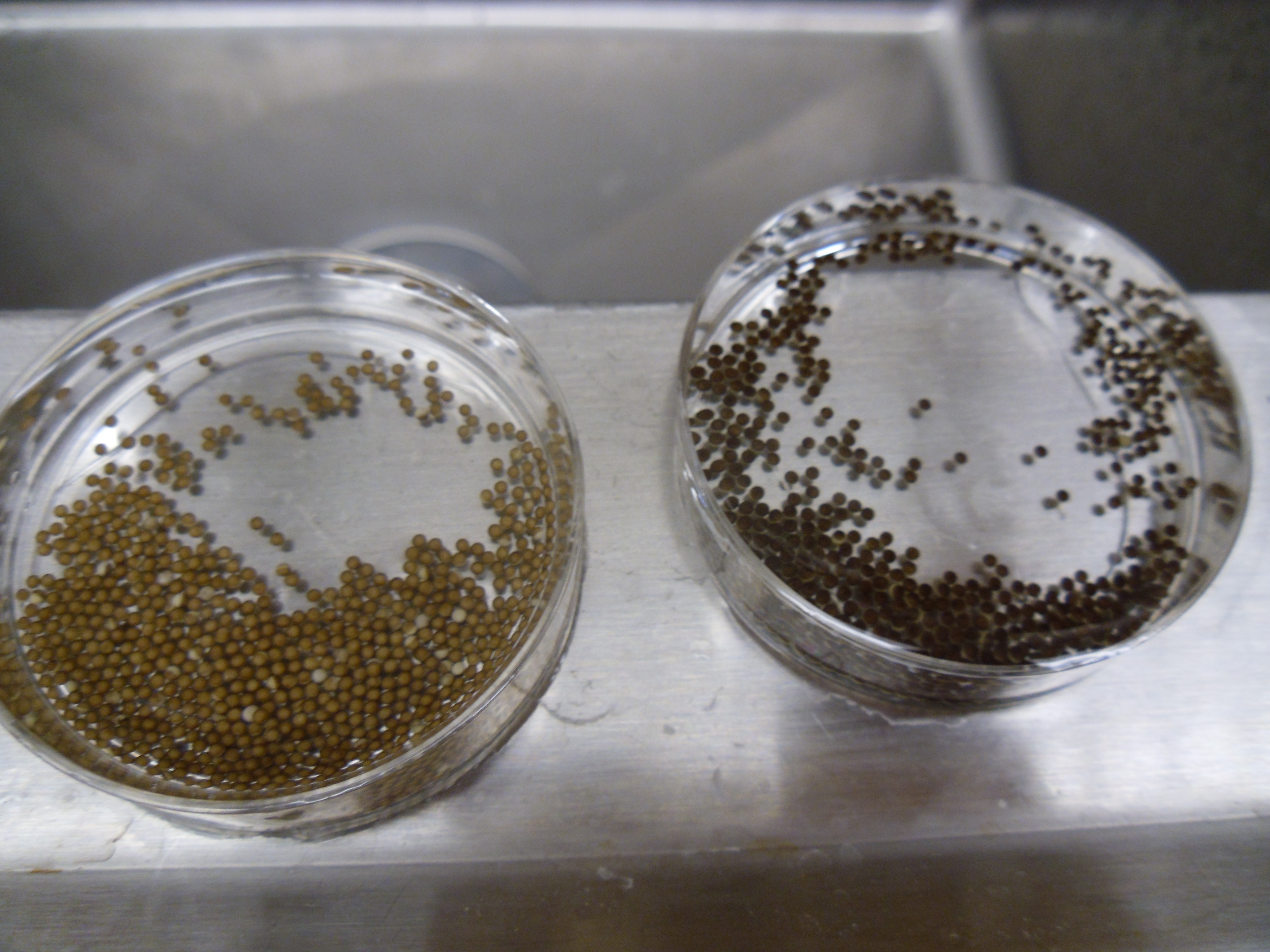
To collect a large amount of eggs at once, however, I have to carry out a procedure rather alarmingly called “squeezing”. Actually, it’s more like a frog massage – I pick up the female and gently stroke her back, which massages out the eggs that fill a large proportion of her body cavity. The eggs are caught in a petri dish of salty solution and fertilised in vitro by the addition of sperm, which is spread over the eggs and then induced to enter the egg by flooding of low-salt solution. The fertilisation of the eggs hopefully results in a visible transformation – eggs are randomly oriented with respect to their pigmented animal hemisphere (PICTURE 3) until fertilisation induces microtubule rearrangement and rotation of the cortex so that the embryos are all oriented with their animal hemispheres pointed skywards (PICTURE 4) (the reason for this is not clearly known, although it is possible that in the murky waters which Xenopus naturally inhabit, dark camouflage may hide the eggs from predators swimming or moving above). One further preparation is required before my work can begin – the removal of the jelly coats that surround the embryos. These thick coats protect the embryos, but will prevent me from microinjecting them or easily lysing them for sample preparation, so they need to go. This requires a brief treatment in cysteine solution, which breaks down the coats and releases the embryos in a more easily manipulated (but also more easily ruptured) form.
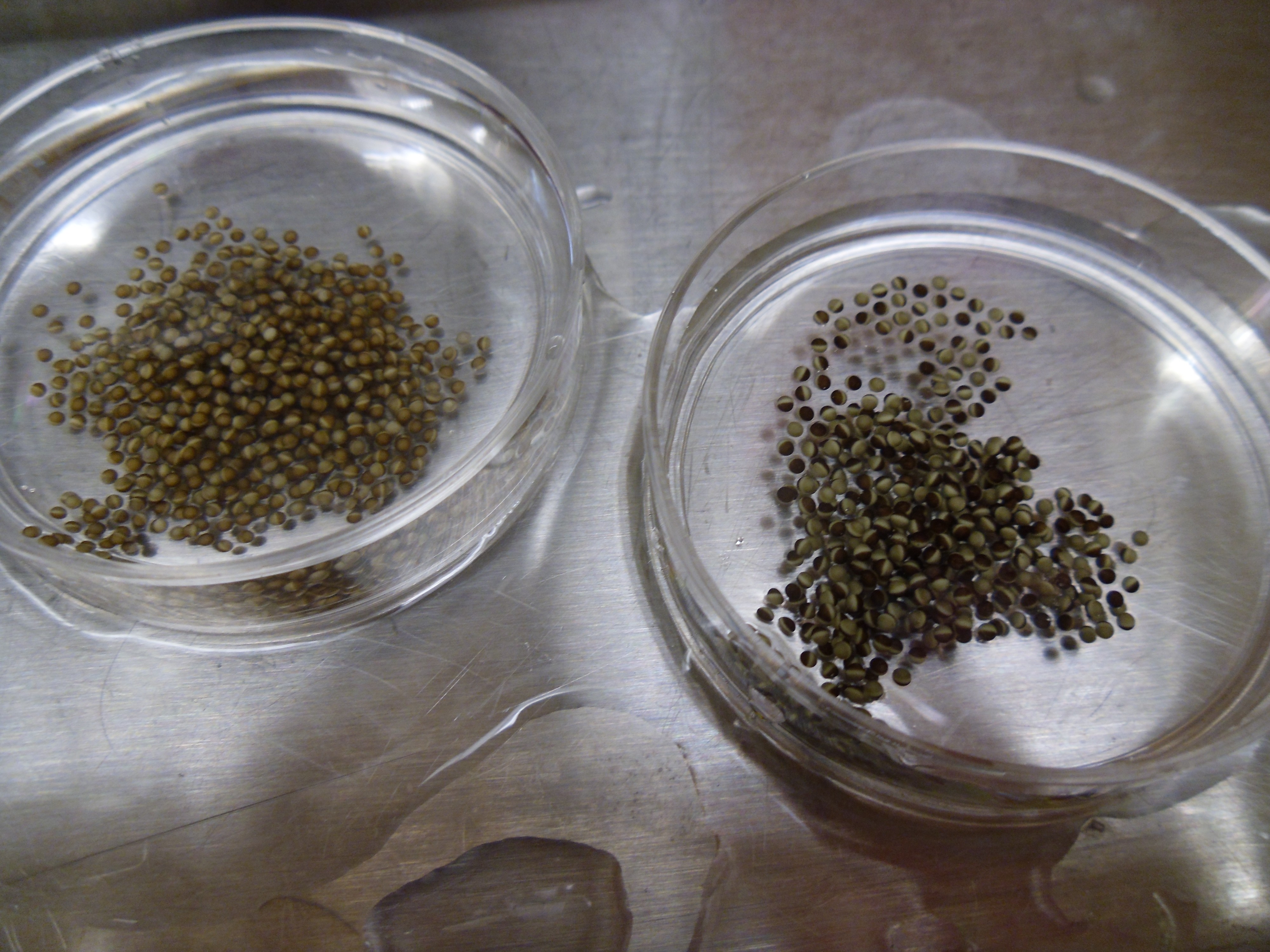
Unfertilised Xenopus eggs
Fertilised Xenopus embryos
The embryos I’ve prepared today are going to be used for two experiments: for the first, I will simply take a few embryos at different timepoints and snap-freeze them in liquid nitrogen. These will then by lysed, and the protein-rich material within will be digested with the protease trypsin, and analysed by mass spectrometry, after the digested peptides are sorted by liquid chromatography. This will give me information about the protein content of the embryos at different stages of development.
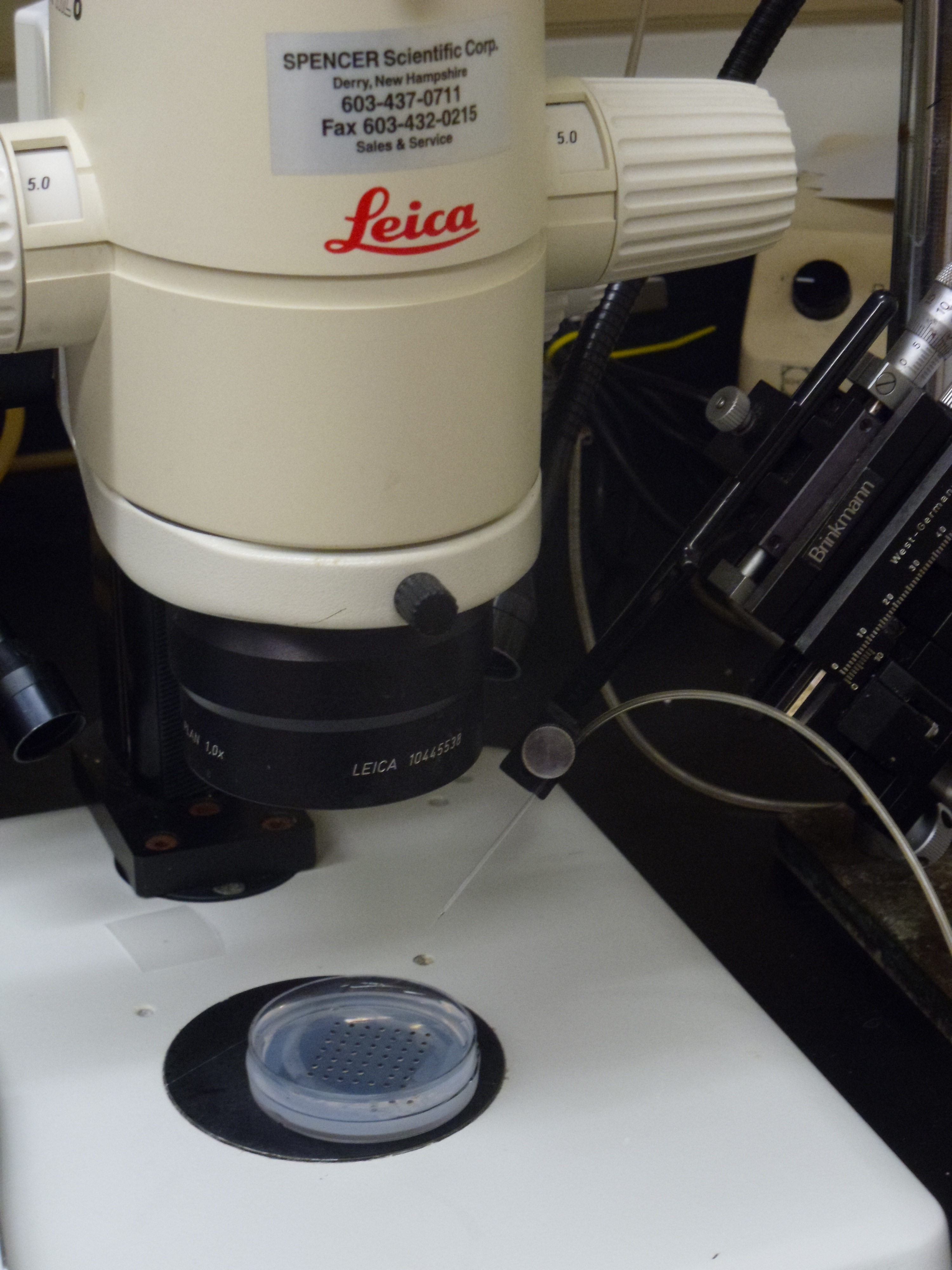
The other experiment I am carrying out is a much more common protocol in the Xenopus field: microinjection of morpholinos. Morpholinos are small synthetic oligonucleotide-like molecules – so-called because they are made using morpholine rings instead of ribose-based sugar rings – that can block mRNA translation either altogether at the point of initiation, or by blocking splicing events. The morpholinos I’m injecting are targets identified from a proteomic screen using differentiating stem cells and our hope is to use Xenopus as an in vivo tool to model the effects of blocking the translation of these targets into protein, and to look at what happens. Morpholinos can be tricky – they are prone to precipitating out of solution at temperatures like that maintained in the frog room! Microinjection itself is a very zen art, that requires great patience and strength of will. Individual embryos are injected using a calibrated glass needle connected to a high-pressure air supply and controlled by a micro-manipulator (PICTURES 5, 6). If the embryos are good, they will be like perky beach balls and the needle will press against the embryo surface, which pops back smartly as the needle enters. An all-too-common problem with novice injectors is breaking the needle, which is fragile. If the embryos are not quite so healthy, they may require some more force to penetrate the surface, resulting in needle breakage.
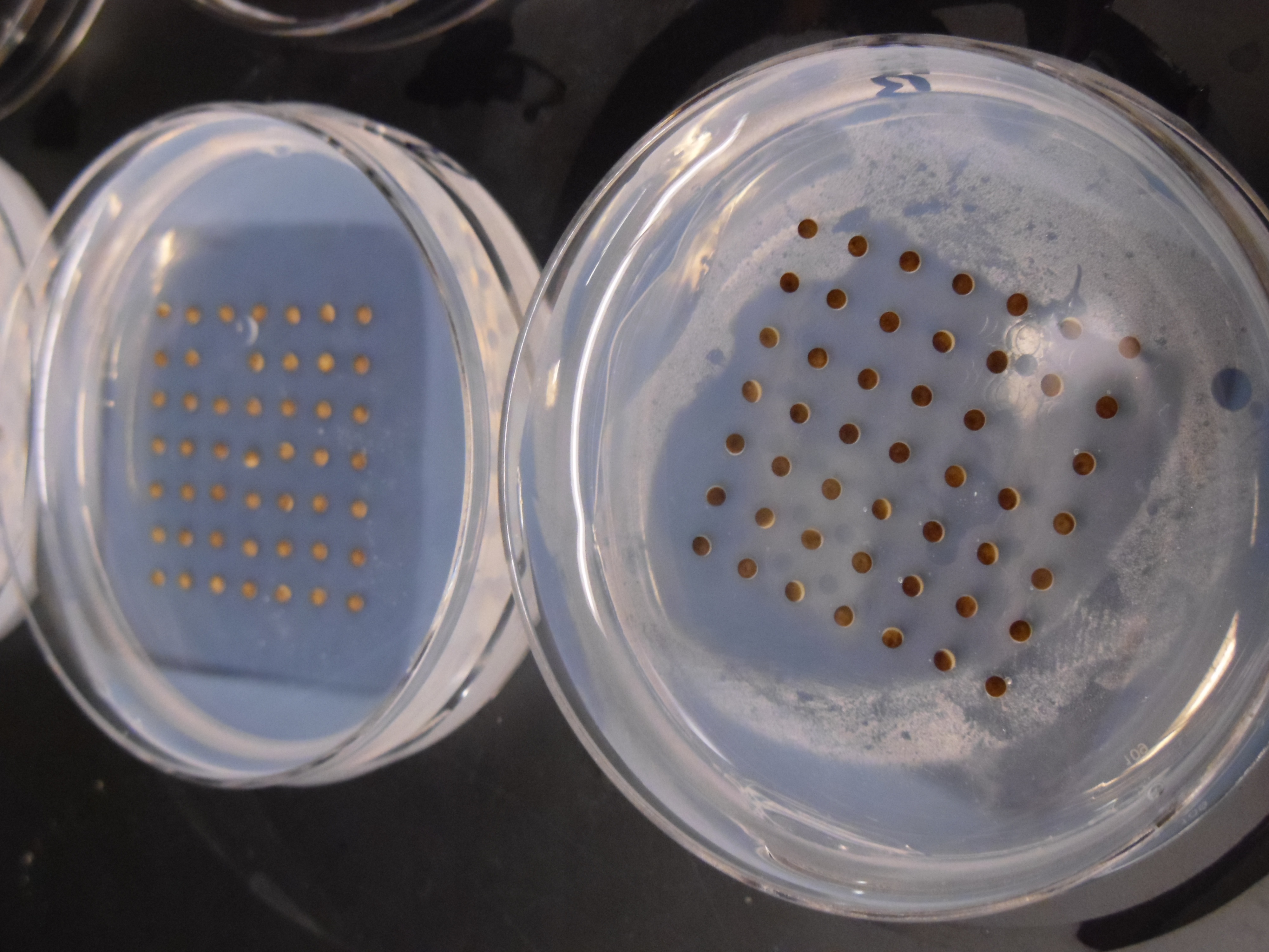
Xenopus embryos ready for microinjection
Microinjection setup
4 pm
At the end, a nice dish of injected embryos was left to develop. I come back later in the afternoon to move the embryos out of the injection dishes, clear out any dead embryos (and today there are not many – just some eggs that clearly were not fertilised – hooray!) and place the embryos in new dishes with fresh buffer.
These are then left to continue development and to be scored for any possible phenotypic changes. Different concentrations of morpholino are injected to look for effects on development and to see whether our targets are having any effect in vivo after being identified by proteomic screening.
The advantage to using Xenopus for this in vivo approach is that they can be used as a screen for large numbers of candidates – provided that you have a hardy frog researcher ready to inject! The embryos are large and easy to manipulate and most importantly they develop very quickly. You can think of an idea at the weekend, start the experiment on Monday and by the end of the week have an answer, far faster than some other vertebrate model systems. And whilst Xenopus may not be understood to be a great model system for diseases in humans, they are a great tool for understanding the mechanisms of these diseases (for a discussion on this topic and why Xenopus is such a useful tool for those of us using it, see Sive, H. (2011) Disease Models and Mechanisms 4, 137-138 ). Certainly as someone who trained initially as a chemist, the discovery of an organism that provides such great in vitro and in vivo tools has revolutionised the way I do science now and hopefully for the years to come.
This post is part of a series on a day in the life of developmental biology labs working on different model organisms. You can read the introduction to the series here and read other posts in this series here.
To uncover the mysteries of development, developmental biologists use an amazingly wide range of systems – from cells in culture to live mouse embryos, and from classical model organisms to unusual critters.
We read the papers and listen to the talks about the work using these organisms, but we only really know the details of the model we work with. So what does an axolotl eat? How do you get a frog to lay eggs? How do you make a transgenic Arabidopsis? Why do fly researchers always talk about collecting virgins?
‘A day in the life of…’ is a new series of posts that will aim to answer these and other questions by giving you an insider’s view of what is like to do developmental biology using different organisms. Our first post is a day in the life of a Xenopus lab, where postdoc Gary reveals the mysteries of doing research on frogs. In the next few months we hope to feature posts on ‘a day in the life of’ many other lab organisms, and we already have a few exciting articles lined up. However, with so many different organisms being used in labs around the world, we need your help! If you are interested in writing about what is like to do developmental biology in your model organism, why not drop us an email? It would be great to have your participation!
We hope you will enjoy this series! You can read all the posts so far here.
 (3 votes)
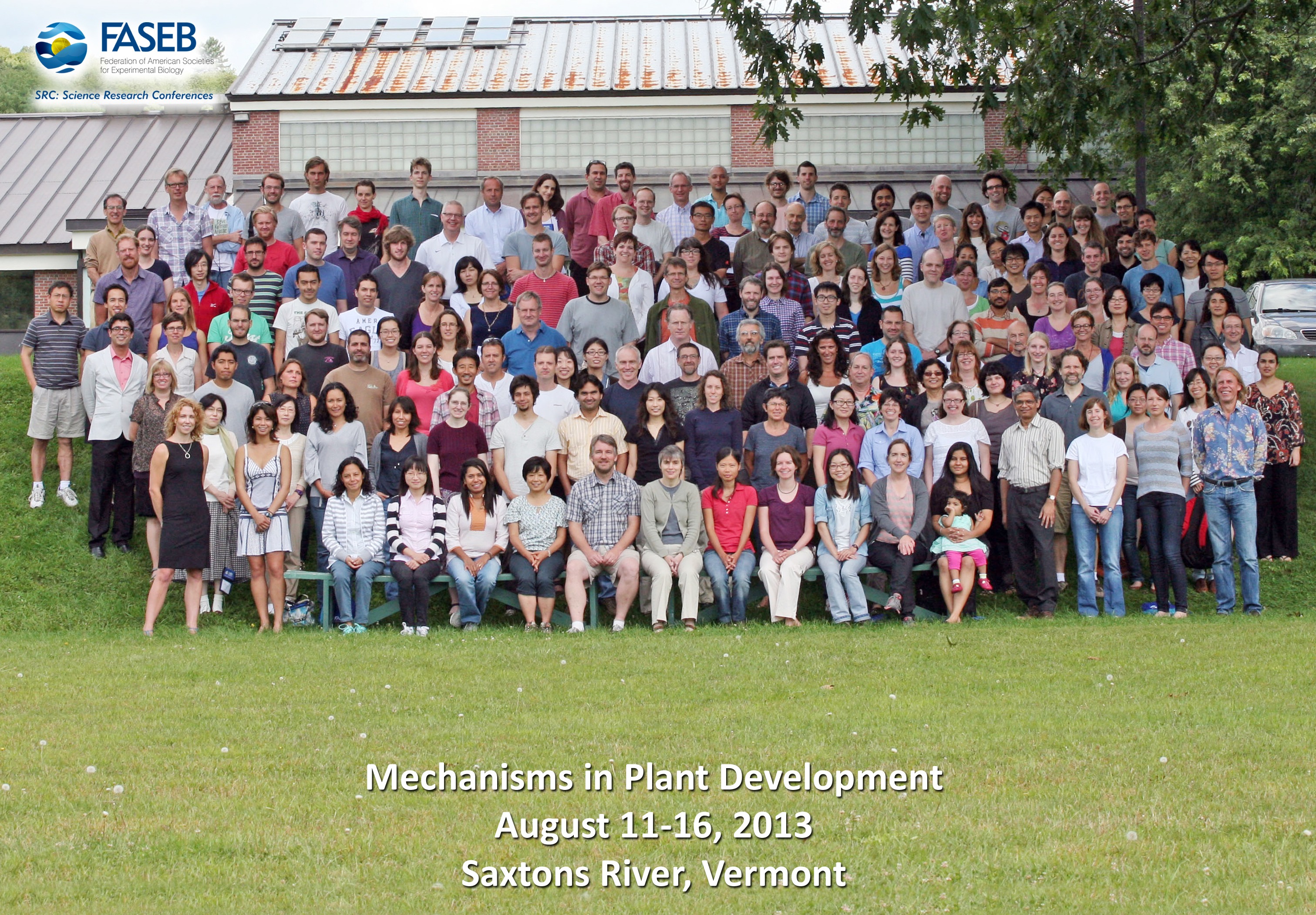

 This post is part of a series on a day in the life of developmental biology labs working on different model organisms. You can read the introduction to the series
  (17 votes)