The community site for and by
developmental and stem cell biologists

Barcelona hosts major European conference on Drosophila research

Posted by , on 14 October 2013

      – On 16-19 October, 700 scientists will meet for the 23 European Drosophila Conference in the Palau de Congressos in Barcelona.

      – Drosophila melanogaster, also known as the fruit fly, is a key model organism in genetics and essential for our understanding of disease.

      – Jules Hoffmann, French researcher and 2011 Nobel Laureate in Medicine, will deliver the opening plenary lecture on Wednesday afternoon.

      – Barcelona hosts a large concentration of biomedical research groups specialized in Drosophila, making the city a European and worldwide reference for this area of research.


From 16-19 October, more than 700 scientists from around the world will attend the biennial European Drosophila Research Conference in Barcelona, in the city’s Palau de Congressos. With 7 plenary lectures, 300 talks, 20 sessions and 400 posters, it is the biggest European event focussed on basic and biomedical research that uses the fly fruit, Drosophila melanogaster, as a model system.

“The European scientific community has been recommending Barcelona as the host site for this conference for some years. The number of participants has nearly doubled since the last edition, testifying to the attractiveness of the field and to Barcelona’s powerful cluster of leading scientists working in this area”, explains the organizing committee, composed of scientists at the University of Barcelona, the Spanish National Research Council (CSIC) and the Institute for Research in Biomedicine (IRB). The 2011 Nobel Laureate in Medicine, Jules Hoffmann, from the University of Strasbourg, will open the meeting with the plenary lecture “Innate immunity: from fly to humans”.

Hoffmann was awarded the Nobel Prize, together with Bruce A. Beutler y Ralph M. Steinman, for their discoveries on the activation of innate immunity, which has allowed scientists to develop new methods to fight disease, including the latest generation vaccines or cancer therapies based on immune system activation. Their discoveries are essential to our understanding of the occurrence of autoimmune diseases –when an organism’s own immune system attacks itself, such as in the case of type 1 diabetes- and has opened new paths for treatment.

“The article by Hoffmann in Cell 15 years ago was one of the catalysts for current biomedical research using Drosophila to study human diseases. This is a growing tendency, which is reflected in the scientific sessions of the conference”, explains Marco Milán, ICREA scientist at the Institute for Research in Biomedicine (IRB), and co-organizer of the conference, along with Cayetano González (IRB), Jordi Casanova (IRB/CSIC), Enrique Martín Blanco (CSIC) and Florenci Serras (UB).

The fly has been used to study basic biology for over a hundred years “and it is still an exceptionally good organism for this kind of research”, adds Milán. “Since Hoffmann’s discoveries, the fly has also proved to be effective for modeling diseases such as cancer, Alzheimer’s, Parkinson’s, diabetes, or drug addiction.” The sequencing of the fly and human genomes has revealed that the species share 70% of genes associated to diseases. Research on Drosophila has yielded no fewer than 6 Nobel Prizes.
 
 
Stem cells and cancer

Topics to be addressed at the conference include stem cells and cancer, which are also the theme of two of the five special workshops scheduled. Organisms need stem cells in order to repair tissues. Their dysfunction is associated with cancer and early ageing of tissues. Stem cells from Drosophila’s nervous system and gut are used to identify new genes involved in tumorgenesis. The plenary lecture “Modeling Cancer in Drosophila”, by scientist and conference organizer, Cayetano González, ICREA scientist at IRB and a recipient of a prestigious ERC Advanced Grant, will address these studies. In 2005 González demonstrated that the abnormal division of stem cells in Drosophila’s nervous system generates malignant tumors.
 
 
Other prominent speakers

Among other scientists of international standing invited to deliver plenary lectures, is Elisabeth Knust, Director of the Max Planck Institute of Molecular Cell Biology and Genetics (Dresden, Germany). Knust identified the CRB1 gene in Drosophila, which is also present in humans. Its mutation is linked to development of Retinitis Pigmentosa, an inherited, degenerative eye disease that causes blindness. Knust’s research has improved the knowledge of essential biologic processes as well as helped to develop new therapies for patients affected by retina dystrophies.

Ginés Morata (Rioja -Almería-, 1945), awarded the 2007 Prince of Asturias Prize for Technical and Scientific Research along with the English biologist Peter Lawrence, will also be speaking at the event.” An international expert in the field, his studies focus on the “biological architecture” of Drosophila. His research, conducted at the Centro de Biología Molecular Severo Ochoa (CSIC-UAM) in Madrid, is also related to tumor generation and ageing.

Link to the press release: http://www.irbbarcelona.org/index.php/en/news/irb-news/corporative/barcelona-hosts-major-european-conference-on-drosophila-research


Thumbs up (3 votes)
Loading...

Categories: Events, News

Navigate the archive

Use our Advanced Search tool to search and filter posts by date, category, tags and authors.

Scientists disclose minute-by-minute details of the biological clock of skin stem cells

Posted by , on 14 October 2013

Researchers lead by Salvador Aznar Benitah describe in detail the cyclic activity of the genes in skin stem cells during the course of a day.

The correct balance of the biological clock of stem cells affects their function, and its disruption causes aging and can lead to predisposition to skin cancer.


Our skin regenerates daily and has to face harmful environmental factors such as sunlight and pathogens. In an article published in the journal Cell Stem Cell, scientists led by ICREA Research Professor Salvador Aznar Benitah, who has recently moved his lab to IRB Barcelona, have described how the circadian rhythm (internal biological clock) modulates the function of human skin stem cells to achieve optimum regeneration and protection against harmful agents.

Thus, during long periods of exposure to pathogens or UV light, stem cells of the human skin protect themselves. In contrast, during the evening and night, they produce new keratinocyte. Found in the uppermost layers of the skin, keratinocytes, which are dead cells rich in keratin, provide an impermeable protective barrier. Over the course of the night the stem cells regenerate tissue and replace keratinocytes that are damaged or that have been lost during the day.

“Stem cells have some genes that control their biological clock and that determine peaks of activity and intervals of inactivity over 24-hour periods. In this study, we describe how the cells manage to perceive what time of the day it is. This precision allows the stem cells to adapt their activity to the time of day and to its environmental conditions,” explains Salvador Aznar Benitah, who conducted this study at the Center for Genomic Regulation (CRG) and who has recently settled his new lab “Stem Cells and Cancer” to IRB Barcelona.

In 2011, Aznar Benitah and collaborators (amongst them Eduard Batlle from IRB) previously reported on the relevance of circadian rhythms in the regulation of skin stem cells. At that time they found that the cells discriminate between day and night. On this occasion, the researchers have managed to monitor the activity of the stem cells minute by minute. “We now know how the cells know exactly what time it is and how, thanks to this information, they regulate their activity accordingly,” adds the head of the study.

The study also demonstrates that a disruption in the internal biological clock deeply affects the correct function of stem cells and leads to tissue aging and potential predisposition to skin cancer.


Reference article:

Human Epidermal Stem Cell Function is Regulated by Circadian Oscillations
Janich P, Toufighi K, Solanas G, Luis NM, Minkwitz S, Serrano L, Lehner B and Benitah SA.
Cell Stem Cell (2013) DOI: http://dx.doi.org/10.1016/j.stem.2013.09.004


This article was first published on the 10th of October 2013 in the news section of the IRB Barcelona website 

Thumbs up (3 votes)
Loading...

Categories: Research

A day in the life of…a mouse lab

Posted by , on 11 October 2013

I work at the National Institute for Medical Research in London. My project is currently investigating the cues governing development of the limb, with an emphasis on cartilage patterning in vitro. I have a PhD in developmental biology and have been working with mice for over eleven years.


The mighty mouse.The mouse is a fantastic model system for several compelling reasons. They have a relatively short generation time, their genome is closely related to ours, and the potential for genetic investigation is virtually limitless. However, working with a protected model organism brings with it ethical and moral considerations, and of course means adhering to strict regulations set by the Home Office. This includes the rigorous application of the 3Rs (replace, reduce and refine), which involves only breeding the mice we need and being vigilant with our experimental design. We are lucky to have the help of trained technicians who cover a lot of the day-to-day maintenance of our colonies, including one long-suffering soul whose job is to process the genotyping of our colony on a weekly basis. Since being a researcher, I’ve done all of these tasks myself at some point or other, and while this gives you a great depth of knowledge, sometimes it’s exceptionally nice to have a helping hand.


mouse_house_copyA bit about our furry friends. To get one mouse from another takes a relatively short 2 months, and gestation period is 19-21 days. Mice are born pink and hairless, deaf and with closed eyes, and require constant attention and feeding from their dam. However, they develop rapidly and are weaned at 3 weeks of age, by which time the pups are fully autonomous and extraordinarily energetic. A friend once coined the phrase ‘popcorn mice’ to describe the astonishing acrobatics a weaner mouse is capable of, which means that nothing sharpens the reflexes like handling weaners at the peak of their game. The litter will reach maturity less than a month after weaning (we breed our males at 8 weeks and females after 6-7 weeks). Our mice are supplied with cosy woodchip bedding with additional nesting material, a sterile cereal-based diet and a clean water supply, which can often put them ahead of our students in creature comforts. Since environmental enrichment is important for rodent wellbeing, each cage is also supplied with a little red house resembling a Lego toy to play and nest in, to which most mice will get exceedingly attached.


Genetics in the morning. As a developmental biologist, my research often focuses on the developmental processes occurring in the embryo prior to birth. For this it is necessary to obtain embryos by caesarean section. Depending on the stage I need, I usually dissect in the morning. For me, the optimal time point for this is between first and second cups of coffee, and after a banana (necessary and sufficient to minimise shaky hands). Dissections take place in specified rooms under low-power microscopes. Dissection can also be an oddly communal process, and some of the best chats are between researchers with their attention directed down their scopes. Genetically altered mice will need to be genotyped and while Mendel and his laws of inheritance allow us to predict genotypic ratios, it is not uncommon for these to be lower than expected. Sadly, this means Mendel comes in for a fair amount of abuse around genotyping time. Some of our lines also harbour fluorescent reporters, so we might also check the embryos under the appropriate filters to detect whether the designated tissue is fluorescing, which can be in a range of colours. As well as being very handy in an actual scientific way, this adds a pleasantly disco feel to the experiment.


Life in the mouse house. Our mice are housed in an exceptionally clean and secure animal facility, and for this reason there are strict barriers in place. My visit to the mouse house begins when I cross the barrier and play a version of not-allowed-to-touch-the-clean-floor to get to my rubber clogs. Clogs on, it’s time to perform a balancing act by changing into restfully green scrubs while keeping on said clogs. Each mouser completes their look with nitrile gloves, a hairnet and in some cases a facemask. Use of a facemask often requires diligent use of what supermodel Tyra Banks calls ‘smeyes’, those smiling eyes that are so essential if one wishes to avoid that dead-eyed look when communicating with other mousers.


Private lives. Once in the facility, my visit might include setting up breeding pairs to generate more mice. Getting to know the quirks of your model system can take a while. For example, females will conveniently cycle through heat, or oestrus, together if housed in the same cage and this can be triggered by adding a handful of the male’s bedding containing his pheromone-laden urine. This is called the Whitten effect, and is handy for when nothing less than a crowd of females in oestrus will do. It is also sometimes difficult to tell the difference between a pregnant mouse and a fat mouse, although some (very popular) users have developed the impressive skill of detecting early embryos by palpating the abdomen. Mice are highly sensitive to noise and smells, and the appearance of a new user, or even a new perfume can put some mice off their breeding.


Business time. Late afternoon is the time to take a final trip to the mouse house to set up an experimental cross to obtain embryos for future dissections. For these experiments, we need to know precisely when our mice have mated so we can count forward to obtain precisely staged embryos. Since mice are nocturnal, we bring our mating pair together in the same cage late in the afternoon. At 7pm the lights go out and Barry White is piped through the facility*. The lights come back on at 7am, completing the 24hr light-dark cycle and theoretically dampening any remaining ardour. During the morning the female is checked for evidence of mating (a waxy solid plug left in the relevant anatomical region by the male to discourage further mating) and the date will be recorded. This early morning pilgrimage to the mouse house to check matings remains one of the enduring memories of my Ph.D. These days though, the luxury of someone to do this for me means that someone else will check the mating for me in the morning, and this evening there is nothing more for me to do.


So as my day ends, life in the mouse house is beginning to wake up, ready for another night at the office. You could say that a day in the life of a mouse researcher is a night in life of a mouse, and alternative office hours aside, it’s a very productive collaboration indeed.


*Barry White may not actually be played in this facility


Node day in the life new doodle squareThis post is part of a series on a day in the life of developmental biology labs working on different model organisms. You can read the introduction to the series here and read other posts in this series here.


Thumbs up (8 votes)
Loading...

Tags: ,
Categories: Lab Life

Outreach activity- Speed-dating with scientists

Posted by , on 10 October 2013

Why is this a good activity?

This event takes the form of a 5-minute conversation between a scientist and a visitor in a relaxed and entertaining space in a way of breaking down the stereotypes of scientists, encouraging scientific careers and involving the public in science-related issues. Researchers who take part range from PhD students and post-docs to group leaders. They can have different backgrounds including biology, physics, bioinformatics, medicine and chemistry. Conversations frequently last more than the 5 minutes estimated for a speed-dating and the visitors have the opportunity to speak with at least three scientists. Topics range from scientific work on genetics, cancer, malaria and the use of computational biology in state-of-the art biomedical research to the day-to-day life of researchers and how it fits in with private life. The questions asked are often insightful, surprising and thoughtful: “Will we have a vaccine against cancer?”, “What degree should I take to be a scientist?”, “Does a scientist also listen to music?”, “Is it safe to eat genetically modified food?”.


Target audience:

Speed dating with scientists at the IGC stand in Optimus Alive 2012Teenagers and adults


Materials needed (requirements):

– Scientists

– Moderator

– Tables

– Chairs or Poufs

– Timer


Step-by-step:

1. The speed-dating should be organized depending on:

– The number of visitors you are expecting;

– The number of scientists you can invite to do the activity;

– The time available.

For example:

If you have 12 tables, each table with 1 scientist, you can organize them in 4 rows of 3 tables. The idea is that each visitor speak 5 minutes with 3 scientists in a row. In this case in 15 minutes 12 visitors can do speed-dating and in an hour you can reach 48 people;

2. It is really important to speak with the scientists before they do the speed-dating and to make sure they will speak in a simple way about their research and to make sure they know the general organization of their institutions and opportunities available.

3. Scientists should take with them some material to show such as pictures, lab material, etc.

Speed-dating with scientists at the IGC space Optimus Alive 2008 

Any tips?

– Make sure you do a briefing with the scientists before the speed-dating and that they do not take important lab materials with them that they can loose;

– Most of the people don’t know how to start the conversation so make sure scientist to do it;

– It is important that people meet more than 1 scientist but it is also important that useful conversations can flow;

– We notice that people tend to stay more than 5 minutes speaking with the scientist.


You can check videos (such as the one below) of the Speed-dating with scientists at Optimus Alive music festival in Portugal, organized by Instituto Gulbenkian de Ciência here:  http://www.youtube.com/user/IGCiencia#p/u/9/RDdY8Pc7hBo. You can also read about this and other activities developed at music festivals by the Instituto Gulbenkian de Ciência in this post.

Speed dating with scientists at the IGC stand in Optimus Alivein 2009


Outreach logo new squareThis post is part of a series on science outreach. You can read the introduction to the series here and read other posts in this series here.


Thumbs up (4 votes)
Loading...

Tags:
Categories: Outreach, Resources

Science powered by music

Posted by , on 10 October 2013

Coldplay were one of the main attractions of the famous Optimus Alive Oeiras music festival, in Portugal. The concert was sold out and, as with any other concert, fans wanted to hear their favourite songs and the band did not disappoint. An explosion of applause came when the audience heard the first chords of “The Scientist”, whose lyrics mix science with daily life:

“Come up to meet you….
Running in circles, Coming in tails
Heads on a science apart….

I was just guessing at numbers and figures
Pulling the puzzles apart
Questions of science, science and progress”

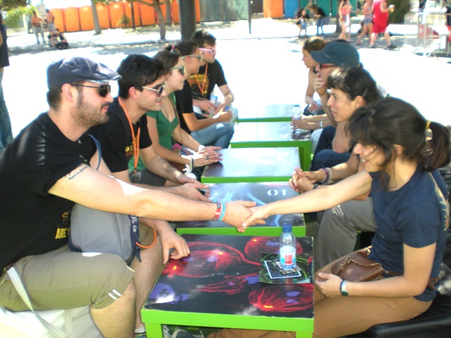
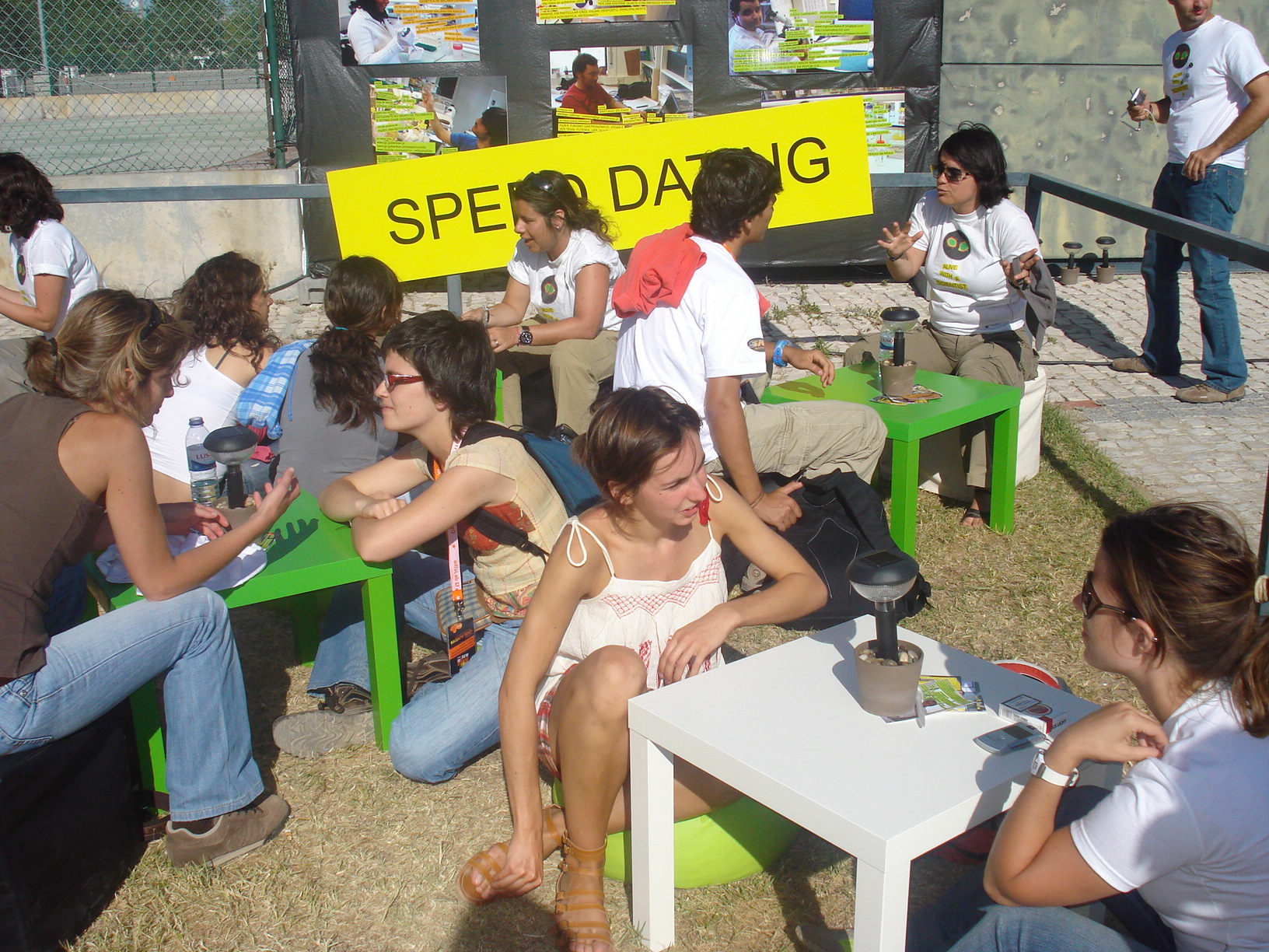
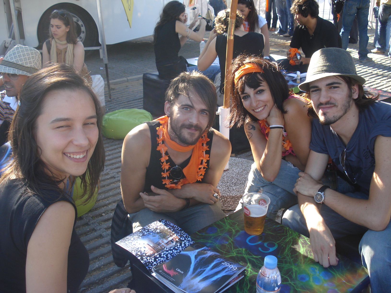
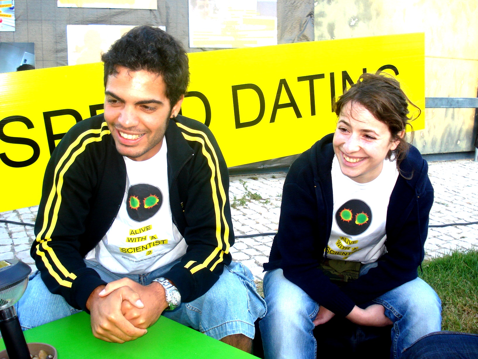
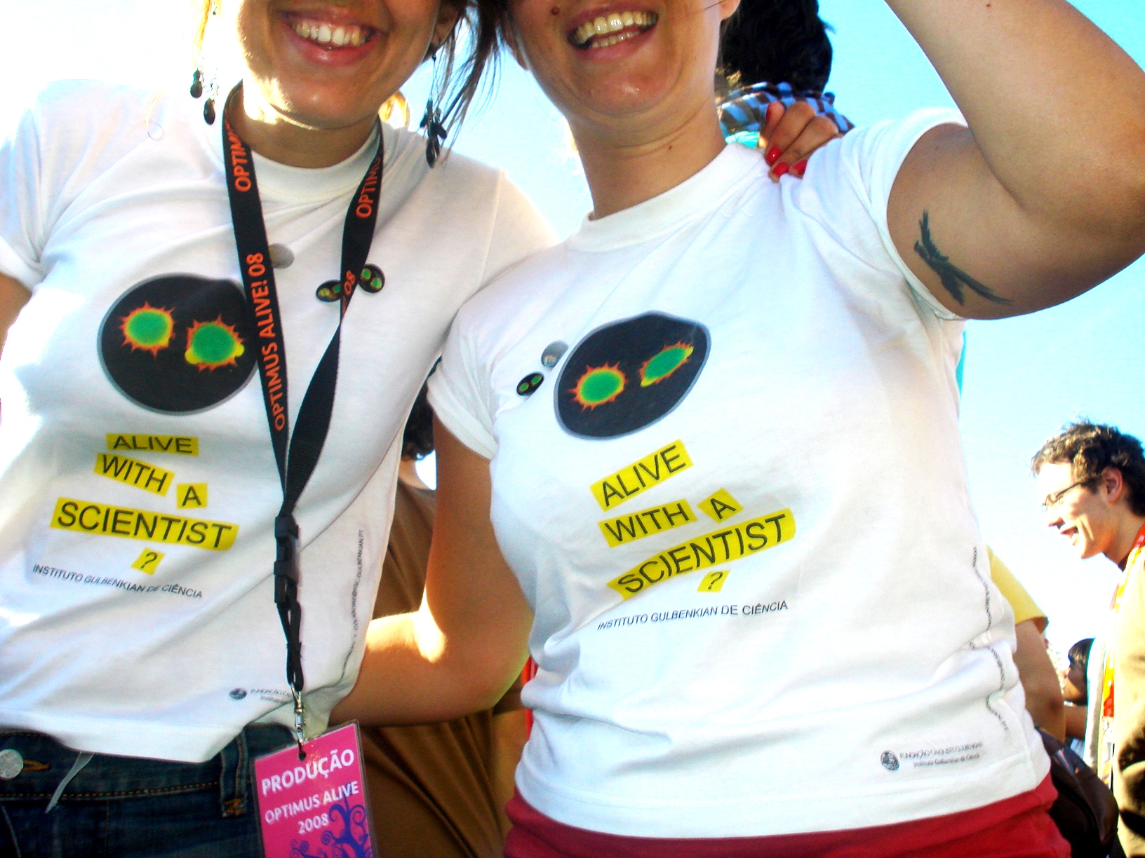
What fans at this festival were not expecting was that very close to the main stage where Coldplay were performing, they could also try “Speed-Dating” with real-life scientists from Instituto Gulbenkian de Ciência (IGC), a Biomedical Research Institute in Oeiras, Portugal, which although just next door, was probably still unknown to them.

IGC stand at the festival in 2012
IGC scientists during speed-dating

The IGC started in 2007 as a Fundraising program, a largely unexplored field in Portugal, aiming at studying and establishing alternative funding opportunities for biomedical research. This unique partnership  between the IGC and Everything is New (the promoter of one of the most important music festivals in Portugal) has allowed us to develop very innovative and successful outreach initiatives of informal science education and public dialogue. It aims to foster interactions between scientists and publics of all ages and socioeconomic groups, in a non-threatening and familiar pace, engaging citizens, patient organizations, companies, politicians and the general public in science and scientific culture, as well as raise funds for scientific research in Portugal. The partnership started at the end of 2007 and since 2008 the IGC has been, for four consecutive years, present at the Optimus Alive Oeiras music festival while the organiser of the festival, Everything is New (under the scope of its corporate responsibility project), has sponsored 8 fellowships for young graduates in Biodiversity, Genetics and Evolution.


IGC stand at the festival in 2008 cropped


IGC Outreach Activities at Optimus Alive Oeiras music festival

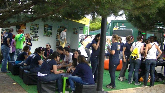
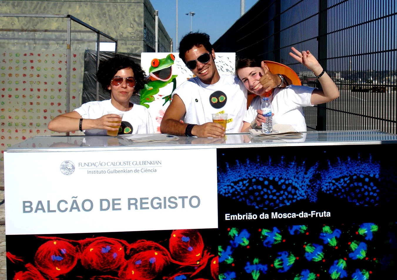
Over the last 6 years, at the IGC stand, very close to the main stages, music and science mix at an unexpected way and different activities have been developed to engage visitors with science and scientists:

“Speed dating” with scientists from different fields of research;

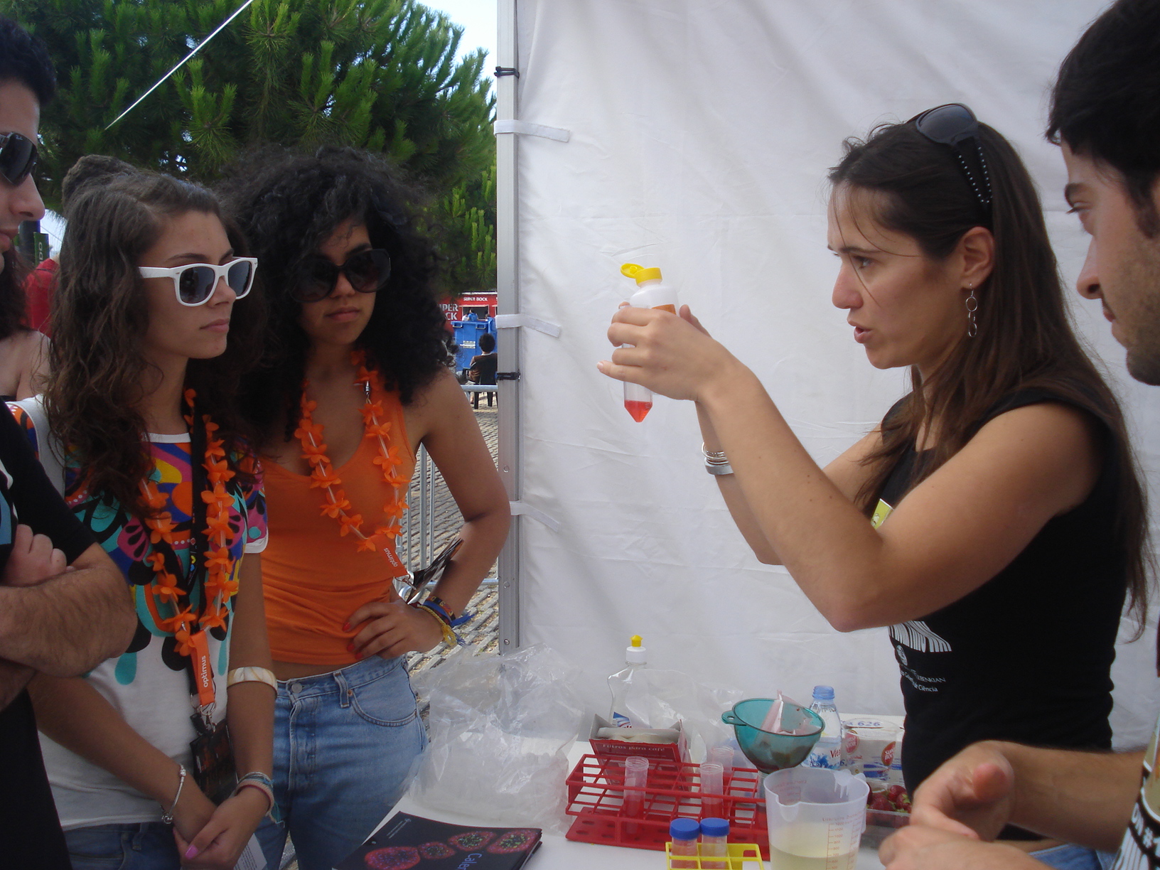
– DNA extraction from strawberries using everyday reagents;

– Molecular cooking by “Cooking Lab” to make flavoured ice-cream frozen in liquid nitrogen;

– Sci-arts installations by the artist Marta Menezes;

– Genetic interactive game, “Find your genes”, to find out how our genes determine eye colour, the alignment of little fingers, ear shape and the ability to roll your tongue;

– Microbiology activity to see phosphorescent bacteria;

– Biodiversity games to show you world distribution of endangerous animal species;

– Photo exhibitions about the research projects developed by young scientists supported through this partnership and about the daily lives of IGC scientists, by IGC members and Roberto Keller respectively;

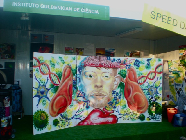
– Graffiti made by “Diálogo em Acção”, mixing music and science topics such as DNA, viruses and human body.

DNA extraction at the IGC stand


Optimus Alive-IGC fellowships in Biodiversity, Genetics and Evolution

This partnership also resulted in the sponsorship by Everything is New of 2 research fellowships per year for recent graduates to start their scientific careers in areas such as Biodiversity, Genetics and Evolution. Each fellowship is for a 12-month period, carried out between the IGC and a foreign institute. Optimus Alive-IGC fellowships are already a reference in Portugal and so far gave the opportunity to 8 young researchers to follow their vocations. Research projects have been developed at different IGC teams with collaborations at foreign countries such as Madagascar, Malaysia and Principe.


Target audience

Surveys of visitors at the IGC space, carried out between 2008 and 2011, revealed that:

– 0.5 % are less than 13 years old;

– 29% are between 13 and 19 years old;

– 51% are between 20 and 29 years old;

– 15 % of the visitors are between 30-39 years old;

– 4.5 % are more than 40 years old.

These data indicate that the majority of visitors at the IGC stand are teenagers and young adults. Although considered difficult to reach in science communication, these age groups are very important target audiences if we aim to promote science-related careers, informed engagement in civic science-related issues, policies and fundraising. Therefore, efforts such as IGC presence at this music festival, must be made to reach these audiences.


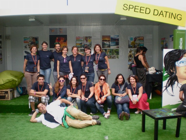
Staff at the IGC stand

Per year, during the 3 days of the event, around 70 volunteers make the outreach activities possible for visitors at the IGC stand. Volunteers have different backgrounds including:

IGC scientists at different stages of their careers (PIs, Pos-docs, PhDs and other graduate students);

– Science communication and Fundraising staff;

– University students from science-related degrees;

– Last degree Ice school students;

– Multi-media staff.

IGC volunteers at the IGC stand


Why to do science outreach at informal environments such as music festivals?

IGC volunteersMusic festivals are interactive venues allowing cognitive and emotional engagement with science and scientists promoting awareness in civic science-related policies and fundraising;

Music festival are excellent to target audiences who would not probably attend science fairs or more formal science outreach activities;

Having scientists at music festivals close to teenagers’ idols allow breaking down stereotypes making science closer to the general public.


The importance of private funding of Science

Scientific research in Portugal is still highly dependent on governmental funding and, contrary to other countries such as the USA and the UK, private funding by individuals and companies is still not very common and fundraising is not taken as a professional practice in most Portuguese scientific institutions. In times of a serious economic crisis in Portugal, the involvement of society in science funding is essential to allow the growth of scientific research that have been observed in the last two decades in Portugal.

Graffiti at the IGC stand 3


Important Links:

Activity protocol (Node post) on how to organise your own ‘speed-dating’ with scientists event


Websites

www.igc.gulbenkian.pt

http://www.optimusalive.com/cienciaambiente/bolsas


Facebook

https://www.facebook.com/BolsasOptimusAliveOeirasIGC


YouTube

http://bit.ly/XZCENd

http://bit.ly/17RKyKh

http://bit.ly/XQPbjy


References

Science and rock

EMBO reports 13, 954 – 958 (12 October 2012) | doi:10.1038/embor.2012.151

http://www.nature.com/embor/journal/v13/n11/full/embor2012151a.html

Science Outreach Through a Music Festival

http://communications.abrf.org/issues/7/science_outreach.html


Outreach logo new squareThis post is part of a series on science outreach. You can read the introduction to the series here and read other posts in this series here.


Thumbs up (3 votes)
Loading...

Tags:
Categories: Outreach

Science outreach- a new series on the Node!

Posted by , on 10 October 2013

Outreach logo new squareResearchers are being encouraged now more than ever to communicate their science to the public. First and foremost science communication is a way to share the enjoyment and excitement of science to others. But in a time of austerity, successfully communicating research is also important to justify to the public that funds it what scientists do.

But science communication can sometimes be daunting. There are so many different ways to do it- giving talks, having a stall at a science festival, collaborating with artists. Where to start? In this outreach series on the Node, we aim to showcase the variety of outreach activities out there, and provide ideas and resources to those interested in getting involved. We have invited various researchers and science communicators to write a series of posts on the different types of outreach that they do, and how they got first involved in science communication. In addition, we have asked them to suggest an easy outreach activity to get you started. From promoting science at music festivals to getting involved in a departmental open day, we hope that you will find something for you in this series, and that you will feel inspired to get involved and communicate your science to others!

Thumbs up (3 votes)
Loading...

Tags:
Categories: Outreach

Call for Papers for DMM Special Issue on Zebrafish

Posted by , on 9 October 2013

Zebrafish_Banner_km


Disease Models & Mechanisms (DMM) is pleased to welcome submissions for a Special Issue scheduled for publication in late 2014. This issue will focus on translational advances made using the zebrafish model, including insights into disease mechanisms and therapeutic targets, new resources and technologies, and drug discovery and development.

The issue will be guest edited by:

James Amatruda (UT Southwestern Medical Center, USA)
Liz Patton (University of Edinburgh, UK)
Lalita Ramakrishnan (University of Washington, USA)

Topics to be covered in invited review articles include advances in regenerative medicine and cancer, mechanisms underlying neurodevelopmental and neurodegenerative diseases, small-molecule screening using zebrafish and more.

We invite you to showcase your breakthrough zebrafish research in this Special Issue. Submissions should describe original research in the form of a Research Article, Resource Article or Research Report. Please read the author guidelines for information on preparing a manuscript for DMM, and submit your manuscript online. For rapid feedback on a paper, send us a presubmission enquiry.

Key benefits of publishing in DMM include:

– Open Access (CC-BY)
– High visibility and impact
– Rapid peer-review
– Author videos featured on Company of Biologists YouTube channel
– Accepted manuscripts online within 1 week
– All articles included in Medline (PubMed), Scopus and ISI Web of Science
– PMC deposition

Submission deadline: Friday 3rd January, 2014

Thumbs up (2 votes)
Loading...

Tags: ,
Categories: News

13th FASEB Plant Biology Conference- Mechanisms in Plant Development

Posted by , on 9 October 2013

The 13th FASEB Plant Biology Conference was held from August 11- 16, 2013, in Saxtons River, Vermont, a modest but beautiful setting.  This was a special meeting, since it marked 25 years since the first FASEB Plant Molecular Biology conference- the theme changed to Plant Development in the mid ‘90’s. Around 160 attendees spent 5 days hearing the latest “developments” in plant development, and enjoying casual discussions in the swimming pond and the bar.


FASEB attendants photo

The meeting kicked off with an inspiring plenary talk by Prof. Liam Dolan (Oxford University).  Liam’s lab has been studying root hair patterning and other aspects of root development in Arabidopsis for many years, but in this talk he described new work that seeks to understand the evolutionary origin of root- like structures in plants. using a recently adopted liverwort model, Marchantia.

The following morning, the first session of the conference focused on the latest research on local signals. The speakers showed their work on plasmodesmata-dependent miRNA movement (Ykä Helariutta, University of Helsinki), endosome-dependent protein movement (Kimberly Gallagher, University of Pennsylvania) and recognition of Calcium signals in roots (Giles Oldroyd, John Innes Centre), sensing of CO2 concentration in leaves (Cawas Engineer, UCSD) and cytokinin levels in secondary meristems (Yuval Eshed, Weizmann Institute). Furthermore, recent findings on the control of Arabidopsis embryo maturation and maize meristem size homeostasis (Michael Pautler, CHSL) were shared with the community.

In the polarity session, several different examples of cell- and tissue-level polarity were discussed. Jeff Long (Salk Institute) presented new work on the genetic specification and maintenance of apical-basal polarity via TOPLESS-dependent and –independent pathways in the Arabidopsis embryo, while Laurie Smith (UCSD) presented insights into the regulation of asymmetric cell divisions during stomatal development in maize. Remko Offringa (Leiden University) described how regulation of the protein kinase PINOID (PID) can influence the pattern of PIN polarity – and hence auxin transport within a tissue. Finally, Xana Rebocho (John Innes Centre) described the importance of the regulation of tissue polarity, and growth rates, for the morphogenesis of the Antirrhinum flower and the insights gained from modeling studies.

Shoot architecture of higher plants is determined by developmental events occurring at the growing tips.  Meristems are the site of organogenesis, giving rise to lateral organs, such as leaves and axillary branches, which themselves have meristematic properties.  A meristem contributes immobile cells to the formation of lateral organ primordia, and it is self-renewing.  A major outstanding question is precisely how cells dynamically accept cell fate at the meristem-incipient organ boundary during organ initiation.  An enthralling session devoted to boundaries featured studies from Rüdiger Simon (Heinrich-Heine University), Marcus Heisler (EMBL) and Klaus Theres (Max Planck Institute for Plant Breeding Research) that honed in on the genetic and molecular dissection of boundary specification and formation.

The meeting then moved on to considering long range signaling in plants. Leslie Sieburth (University of Utah) presented new findings on the bypass signal that coordinates the development of shoots and roots. David Braun (University of Missouri) discussed the Tie-Dyed (TDY) genes of maize; mutations in which show defects in phloem differentiation and sucrose trafficking. Micro-RNAs can also be transported long distances in the plant, and Tzyy-Jen Chiou (Academia Sinica, Taiwan) presented her research on the regulation of phosphate uptake and translocation, which involves miR399 acting as a systemic signal. In the last talk of the session, Catherine Rameau (INRA Versailles) discussed how Strigolactones and other long-range signals regulate shoot branching in pea.

Compared to Arabidopsis, many crops exhibit unique inflorescence structures requiring more coordinated transition from stem cell maintenance to floral organ formation. Inflorescence architecture is largely determined by the fate of meristems, population of stem cells under specific genetic control. In the session on switches, novel molecular regulators of this inflorescence development were presented in three major crops: tomato (Zachary Lippman, CSHL), rice (Junko Kyozuka, University of Tokyo), and corn (Paula McSteen, University of Missouri).

The signal integration session focused on the integration of molecular signals that coordinate complex environmental inputs and govern developmental outputs. The session highlighted an exciting array of developmental responses to the environment, from the phototracking of sunflowers to root patterning in heterogeneous soils. Jennifer Nemhauser (University of Washington) started out the session by introducing the hypocotyl as a model system for measuring the role of complex environmental input on developmental output. In the next talk, José Dinneny (Carnegie Institution) elucidated the role of heterogeneous water availability on lateral root patterning, a phenomenon referred to as “hydropatterning”. Stacey Harmer (UC Davis) was the next speaker. She emphasized the impact of the circadian clock on transcriptomic patterns. The Harmer lab harvested tissue every 4 hours for 2 days and found that half of all genes are differentially expressed in a time dependent manner. Harmer also introduced sunflower solar tracking as a model system to study circadian clock entrainment. The final speaker for the session was José Alonso (North Carolina State University), who presented a very clever mutant screen that was performed to fish out genes coordinating the relationship between auxin signaling and ethylene response.

Talks then turned to regulation of morphogenesis and shape. Olivier Hamant (Lyon) presented research on mechanical signals in plant morphogenesis, and the proposal that coupling between mechanical cues, microtubule (MT) alignment and PIN1 orientations plays a key role in morphogenesis and the generation of phyllotactic patterns. Uptal Nath (Indian Institute of Science) focuses on the molecular basis of polar leaf growth. Leaf growth is often allometric, where leaves experience polar growth – more growth at the base and progressively less towards the tip. His work has shown that the polarity of leaf growth can be diverse, and that this may be related to the expression of miR396. Next, Elliot Meyerowitz (Caltech) presented work describing how physical and chemical signals control morphogenesis at the shoot apical meristem. He suggested an approach in which all developmental biologists should consider the physical stresses acting in tissues and study morphogenesis as a whole using all parameters. Adrienne Roeder (Cornell) discussed how coordination of cell division and cell type control the morphogenesis of the Arabidopsis sepal, while Siobhan Braybrook presented data on how cell wall mechanics and pressure affect the overall shape of hypocotyl extension.

In the last session, participants returned to the Vermont Academy auditorium after an afternoon of outdoor activities. Refreshed by a game of volleyball, a jog through the woods, or a swim at the local pond, attendants were pleased to hear three fantastic stories outlining recent discoveries in developmental phenomena related to species evolution. Vincent Colot (Institut de Biologie de l’Ecole Normale Supérieure) presented his group’s recent work exploring the link between genome sequence, DNA methylation status, and plant phenotype. Claudia Köhler (Linnean Center of Plant Biology, Uppsala, Sweden) presented her group’s recent breakthroughs in understanding the mechanisms behind the ‘triploid block’ that causes the post-zygotic isolation of newly formed polyploid progeny. George Coupland (Max-Planck-Institute for Plant Breeding Research) finished off the session by presenting his group’s recent findings on the molecular mechanisms behind vernalization (induction by cold treatment) and age requirements for flowering in the genus Arabis.

The conference ended with a thoughtful overview by Scott Poethig (U. Penn). He reminded us of how our field has come full circle, from a situation where everyone studied a different plant, through a bottleneck where almost everyone worked on Arabidopsis or one or two other model systems, to a new era where genomics allows us to again use diverse species to tackle important problems in plant development.  He also reminded us how some concepts discussed at the meeting had been discussed much earlier, in particular reminding us of the seminal ideas of Paul Green in thinking about the role of mechanical forces in plant morphogenesis.  Overall it was a truly memorable meeting, and we look forward to the next FASEB Mechanisms in Plant Development meeting in 2015, which will be organized by Dominique Bergmann and Rudiger Simon.


This report was written by several of the students who attended the meeting. If you’re interested in finding out more, you can read their full report here (pdf)


Thumbs up (2 votes)
Loading...

Tags:
Categories: Events

A Day in the Life of… a Zebrafish lab

Posted by , on 8 October 2013

My name is Andrew Mathewson and I am a fourth year graduate student in the University of Washington’s Molecular and Cellular Biology program. Whereas most my friends from college are pursuing productive careers in medicine, social justice, or environmental preservation (or just trying to get by), I spend most my days deep in a basement encouraging small fish to mate so that I can steal their babies for science. I am currently pursuing a PhD in the lab of Dr. Cecilia Moens in the Basic Sciences division at the Fred Hutchinson Cancer Research Center (FHCRC) in Seattle, Washington. The Moens lab exclusively studies the zebrafish as a model for vertebrate development, focusing primarily on hindbrain patterning and cell migration. My project investigates the interplay between basic cell polarity signaling and the ability of neurons to move throughout the developing brain.
 

A tank of transgenic zebrafish

A tank of adult zebrafish expressing a red fluorescent protein in all muscle tissues bustles with excitement during their morning feeding.


Zebrafish are small tropical freshwater fish species native to the Indian subcontinent that were popularized as aquarium fish many years ago. They were pioneered as a model system for scientific research by George Streisinger and colleagues at the University of Oregon, where the Zebrafish International Resource Center (ZIRC) is now located. Many developmental biologists have adopted these robust little critters for their research in the last several decades due to the fact that their biology is very amenable to experimentation and observation. Zebrafish develop externally from a single cell to a rather complex larval fish in just a couple of days, meaning that in just a few hours, zebrafish speed through developmental processes that take weeks or months in other vertebrate models. Most incredibly – they remain entirely transparent while doing so.  A single mating pair of zebrafish can produce upwards of a thousand embryos in a single morning, making zebrafish a powerful system for generating large experimental datasets. Zebrafish has triumphed as a system for forward genetic screens and researchers have developed innumerable transgenic lines for the scientific community in recent years. It is the unique combination of developmental speed, optical transparency, and the rapidly growing array of genetic tools that have made zebrafish a popular system for modern developmental research.

If you have never worked in a fish lab you might wonder what goes on behind the locked doors of our facility deep in the bowels of the FHCRC. In the evening before most of my experiments I set aside a lucky few male and female fish with the mutations and/or transgenes I am using and place them into crossing tanks that allow the fish to see and smell but not touch one another. At 9AM the next morning the facility lights turn on and the fish are frisky after a long night’s rest. One of my favorite parts of my job is when I get to remove the transparent dividers from crossing tanks, allowing the fish to finally interact after a long night of frustrated solitude. Healthy males will almost immediately begin aggressively courting any willing (or not) female in sight. This courting mostly looks like a game of tag, where the female is “it”. Usually in a matter of minutes the females will begin to release eggs that the males dutifully fertilize and I patiently collect for my morning experiments.


The Moens Lab main fish facility.

The Moens Lab main fish facility


While any research demands practice and patience, zebrafish work in particular demands exceedingly steady hands, sharp eyes, and an even sharper pair of forceps. A typical day can involve anything from molecular cloning and sequence analysis, confocal microscopy and imaging, visual or genetic screening of mutant embryos, all the way up to actual wrangling of adult fish. Most of my current work involves studying developmental processes that occur within the first two days of life. This means that I spend the first half of most work weeks crossing fish lines and manipulating embryos and the second half imaging and analyzing my results. One technique I’ve tried to master is cell transplantation between embryos of different genotypes in order to ask questions about cell-specific gene function. Zebrafish is a great system for this type of work since embryos develop externally, are produced in large numbers, and can tolerate a lot of manipulation. However, most of my current experiments revolve around cloning interesting bits of DNA together and injecting them into newly laid single-cell embryos with infinitesimally small glass needles. I can efficiently create mosaic embryos (where some cells express my DNA and others do not) by mixing my DNA with transposase to increase genomic integration. Most people can comfortably inject hundreds of embryos per hour making it possible to rapidly conduct multiple experiments in parallel. Once I’ve injected my embryos, I spend the rest of my days in dark rooms peering through dissection scopes trying to discern if my manipulations change how cells behave during development. Even though these cells tend to be labeled with at least one type of fluorescent protein, this process can be difficult because zebrafish embryos are quite small. Therefore, once I’ve identified embryos that express my injected DNA or contain successfully transplanted cells, I’ll typically anesthetize the embryos, mount them in agar, and image them on one of our confocal microscopes for closer analysis. The coolest part of this is that I do most of my imaging on living embryos, so I can see how cells behave in their natural environment as the fish continue to develop. This also allows me to use the fish in further experimentation or I can raise them to generate new transgenic lines. Depending on the type or combination of transgenes I want for my future experiments, it can sometimes take several generations and many months to actually generate a new stable transgenic line.

Not surprisingly, it can take a little bit of work keep my experimental stocks of hundreds (or thousands) of fish alive and happy throughout my experimental timeline which in some cases can extend for years. Fortunately the Moens lab is able to hire a full-time fish technician that cares for our facility and generally keeps things running smoothly. She maintains cultures of brine shrimp (aka sea monkeys, a zebrafish’s favorite snack) and rotifers (microscopic water creatures that baby fish love to munch on), feeds the entire facility both live and dry food several times a day, cleans used tanks and nets, helps keep track of our stocks as they mature and age, and euthanizes aging or diseased fish, to name just a few of the amazing things she does for us. Occasionally our technician takes a day off work (like any normal human being), leaving us researchers responsible for our own fish. It’s usually not a big deal—everyone in the lab is at least nominally trained in fish facility maintenance for our own work, and ever since the funding drop, everyone has gotten used to sacrificing the odd weekend to keep our stocks alive.  Yet when things go wrong it forces one to recognize the sheer amount of effort required to generate the massive amounts of data produced by the zebrafish research community. Just the other day it was my turn to feed our baby tanks but it turned out that our rotifer cultures had crashed. Our babies went hungry that day—slowing their growth and decreasing their chances of survival, meaning that we can’t count on them for future experiments. Fortunately these small disasters are pretty rare because we have such an awesome fish technician and everyone in lab pays attention to the needs of the fish.

Disease and parasites are a major concern when running a large facility of animals. We try to minimize introducing unwanted pests into our facility by keeping an entirely separate “quarantine” facility for housing fish sent from other labs as well as regular sterilization of media and equipment. Even with these precautions it is impossible to prevent the occasional unwanted pest from getting in.  It’s not uncommon to discover an odd arthropod swimming in one’s dishes while sorting embryos. Though tiny creatures like these are usually a normal part of a complex water environment in nature, they can devastate young (or even adult) zebrafish populations in lab. Instances like these remind us that the hard work we do to keep our facility clean and our fish happy isn’t just because the FHCRC requires us to—it’s so we all can continue to do our research.


The Moens Lab

Lab mates cluster around our central work bench most afternoons. Adam Miller and Crystal Davey inspect injection needles (on the left) for cell transplantations while Daniel Berman and Arish Shah (on the right) sort zebrafish embryos.


I like working with zebrafish. While I find the pace of yeast and tissue culture almost frenetic, and the speed of Arabidopsis growth to be at times overly meditative, zebrafish grow and live at a rate that I can relate to. Most experiments fall into a natural daily rhythm in sync with our fish facility’s diurnal cycle. From my experience, zebrafish labs tend to foster a friendly and collaborative lab atmosphere due to the tiny bit of mutual dependence required for keeping stocks alive and experiments running (or maybe zebrafish work just attracts friendly people). Given that we usually want our fish to stay healthy, we have a pronounced lack of the toxic chemicals one usually associates with this type of research, which means the lab can be a comfortable space for everyone. One nice thing about zebrafish work in general is that the fish do most of the work. Once I’ve done my manipulations early in the week I place my embryos in a water filled dish in an incubator and don’t touch them until they are old enough to study. Each fish has its own yolk sac meaning that they don’t need food (or much of anything) for the first week of development. Though zebrafish is a great system for cell transplantation and other active manipulations, the fish really shine as a model organism once they are old enough to look at. Whether you are tagging an interesting protein with GFP, tracking transplanted cells with viable dyes, screening for mutant phenotypes, or just watching the precise choreography of development, zebrafish is the system to turn to. The fact that I can mount living transgenic embryos and image their (often glowing and multicolored) cells as they grow and interact with one another just blows my mind. This aspect is what caught my attention early in graduate school and is a big part what keeps me working towards my Ph.D. It’s hard to describe the beauty of a living, glowing brain as seen through a powerful confocal microscope, and even after several years of works it never fails to leave me in awe.
 

Transgenic zebrafish hindbrain

Dorsal view of a live transgenic zebrafish embryo’s hindbrain at 24 hours post fertilization. Facial branchiomotor neurons express membrane red fluorescent protein and migrate posteriorly through hindbrain segment rhombomere 5 while leaving behind trailing axons. Rhombomeres 3 and 5 expresses a red fluorescent protein marker as well as a cytoplasmic protein tagged with green fluorescent protein.


Node day in the life new doodle squareThis post is part of a series on a day in the life of developmental biology labs working on different model organisms. You can read the introduction to the series here and read other posts in this series here.


Thumbs up (17 votes)
Loading...

Tags: ,
Categories: Lab Life

Matthew H. Kaufman (1942-2013) – mouse developmental anatomist

Posted by , on 7 October 2013

This post was first published in Development.


Jonathan Bard, long-time colleague and friend in Edinburgh, recalls the life and work of the mouse developmental anatomist Matt Kaufman, who died in August 2013.


Matt- pictureMatt Kaufman was the leading mouse developmental anatomist of his generation and played key roles in three major aspects of mouse research. He was the person who initially dissected out the blastocysts from which he and Martin Evans went on to culture embryonic stem cells. He wrote The Atlas of Mouse Development that taught mouse developmental anatomy to a generation of molecular biologists and he subsequently worked with his colleagues in Edinburgh to produce the anatomical infrastructure of mouse informatics. In all, he wrote more than 200 papers and a dozen books on mouse development as well as on Scottish medical history.


Matt grew up in a very orthodox and poor Jewish family in London. His parents saw him as a future scribe, writing the five books of Moses on parchment. One result of this early training was his exquisite handwriting; another was some formal training in Jewish learning and a long-lasting love of scholarship. This plan fell foul of a grammar school education, medical training at the University of Edinburgh and a taste of research in reproductive physiology there with Professor Anne McLaren. He decided on an academic career and moved to the mouse lab of Professor C. R. (Bunny) Austin in Cambridge in 1970 to do a PhD on mouse parthenogenesis. After this, he spent two years at the Weizmann Institute in Israel, and returned to Cambridge as a demonstrator, then lecturer in anatomy. By 1980, he had published almost 30 mainly first-author papers – his capacity for hard work and rapid writing was extraordinary!


Around then, he started his collaboration with Martin Evans who had been working on embryonal carcinoma (EC) cells in Cambridge. Matt’s experience with isolating early mouse blastocysts and Evans’ knowledge of culture enabled them to obtain cell cultures from the inner cell masses: originally called Evans- Kaufman (EK) cells but now known to the world as embryonic stem cells (ESCs). The five papers that they published together in 1981-1984, together with those of Gail Martin in the USA at around the same time, provided the baseline knowledge for all subsequent work on the genetic manipulation of ESCs, their use in making transgenic mouse strains and their potential for regenerative medicine. It has to be said that Matt took no part in this later work because he was really interested in the anatomy of the developing mouse.


In 1985, Matt was awarded the chair of anatomy at the University of Edinburgh, a post first established in 1705, and remained there until he retired in 2007. The University did not come to view this as an entirely successful appointment for two main reasons. First, they had hopes that he could introduce molecular biology into the department, but he was not really equipped to do this. Second, they wished to reduce the amount of anatomical teaching in the curriculum to make room for genetics and social medicine; this shocked Matt because he believed that all of medicine stemmed from the cadaver and he fought the inevitable changes at every step. In hindsight, Matt lost more battles than he should have – probably because, for all his academic strengths, he was a dreadful university politician! Things were not helped by his ill health: in 1995 or so, he was diagnosed with polycythaemia (a blood malignancy) and spent the rest of his life on the maximum dose of methotrexate his body could tolerate – he lived in a state of continuous pain and discomfort.


Despite his political and medical problems, he continued research on the developmental anatomy of the mouse and on how it was affected by polyploidy and by external factors such as alcohol and anesthesia. These essentially minor papers were part of a much bigger project (on which he worked every evening): to put on paper everything that he knew about mouse development. This work all came together in 1992 when he published The Atlas of Mouse Development, a book that turned out to be, by the standards of mouse development, a major bestseller and is still in press (a supplement is being planned that will be part of his legacy). It contained around 1500 micrographs of sections from all 26 Theiler stages of mouse embryo development, each labeled to show the tissues, together with detailed explanations of what was happening at each stage. The book explained mouse development to a generation of molecular biologists making and analyzing transgenic mice, who had the shock of realizing that doing the molecular genetics was far easier than understanding the resulting phenotypic changes!


About then, mouse informatics was getting off the ground both at the Jackson Laboratory in Maine and at the MRC Human Genetics Unit in Edinburgh and it was soon clear that more was needed than just the basic genomics. I approached Matt and suggested that, for each Theiler stage, we should integrate all of the tissues into a parts-of hierarchy. One benefit of this would be that his beloved Atlas would get a proper index; another, of course, would be that the hierarchy (which soon became an ontology) could be used as the anatomical core for a database to which could be added tissue-associated data (e.g. gene expression). This was implemented by the Jackson Laboratory and is now a key feature of the mouse informatics resource there.


At the MRC Human Genetics Unit, Richard Baldock, Duncan Davidson and I decided to go further and to make 3D reconstructions of mouse embryos from the slides that Matt had used for his Atlas and to include all the tissue boundaries so that gene expression could be shown accurately. This led Baldock and Davidson to produce the first online graphical atlas for capturing gene expression data, to which Matt continued to provide detailed anatomical input up until about 2011.


This is not to say that he had stopped doing other research. Apart from mouse work, he had a long interest in the medical history of Scotland in general and the University of Edinburgh in particular. Starting in 1992, he produced a series of articles and books on medical history, including an important history of the chair of anatomy at Edinburgh that was part of the exhibition he organized to celebrate the 300th anniversary of its initiation. By this time, Matt was near retirement and there was some degree of reconciliation between him and the University. Indeed, there was no argument when, in 2007, he was finally and properly elected to the Royal Society of Edinburgh.


Matt was fortunate in having a very happy home life. Claire, his wonderfully tolerant wife, put up with his never-ending work, his 1930s Lagonda car that was always breaking down and the sheer quantity of working papers, historical documents and research publications, not to mention histological slides, that littered the house as well as his very large office. Indeed, it was only those who had to deal with Matt’s non-research, academic life who did not get on well with him! Everyone else appreciated his somewhat eccentric interests, his kindness, his ability and his old-fashioned courtesy. For me, he was a very good colleague and friend – I miss him

Thumbs up (2 votes)
Loading...

Tags:
Categories: News, Research