The community site for and by
developmental and stem cell biologists

The profession that isn’t

Posted by , on 10 September 2011

This post was my contribution to Science is Vital’s latest campaign on science careers. If you haven’t done so yet, I warmly encourage to get involved with the movement.

THE PROFESSION THAT ISN’T

As I enter the last 6 months of my first postdoc, I am confronted by a number of issues with having chosen (and continuing to choose) science as a career that I suspect apply quite broadly:

1. The UK has cut science funding in real terms significantly, and will likely continue to do so. Obviously, I think this is misguided, and the economic arguments in a knowledge economy in 21st century Western Europe are well worn, so I will not re-state them. It is, though, worth contemplating that not every country is adopting this approach, and I fear that our medium and long term competitiveness as an economy, not to mention as a health service, will struggle in comparisons with countries such as Germany that are taking the opposite approach to their science budget.

2. I am an average, middle class, married, soon-to-be 30-year-old, and have a close-knit family. My regular 12-hour days and weekend working are not very sustainable.

The arguments surrounding about point 1 are well worn, as I say, but point 2 needs significant re-statement in the corridors of power. Elaborating on my, I suspect typical, situation I hope will be informative.

I am fairly normal. In my more self-confident/self-indulgent moments I think I might actually be quite bright, with much to contribute to science, though I should point out that I am more often of the opposite opinion. But then, it isn’t like I spend my professional life on the lookout for incorrect conclusions not based on reasonable evidence or anything.

Seriously though, whether or not I as an individual am any good, I do not bear comparison as a scientist with people in other professions at my stage of career. The contrast with my wife is stark. She is a civil servant working for her majesty’s government in London. She is better paid than me, has a permanent contract, and is encouraged to pursue a balance in her life between work and leisure/family. I also like to remind her that she is less well educated than me, but I will humbly ignore that for now. In contrast, I know academics (and quite a few of them) who are of the opinion that a good work-life balance involves having a weekend off. Sometimes.

For those of us not in a position to drop everything and move to Boston for 5 years, it seems that science and life are not compatible, and are becoming even less so. At best it is like we have all the negatives of the private and public sectors with none of the benefits of either. At worst it is as if we are participating in a career structure that is fundamentally undermining of one of the UK’s truly outstanding endeavours. It is an issue that needs addressing.

Thumbs up (9 votes)
Loading...

Categories: Careers

Navigate the archive

Use our Advanced Search tool to search and filter posts by date, category, tags and authors.

An Informative Career Day at The EMBO Meeting in Vienna

Posted by , on 10 September 2011

What shall I do, and once decided, how can I get my dream job after completing my PhD or postdoc, especially if I don’t want to become a group leader? Questions many of us ask ourselves sooner or later, and there are more options than the pessimistic among us might be able to imagine. These issues were addressed at today’s EMBO Career Day, taking place before the start of The EMBO Meeting, from which I’ll be blogging during the next few days.

There was a choice of four different workshops, of which one could pick two. The first I attended covered the process of applying for a job – “Cover letters, CV writing & interview skills”. Barbara Janssens, PhD career advisor at the German Cancer Research Center (DKFZ) in Heidelberg, Germany, started her workshop by telling us how she ended up in her current job. After having produced “no papers but two kids” during her postdoc, she decided that she would rather have a career outside of academic research. Barbara talked to many people about their experiences, and finally came across someone who suggested her to try at Wiley, where she started as a trainee and even ended up setting up a new journal! From there she went into teaching scientific writing, before finally becoming the PhD Career manager at the DKFZ.

Barbara and EMBO’s Gerlind Wallon both did a great job in giving useful advice, such as to always have an up-to-date CV (you never know when someone might ask for it), to network in an intelligent manner (know who has the power to hire you – it won’t be HR!), and stand out from the crowd by being involved in relevant extracurricular activities (as everyone applying for the job will have a PhD – that’s not enough).

Several of the participants had sent in their applications for a mock job advertisement we had received a while before the workshop took place. We evaluated these (anonymised) cover letters and CVs, and one brave volunteer even did a practice interview in front of the whole group. These exercises resulted in two precious “Dos and Don’ts” lists; the strongest advice I extracted from this was to always be aware of what’s relevant and what isn’t when presenting yourself.

After this excellent workshop I attended the Expanding Career Options lunch, where in an informal setting, I learned about careers in science policy, intellectual property and non-governmental organisations.

Finally, EMBO reports’ Sam Caddick guided us in explaining research in simple terms in the very interactive “Make Science Make Sense” workshop. Avoiding jargon and deciding on a single, simple message to describe your work turned out to be a lot harder than one might imagine, and it had the beneficial side effect of making me think more carefully about the relevance of my research. All in all, the EMBO Career Day gave me a lot of information and I can definitely recommend attending it next year in Nice!

Thumbs up (6 votes)
Loading...

Tags: ,
Categories: Careers, Events

Satellite cells muscle their way into the stem cell spotlight

Posted by , on 8 September 2011

Researchers have long known about regeneration of injured muscles, and have debated about the exact source of the muscle stem cells that perform this amazing feat.  A group of papers in a recent issue of Development shine a stem cell spotlight on satellite cells.

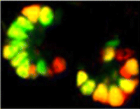
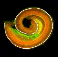
Following injury, skeletal muscles are regenerated by muscle stem cells, but the exact identity of these stem cells has been unclear.  Recent research on muscle repair has focused on satellite cells, and clarifies their role as muscle stem cells.  Satellite cells are found sandwiched in between the membranes of individual muscle fibers and the basement membrane, and can be identified by the presence of Pax7.  Lepper and colleagues genetically ablated Pax7+ satellite cells in the muscles of mice using an inducible system and found that ablation of these cells prevented regeneration following cardiotoxin-induced injury.  These results show that satellite cells are absolutely necessary for muscle regeneration.  Images above show degenerated muscle grafts transplanted into healthy muscle tissue in nude mice.  Lepper and colleagues used this technique to circumvent the lethality of their induced system after satellite cell ablation, allowing for the longer-term study of muscle regeneration.  Control tissue (left, green) grafted into a healthy muscle bed was able to regenerate, as seen as the presence of myosin (red + green = yellow).  After satellite cell ablation, grafted tissue (green) was unable to regenerate.

For a more general description of this image, see my imaging blog within EuroStemCell, the European stem cell portal.

Check out a summary of all four papers on muscle regeneration in Development here.

ResearchBlogging.orgLepper, C., Partridge, T., & Fan, C. (2011). An absolute requirement for Pax7-positive satellite cells in acute injury-induced skeletal muscle regeneration Development, 138 (17), 3639-3646 DOI: 10.1242/dev.067595

Thumbs up (3 votes)
Loading...

Tags: ,
Categories: Images, Research

Optical clearing with Scale

Posted by , on 8 September 2011

Transparency.
A desirable virtue in many walks of life, and a particularly useful trait in developmental biology.  Model organisms that are see-through offer unique advantages, especially when it comes to detailed 3D imaging.

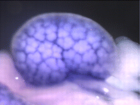
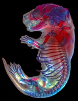
A new report in Nature Neuroscience offers a potential advance in this area. Researchers from Japan have stumbled upon a novel aqueous solution that renders tissues transparent with less of the non-desirable side effects of other methods.  Their results look impressive: whole mouse embryos are presented which are completely see-through. The new clearing solution has the considerable benefit of not quenching fluorescent proteins – extremely useful for imaging transgenic animals.

Image: RIKEN, BSI

The benefits of this reagent are clear (excuse the pun); it’s cheap to make, effective, and produces great imaging conditions.  The authors demonstrate some potential applications by performing 3D reconstructions of mouse commissural axons from intact brain samples, and by visualising the intricate relationship between neural stem cells and blood vessels from dissected hippocampus.

One drawback of the procedure (for me at least) is the speed at which the reagent works.  The beautifully clear whole mouse embryo highlighted above was treated for 2 weeks.  The variant reagent ScaleU2 (optimised to limit swelling of soft tissues) is slower still; the transgenic mouse embryos in figure 6 of the paper were treated for 6 months! In contrast, the organic reagent BABB (benzyl-alcohol/benzyl-benzoate mix) works in a matter of minutes, although it does harden tissues, and also reduces fluorescent signals.

For 3D imaging of traditionally opaque embryos, the new Scale reagent could be a key innovation – you’ll just need a little bit of patience to use it.

Hama, H., Kurokawa, H., Kawano, H., Ando, R., Shimogori, T., Noda, H., Fukami, K., Sakaue-Sawano, A., & Miyawaki, A. (2011). Scale: a chemical approach for fluorescence imaging and reconstruction of transparent mouse brain Nature Neuroscience DOI: 10.1038/nn.2928

Thumbs up (9 votes)
Loading...

Categories: News, Research

Turtles in a nutshell

Posted by , on 7 September 2011

Turtles are peculiar vertebrates. They have a compact skull with no temporal openings, a beak instead of teeth, a contractible neck, and a shell covering its trunk. The famous turtle shell is composed of two halves, a plastron (ventral) and a carapace (dorsal). The latter is an exquisite arrangement of vertebrae and fan-shaped ribs with secondary ossification forming a rib cage that encloses the limb girdles.

Side view of turtle skeleton
Side view of a turtle skeleton. Illustration by M A Smith.

Please, take a moment to imagine your shoulder inside your rib cage. How such intriguing anatomy has evolved from a standard external-to-the-ribs configuration? As drastic as this change may seem, researchers from the Japanese RIKEN Center for Developmental Biology provided an elegant and straightforward explanation.

Dorsal view of chicken and turtle scapula and ribs
Scapula (sc) position and ribs (r) in birds and turtles. Modified from Figure 2 of Kuratani et al. (2011) [1].

A basic step for evolutionary studies is to understand the history of the group. Who is it related to? The evolutionary relationships of turtles within the amniotes (mammals, reptiles, and birds) are not well understood. The traditional view based on skull morphology places them as basal reptiles while more recent molecular data group them with birds and crocodilians. A robust phylogenetic hypothesis allow us to infer evolutionary processes by the arrangements of nodes and characters. However, it is generally difficult to extract more fine-grained insight about how the changes occurred. Fossils do provide direct evidence of ancient forms, but they are rare. Can we do better?

Tortoise hatchling
A turtle hatchling. Photo by Mayer Richard.

All that we call phylogeny is today, and ever has been, ontogeny itself. (…) Phylogeny is but a name for the lineal sequences of ontogeny, viewed from the historical standpoint.” (Whitman, 1919; via Hall 1999)

Embryonic development is the process that builds the morphology of multicellular organisms. Scrutinizing development help us understand how structures are formed and regulated during ontogeny, and thus, how variant patterns can arise. Comparing the developmental sequence of a structure in different organisms may provide insightful information about evolutionary changes and the origins of morphological characters; given that you have a working phylogenetic hypothesis and a certain amount of care, since development also evolves.

To investigate the developmental changes related to the turtle unique body pattern Nagashima et al. (2009) [2] did exactly this: followed the developmental sequence of the turtle Pelodiscus sinensis while comparing to the correspondent stages in chicken and mouse embryos. This comparative approach allowed to pinpoint the exact moment in time when the morphology of the embryos began to differ during development and correlate the data with adult body patterns. And what they saw was simply awesome.

Chicken and turtle early development

The authors observed that the scapula appeared lateral to the body wall, near the limb bud (see above, ribs: white; scapula: green), and with similar muscular connections in the three embryos. The ribs of the turtle embryo were shorter in length while the ribs of the mouse and chicken had grown ventrally into the lateral body wall. At this stage, except for minor positional differences and rib length, the musculoskeletal pattern of the embryos was quite similar. But from this point on the turtle embryo began to differ from the other two.

In the mouse and chicken embryos the scapula simply grows posteriorly, above the ribs. Instead, in P. sinensis the scapula does not grow a posterior blade-like portion and is held inwards by a lateral folding of the body wall. It is positioned over the first rib and is encapsulated by the second rib which grows laterally and anteriorly. The trunk muscles connecting the scapula to the back retained their ancestral connections only adjusting to the “new” position. Limb muscles, on the other hand, formed new connections that are turtle-specific.

Chicken and turtle late development

The process is much easier to understand with 3D animated embryos:

Chicken animation
Turtle animation
Movies provided by the Laboratory for Evolutionary Morphology, RIKEN Center for Developmental Biology and available here.

It is interesting to note that rib development is somehow refrained from moving ventrally and the ribs remain in the axial region (unlike other amniotes whose ribs grow ventrally). This might inhibit the posterior growth of the pectoral girdle and is possibly the reason why ribs grow laterally in the turtle. This lateral growth and the body wall folding enclose the limb girdles.

Section of chicken and turtle ribs
Chicken (left) and turtle (right) ribs (arrows) positioned ventrally and laterally, respectively. Image by Shigeru Kuratani.

What could be regulating these ontogenetic movements? Right above the folding of the body wall there is a thickened ectoderm with undifferentiated mesenchyme forming a longitudinal ridge which is unique to turtles. This Carapacial Ridge (CR) is coextensive with rib growth and shares histological and molecular characteristics with the Apical Ectodermal Ridge, responsible for the patterning of limb buds. For these reasons it is believed to have a role in the turtle shell formation, although its functions remain unclear. Implanting CR grafts more dorsally or ablating the ridge did not alter rib growth, although in the latter the tips of the ribs joined distally at the site of the wound. These experiments suggest that the CR does not induce rib growth, but may regulate the fan-shaped pattern of the ribs.

Turtle Carapacial Ridge
Carapacial Ridge of the turtle P. sinensis (arrowheads indicate the lateral of the embryo, where the longitudinal ridge begins). Modified from Figure 1 of Nagashima et al. (2007) [3]

The development of a modern turtle does not reconstitute the evolutionary history of its kinds, but it can provide clues to the underlying mechanisms of the turtle body evolution. For instance, if the CR indeed is responsible for the maintenance of the fan-shape pattern of the ribs, it is likely that ancient turtles with fan-shaped ribs had a CR during embryonic development. It is also possible to make predictions based on the observed developmental processes; for example, that the arrest in rib growth preceded the enclosure of the scapula during evolution. Ribs encasing the lateral body wall ventrally would pose a physical barrier to the displacement of the scapula, so it is more likely that rib growth was altered first.

Fact checking these predictions is complicated and we must rely on fossils to get a glimpse of the past.

Before 2008 the oldest known turtle fossil was Proganochelys, a creature that had a complete shell and internal girdles. Since it already had a turtle body pattern, not much information could be extracted to understand the onset of the group’s evolution. But in 2008, Li et al. [4] found a 220 million years old fossil of an ancient turtle with many interesting (read intermediate) features, named Odontochelys.

What does it looks like? It had a plastron, but not a carapace; its ribs were not fan-shaped, but were short and not bended ventrally; and the scapulae are ahead (rostral) of the ribs, but not underneath. It also had teeth and many other specific anatomical details.

Odontochelys artistic representation
Illustration of Odontochelys by Marlene Donnelly.

Not only Odontochelys morphology is compatible with Nagashima et al. (2009) observations, but the shoulder anatomy even roughly paralels an early stage of P. sinensis development, although muscle connections were not clear. The authors speculate that the CR of Odontochelys was reduced and did not form a complete carapacial margin because the ribs do not exhibit a fan-shape pattern and there is no carapace.

Odontochelys could be interpreted as an intermediate stage for the turtle shell evolution, unless, of course, the apparent absence of a carapace is the result of a secondary reduction. Reisz & Head (2008) [5] argue that some modern turtles have greatly reduced the ossification of dermal components of the carapace, commonly associated to aquatic environments. Truncation of the carapace ossification is a plausible developmental mechanism that would lead to Odontochelys morphology. And the debates go on…

Anyway, developmental and fossil evidence insinuate that the once thought dramatic body pattern transformation, might well have occurred by a series of gradual developmental changes during turtle evolution. Nagashima and colleagues also made an animation about their take on the turtle evolution:

Turtle evolutionLaboratory for Evolutionary Morphology, RIKEN Center for Developmental Biology

Things I wonder: how large is the variation of scapular positioning pattern within the turtles? Does their ecological habits influence shoulder anatomy? Does the secondary loss of the carapace (leatherback) affect girdle positioning?

Literature

1. Kuratani, S., Kuraku, S., & Nagashima, H. (2011). Evolutionary developmental perspective for the origin of turtles: the folding theory for the shell based on the developmental nature of the carapacial ridge Evolution & Development, 13 (1), 1-14 DOI: 10.1111/j.1525-142X.2010.00451.x

2. Nagashima, H., Sugahara, F., Takechi, M., Ericsson, R., Kawashima-Ohya, Y., Narita, Y., & Kuratani, S. (2009). Evolution of the Turtle Body Plan by the Folding and Creation of New Muscle Connections Science, 325 (5937), 193-196 DOI: 10.1126/science.1173826

3. Nagashima, H., Kuraku, S., Uchida, K., Ohya, Y., Narita, Y., & Kuratani, S. (2007). On the carapacial ridge in turtle embryos: its developmental origin, function and the chelonian body plan Development, 134 (12), 2219-2226 DOI: 10.1242/dev.002618

4. Li, C., Wu, X., Rieppel, O., Wang, L., & Zhao, L. (2008). An ancestral turtle from the Late Triassic of southwestern China Nature, 456 (7221), 497-501 DOI: 10.1038/nature07533

5. Reisz, R., & Head, J. (2008). Palaeontology: Turtle origins out to sea Nature, 456 (7221), 450-451 DOI: 10.1038/456450a

Thumbs up (10 votes)
Loading...

Tags: , ,
Categories: Research

Leaving the lab: career development for developmental biologists

Posted by , on 6 September 2011

Over the past months, we’ve heard from several people who left research for a career away from the bench. Now, a summary of all these posts appears in Development, followed by some tips for graduate students, postdocs, and their supervisors. Below is the full text of the article, but it’s also free on Development, and you can get it as a PDF from there.

Leaving the lab: career development for developmental biologists

Let’s face it: not all PhD students and postdocs will become lab heads. Every few years, the National Science Foundation surveys doctorate recipients in the USA about their career progression, and their latest published data (collected in 2006) show that only about one quarter of biomedical science PhDs held tenured or tenure-track positions (see the links at the bottom of this post). If graduate and postdoctoral training are merely apprenticeships for tenure-track jobs, these numbers suggest that there are too many people being trained for the number of research jobs that are available. But if trainee positions are more than a stepping stone to running a research lab, what value does a PhD in the life sciences have outside of the lab, and what types of job do the remaining three quarters of PhD graduates go on to have?

In July 2010, I asked the following questions on the Node: `Should there be fewer postdoc and PhD positions? Or different kinds of [research] trainee positions, where some include training for scientific careers outside of the lab?’

The ensuing discussion suggested that the PhD degree and the postdoc system are not in need of reform, but that attitudes towards these positions should change. Greg Dressler, a professor at the University of Michigan, wrote in a comment on the Node post, `I do think we need to get over the idea that nothing short of an academic career fulfills the ideal goal of our students and post-docs. Most of the folks I went to graduate school with are not in academia anymore, yet they have meaningful and successful careers.’ In the same discussion, James Briscoe, a group leader at the MRC National Institute for Medical Research suggested that we need `the acknowledgment and encouragement of a diversity of career routes and development paths’.

These are good suggestions. There are a number of jobs outside of research or academia that are suitable for PhD graduates. A research job in industry, for example, connects seamlessly to research experience gained during PhD and postdoctoral training. But not every PhD graduate wants to continue in a research career, academic or otherwise. What kind of non-research jobs are available and how do PhD graduates get these jobs? And how is scientific training useful to people in a non-research career? To answer these questions, I invited a number of people to write a post on the Node to explain how they moved away from a career in research after their PhD. These posts can be found on the Node, but it’s worth discussing here the trends they raise collectively, and distilling some of the advice from those people who have left the life of the lab bench behind them.
(more…)

Thumbs up (5 votes)
Loading...

Tags:
Categories: Careers

In Development this week (Vol. 138, Issue 19)

Posted by , on 6 September 2011

Here are the research highlights form the current issue of Development:

Modelling liver development with ES cells: HNF4A is key

Human embryonic stem cells (hESCs), via their ability to differentiate into a plethora of cell types, offer an attractive approach for regenerative medicine, but they also offer a means of studying cell differentiation, and hence development, ex vivo. Here, Stephen Duncan and co-workers analyse the differentiation of hESCs to probe the molecular mechanisms that underlie human hepatocyte differentiation (see p. 4143). Using a protocol in which hESCs differentiate into hepatocytes in a stepwise manner, the researchers show that each stage of the differentiation process is associated with a characteristic mRNA profile, as shown by microarrays. Importantly, they show that the transcription factor HNF4A, which has been implicated in liver development, is essential for specifying hepatic progenitors; the onset of HNF4A expression is associated with specification of the hepatic lineage from hESCs, and shRNA-mediated knockdown of HNF4A prevents hESC differentiation into hepatic progenitors. These and other studies demonstrate that HNF4A establishes the expression of a network of transcription factors that promote hepatocyte cell fate.

Gcm/Glide-ing to a glial fate

Neurons and glia originate from a common precursor, the neural stem cell (NSC). These multipotent precursors display a high degree of plasticity in vitro, but the basis of this plasticity and the mechanisms underlying the neuron-glial switch in vivo are unclear. Now, Angela Giangrande and colleagues (see p. 4167) show that the transcription factor Glial cells missing (Gcm, also called Glial cell deficient; Glide) triggers a conserved chromatin signature that converts Drosophila NSCs to a glial fate. The researchers show that overexpression of Gcm in fly NSCs produces glia at the expense of neurons. This gliogenic potential of Gcm decreases with time and does not affect quiescent NSCs, suggesting that it is dependent on temporal cues rather than on the mitotic potential of NSCs. Finally, the investigators demonstrate that the glial fate switch is associated with a chromatin signature, which includes low levels of histone H3 lysine 9 acetylation and is similar to that observed in vertebrate glia, suggesting that this epigenetic mechanism for specifying glia has been conserved throughout evolution.

Paired up: histone methylation and HP1γ

The pairing of chromosomes during meiosis requires histone modifications, such as histone H3 lysine 9 di- and tri-methylation (H3K9me2 and H3K9me3, respectively), at pericentric heterochromatin (PCH) regions. But how do these epigenetic marks control chromosome interactions? Haruhiko Koseki and colleagues demonstrate that heterochromatin protein 1γ (HP1γ) regulates chromosome interactions by recognising histone methylation marks during meiosis in mice (see p. 4207). The researchers show that, in meiotic spermatocytes, H3K9me2 by the G9a histone methyltransferase requires pre-existing H3K9me3 marks, which are deposited by the Suv39h histone methyltransferase. They further show that HP1γ recognizes H3K9me3 marks and localises to PCH regions in an H3K9me3-dependent manner, where it then recruits G9a. Importantly, the loss of HP1γ results in defective spermatogenesis, aberrant centromere clustering and impaired homologous chromosome pairing. The authors thus propose that HP1γ acts as an important link between the cascade of H3K9me3 and H3K9me2 modifications, acting to align homologous chromosomes and facilitate their pairing during meiosis.

A new look into Sfrps and Wnt

Secreted frizzled-related proteins (Sfrps) are classified as Wnt antagonists, but recent studies have shown that some Sfrps can positively modulate Wnt signalling. Is this a general property of all Sfrps and, if so, how do Sfrps regulate Wnt signalling? Here, Paola Bovolenta and colleagues (see p. 4179) show that Sfrp1 and Sfrp2 positively regulate Wnt signalling, and are required for Wnt-mediated development of the mouse optic cup. The researchers show that specification of the peripheral optic cup (OCP), which is known to be dependent on Wnt signalling, is grossly defective in mice lacking both Sfrp1 and Sfrp2. In these mutants, Wnt spreading across the OCP is impaired, suggesting that Sfrps can influence the diffusion of Wnts. In support of this, the researchers demonstrate that Sfrp1 overexpression flattens the gradient of Wingless (a Drosophila Wnt homologue) across the Drosophila imaginal disc. These studies highlight a new and unexpected role for Sfrps in regulating the levels and distribution of Wnts during development.

Kidney development: a Notch above the Wnts

The kidney comprises functional units known as nephrons, which are made up of specialised epithelial cells. During development, each nephron arises from a pool of stem cells that undergo mesenchymal-to-epithelial transition (MET) in response to signals such as Wnt4 and Wnt9b. Here, Raphael Kopan and co-workers show that Notch pathway activation can replace inductive Wnt signals during this process (see p. 4245). Using gene manipulation in cultured kidneys, the researchers show that Notch pathway activation can induce epithelialisation in nephron stem cells but not in the closely related stromal mesenchymal cells. Continued Notch pathway activation following MET directs cells towards a proximal tubule fate. Finally, they report, Notch-induced MET can occur in the absence of Wnt4 and Wnt9b, suggesting that nephron stem cells are poised to undergo MET, which requires a permissive signal that can be provided by Wnts or by Notch pathway activation. These studies shed new light on our understanding of the early cell fate decisions that are made during kidney development.

Ngn2 phosphorylation links neurogenesis to the cell cycle

Cell cycle length influences the balance between progenitor maintenance and differentiation in the nervous system, although the mechanism for this is unknown. Here, Anna Philpott and co-workers show that multi-site phosphorylation of neurogenin 2 (Ngn2), a master regulator of neuronal development, controls neuronal differentiation in response to cell cycle lengthening in Xenopus embryos and in mammalian P19 cells (see p. 4267). The researchers show that, in Xenopus extracts, Ngn2 phosphorylation is regulated by the cell cycle, and analyses of HeLa cell extracts show that Ngn2 is phosphorylated on multiple sites by cyclin-dependent kinases (cdks). The phosphorylation of Ngn2, they report, reduces its ability to induce neuronal differentiation in vivo, and this is due to the decreased ability of phosphorylated Ngn2 to bind to its target promoters. The authors thus propose a model in which multi-site phosphorylation of Ngn2, which is quantitatively sensitive to cell cycle length, is used as a way to interpret cdk levels in order to control neuronal differentiation in response to cell cycle lengthening during development.

Plus…

The control of developmental phase transitions in plants

Plant development progresses through distinct phases, each controlled by genetic pathways that integrate endogenous and environmental cues. Recent studies, reviewed by Huijser and Schmid, show that the genetic networks underlying the transitions between these phases share some common factors.

See the Review article on p. 4117

Modeling new conceptual interpretations of development

As reviewed by Julien Vermot and Markus Affolter, the EMBO workshop on Biophysical Mechanisms of Development (organized by Ana Borges, Ana Certal, Ana Tavares, Filipa Alves and Beatriz Garcia Fernandez) brought together scientists in the field of developmental biology for whom interdisciplinary and quantitative approaches are central to the issues they are investigating.

See the Meeting Review, p. 4111

Thumbs up (1 votes)
Loading...

Categories: Research

Group Leader position available at MRC-NIMR London

Posted by , on 4 September 2011

Closing Date: 15 March 2021

Applications are invited for a Principal Investigator position to lead a new research group in Cell and Developmental Biology. We are particularly interested in candidates using quantitative approaches to study any aspect of the cellular and molecular dynamics of developing tissues.

Candidates should have an outstanding track record and an ability to lead a team pursuing original long-term research goals. Core support, including laboratory and animal staff will be provided by the Medical Research Council. MRC-NIMR offers an exciting, supportive and collaborative environment, comprising state-of-the-art imaging and mass spectrometry, high throughput sequencing and computational facilities, FACS, and excellent transgenic fish, frog, mouse and Drosophila facilities

For an overview of research at NIMR, see www.nimr.mrc.ac.uk

Informal enquiries to J-P Vincent (jvincen@nimr.mrc.ac.uk) or James Briscoe (jbrisco@nimr.mrc.ac.uk)

Applications should include a cover letter, full Curriculum Vitae, the names and addresses of three referees, an outline of current research interests (1 page) and a proposal for future research (1-2 pages).

The level of appointment will be determined based on expertise and experience. Benefits include Membership of MRC Final Pension Scheme and 30 days annual leave.

Closing date: 31 October 2011 Interview date: to be confirmed

Applications are handled by the RCUK Shared Services Centre; to apply please visit our job board at https://ext.ssc.rcuk.ac.uk and complete an online application form. Applicants who would like to receive this advert in an alternative format (e.g. large print, Braille, audio or hard copy), or who are unable to apply online should contact us by telephone on 01793 867003, Please quote reference number IRC 27831.

The MRC is an Equal Opportunities Employer
Final appointments will be subject to a pre employment screening

Thumbs up (1 votes)
Loading...

Tags: , , ,
Categories: Jobs

Node updates

Posted by , on 31 August 2011

Survey
As you know we carried out a survey about the Node this summer. Thank you to those who answered our questions! It was very helpful. We’re currently analysing the results, and you can expect a report on the Node soon.
One lucky survey participant, Greg Shanower of The Commonwealth Medical College in Scranton, Philadelphia (US), won the prize draw, and has been sent some gifts from Development and the Node. Congratulations!

Technical updates
We’ve been battling some technical glitches on the Node. At the moment, commenting is not working properly. We’re trying to find a solution for this as soon as possible. We may need to take the site down for a short while later this month to work on this and general site updates, but will let you know about that beforehand. You can always find us on Facebook and Twitter to keep up to date on any site downtime or to leave us comments, or you can email us directly via thenode[at]biologists.com .

Thumbs up (No Ratings Yet)
Loading...

Categories: News

Better late than never… 70th SDB meeting report

Posted by , on 31 August 2011

Thanks to the support from the Company of Biologists I had the opportunity to attend the 70th SDB meeting that was held last month in the hot but wonderful city of Chicago. During three days we enjoyed more than 60 excellent talks and about 500 really good posters that were focused on different aspects of developmental biology …

We had the opportunity to hear many outstanding talks but two of them particularly caught my attention, the one given by Tom Kornberg during the Presidential Symposium and the one given by Peter Reddien during the Stem Cell Biology Session. Tom Kornberg lectured us about the specificity of a long distance signalling mechanism that operates during Drosophila imaginal discs development and involves the use of cytonemes, specific types of filopodia that work as channels through which  morphogen signalling proteins move from a producing cell to a target cell.  Peter Reddien gave a remarkable talk about the molecular basis of regeneration in planarians, in which he explained to us how a single transplanted neoblast (adult planarian stem cell) can restore the regenerative capacity of a lethal irradiated worm.

Among the sections I enjoyed the most was the Hilde Mangold Posdoctoral Symposium where eight SDB postdoctoral members gave short talks that were judged by a committee who selected the best speaker to receive an Award. The winner of this year was Lena Ho from the Institute of Medical Biology in Singapore.

During the closing session we had the pleasure of listening to the Awards lectures where Gail Martin and Ruth Lehmann gave two extraordinary talks in which they made a summary of their successful scientific careers.

The meeting was exceptionally well organized, everything from the welcome talk to the Closing banquet went perfectly.

Thumbs up (1 votes)
Loading...

Categories: Events, Research