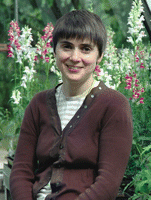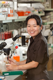
I am Giovanni Povero, a 4th year PhD student in Agrobiosciences at the “Plant Lab” Department of Prof. Perata (http://www.plantlab.sssup.it/) in Pisa, Italy. My PhD research project is mainly focused on the study of the role of genes and transcription factors in the regulation of pigmentation in different plant tissues and species. One of my interests is also the study of the evolution and development (“Evo-Devo”) of pigmentation patterns in plants, in order to gain new insights into this completely new field. The final goal consists also in comparing my results with similar/different demonstrations already found in studies about shape evolution in animals and plants.
I really have to thank the “Development Travelling Fellowship” recently received, because it gave me the opportunity to join the “Genetics” Department of Prof. Ronald Koes and Dr. Francesca Quattrocchio in Amsterdam (The Netherlands). I knew them because I studied their very interesting publications. Therefore I contacted the Dutch group, and together we decided to prepare a project that can be carried out in a short period but at the same time can fit with our reciprocal interests. The project regards a very new and fascinating topic: the study of the relationships between the genes conferring petal identity (in the flower) and genes for pigment biosynthesis. The project was luckily approved from the “Travelling Fellowship” commission, and the result is a nice continuation of my PhD project in Netherlands. In particular, I had the opportunity to spend a 4 months period at the “Genetics” Department in Amsterdam. People in the lab are very kind with me and they prepared all the material I needed before my arrival. We work together, have lunch together, and we go often outside (when the weather is nice) to have picnics in the parks of this fantastic town. Moreover, the lab people help me every time I need some help or I have some technical (or any other kind of) difficulties. I am learning a lot about Dutch habits and culture, and I find it very interesting. Here in Holland they speak a very good English, and this gives me the possibility to train this language. On the other hand, learning some words of Dutch will be a “mission impossible”. There are many differences between Italy and Netherlands, and between Italians and Dutch… but I am fascinated from the way in which this country is organized/administrated, and the way people spend their free time (cycling, jogging, practicing sports and meeting people in the bars, chatting and drinking something together).
I am almost at the end of my period in the Dutch Lab, but I can say already that during this months I have learnt a lot of things: techniques like plant transformation (petunia, tomato, Arabidopsis), different cloning systems and methodological approaches. This makes me really much self-confident, and now I am more than ever sure that an experience abroad is really important; it’s something that people working in a laboratory should do at least once in their life! And now I have also the possibility to strengthen the collaboration with the Amsterdam group. In conclusion, it is a very interesting international experience…
Meeting Report: First Tunicate Information System Meeting, Nice, France November 11-13 2010
Ken Hastings
Montreal Neurological Institute and Biology Department
McGill University
Approximately 50 scientists, including members of the international tunicate research community and representatives of major bioinformatics databases, gathered in Nice, France, November 11-13, 2010 to consider the future development of tunicate informatics. This meeting, termed the First Tunicate Information System Meeting was the inaugural meeting in what is expected to be a regular series devoted to this subject.
First Tunicate Information System Meeting participants on the beach at Nice, November 13 2010
Some meeting participants on the beach at Nice, November 13 2010
Tunicates – a diverse group consisting of ascidians, thaliaceans and larvaceans – are research model organisms that share with vertebrates a common ancestry [1] that is reflected in a common basic chordate body plan [2,3,4]. Compared with vertebrates, development is stereotyped and much more rapid, with far fewer cells [5,6], and is driven by a smaller genome [7,8], all of which make tunicates an ideal experimental system for multiscale molecule-to-cell-to-organism investigation of chordate development [3,9,10,11].
Evolutionarily, tunicates are an extremely successful and diverse group and this diversity is a further asset in their application as research model organisms. In some lineages different species show very similar development and morphology despite having genomes that have diverged greatly at the level of nucleotide sequence [12]. This provides great opportunities for comparative understanding of how genomic information is biologically processed during development. In addition, different tunicate lineages exhibit a remarkable range of lifestyles, adult morphologies, and biological features such as the extreme genome reduction and short lifecycle time of the larvaceans [8], and the amazing regenerative capabilities of the ascidians [13,14], especially the colonial ascidians which can generate identical adult forms either through gametogenesis/fertilization/larval development/metamorphosis, or by asexual direct development by budding or by regeneration from cells of the vascular system [13,15,16]. This great biological diversity promises insight into a wide range of fundamental biological mechanisms, and coupled with the solid platform provided by the great depth of existing molecular, cellular, and gene regulatory data for the intensively studied solitary ascidian Cionaintestinalis, and for the larvacean Oikopleura dioica, makes tunicates a very attractive group in which to develop an integrated database system. Given the recent explosion of capabilities for genome and transcriptome sequencing, now being applied in ongoing projects for several solitary (Halocynthia roretzi, Phallusia mammillata) and colonial (Botryllus schlosseri, Didemnum vexillum) ascidian species, this was the right moment for tunicate researchers, a collegial and interactive world-wide community, to initiate an ambitious plan for a multi-species, multi-class, multi-system, and multi-scale database organization.
The meeting, co-organized by Patrick Lemaire (France), Kazuo Inaba, Yutaka Satou, Toshinori Endo, Kohji Hotta (Japan), and Tony de Tomaso (USA) with funding from AVIESAN and DOPAMINET, drew together tunicate researchers from Europe, North America, Israel, and Japan, and informatics experts from Ensembl UK (Ewan Birney, Fiona Cunningham, Daniel Sobral), DDBJ, NIG Japan (Kazuho Ikeo), NIAIST Japan (Tadashi Imanishi) and Chado USA (Joshua Orvis) (see participant list on Meeting website). In the weeks before the meeting eight working groups carried out email surveys of tunicate researchers to probe their hopes and expectations for a community database. At the meeting these survey results were presented by each working group and discussed in plenary session. In addition, overview presentations of current capabilities and future prospects of the major existing Ciona intestinalis databases were given by Satou (Ghost [17]), Lemaire (ANISEED [11]), Endo and Inaba (CiPro [18]), Hotta (FABA [19]), and Takehiro Kusakabe (DBTGR [20]). Additional presentations by Birney on the relationships of “community” databases to the general databases (e.g. Ensembl), by Cunningham on Ensembl informatics pipelines, by Imanishi on automatic maintenance of hypertext cross-links between databases, and by Orvis on the Chado system of data architecture, provided perspective and insight into the organization and methods of large-scale bioinformatics efforts. Participants broke into roundtable discussion groups to define informatics objectives in the areas of Gene Expression/Transcription, Phenotypes/Anatomy, and Proteins/Cell Biology and discussion outcomes were reported back for further comment in plenary session.
From these proceedings there emerged consensus on standards and priorities on a wide range of issues, and an overall plan for the rationalization and improvement of existing databases in a setting that would foster the emergence of a comprehensive “Tunicate Community Database” into which existing and future data could be functionally integrated (additional details in Meeting summary conclusions). Single-species and themed interest databases currently maintained by individual laboratories were deemed extremely useful and should be further developed going forward. As a first step in their coordinated development, overlap/duplication among the various existing Ciona databases will be reduced. Ghost will retain its focus on the gene and genome and will maintain the principal community genome browser with annotation support from CiPro, CiPro will focus on the level of the cell and its constituents, and ANISEED and FABA will concentrate on multicellularity/development/morphology. Looking forward, it was thought vital that there be uniform standards of data vocabulary and architecture to permit integration with the Tunicate Community Database which will serve as a central access point. The Chado data architecture and a wide range of specific standards were adopted. An additional important aspect of the plan is the development of a single reference data store, a “Tunicate Data Repository” with which the various individual “client” databases would be synchronized to ensure a common set of basic data.
Additional objectives include the comprehensive incorporation of existing gene expression data, and of experimental information regarding cell biology, including experimental protocols, videos, and collections of data on useful tools/reagents, and extending to the published literature, including the difficult-to-access classical literature of past centuries, and also unpublished information, e.g., reports of negative results. Assembly of some of these data, and further discussion of database issues, are to be through Wiki-type community-based input.
In accordance with the general plan for successful community databases outlined by Birney, the Tunicate Community Database would occupy an intermediate level between the individual laboratory databases and the general global databases such as DDBJ, Ensembl, and NCBI. The desire was clearly expressed that the Tunicate Community Database be a real integrator, and not merely a collator, of individual databases. A common data architecture and common controlled vocabularies introduced early can form the basis for joint, rather than parallel, future growth, and can permit true value-added integrative analysis. Such a database system will be ready to serve the expanding need for informatics, and especially integrated informatics, as the number of characterized species increases and the full impact of high-throughput sequence data is felt and exploited.
A philosophical principle governing the future development of the tunicate database system is that it should strive to go beyond the gene-centred approach of current model organism databases and provide an integrated view of development. Because of the stereotyped lineage-based development of tunicates [5, 6,10, 21], the anatomical aspects of development are readily formalized. Such formalization, expressed in terms of controlled-vocabulary ontologies, permits computers to manipulate and understand data on the basis of a semantic web of defined relationships. Ontologies for gene-based molecular data are well-developed and widely used (e.g. Gene Ontology [22]) but this is not yet the case for developmental biology. The stereotyped development of ascidians should permit the creation of highly precise cellular and anatomical ontologies. The simplicity of tunicate development offers the opportunity to create a novel type of integrated database, that may foreshadow future developments in more complex model organisms.
To guide the development of the tunicate database system two committees are being organized. One, a Scientific Steering Committee (SSC), will be composed of representives of the major existing Ciona databases and additional members representing the major tunicate groups – solitary ascidians, colonial ascidians, larvaceans, and possibly thaliaceans, selected by researchers working in each of those communities. This committee will provide overall leadership and will coordinate grant applications to fund the development of the system. In addition, a Scientific Advisory Committee, made up of world-class scientists from outside the tunicate community who are experienced in developing/managing large database systems, would advise the SSC on strategic issues. With such leadership, and the enthusiastic participation of the tunicate research community, this enterprise promises to create a most useful research tool whose development and implementation may provide a model for multi-scale, multi-level integrative informatics in other model organism research communities.
References
1. Delsuc F, Brinkmann H, Chourrout D, Philippe H (2006) Tunicates and not cephalochordates are the closest living relatives of vertebrates. Nature 439: 965-968. Pubmed link
2. Katz MJ (1983) Comparative anatomy of the tunicate tadpole, Ciona intestinalis. Biological Bulletin 164: 1-27. Link to article
3. Satoh N, Satou Y, Davidson B, Levine M (2003) Ciona intestinalis: an emerging model for whole-genome analyses. Trends Genet 19: 376-381. Pubmed link
4. Passamaneck YJ, Di Gregorio A (2005) Ciona intestinalis: chordate development made simple. Dev Dyn 233: 1-19. Pubmed link
5. Satoh N (1994) Developmental biology of ascidians. Cambridge UK, New York USA: Cambridge University Press.
6. Lemaire P (2009) Unfolding a chordate developmental program, one cell at a time: invariant cell lineages, short-range inductions and evolutionary plasticity in ascidians. Dev Biol 332: 48-60. Pubmed link
7. Dehal P, Satou Y, Campbell RK, Chapman J, Degnan B, et al. (2002) The draft genome of Ciona intestinalis: insights into chordate and vertebrate origins. Science 298: 2157-2167. Pubmed link
8. Denoeud F, Henriet S, Mungpakdee S, Aury JM, Da Silva C, et al. (2010) Plasticity of animal genome architecture unmasked by rapid evolution of a pelagic tunicate. Science 330: 1381-1385. Pubmed link
9. Imai KS, Levine M, Satoh N, Satou Y (2006) Regulatory blueprint for a chordate embryo. Science 312: 1183-1187. Pubmed link
10. Nishida H (2008) Development of the appendicularian Oikopleura dioica: culture, genome, and cell lineages. Dev Growth Differ 50 Suppl 1: S239-256. Pubmed link
11. Tassy O, Dauga D, Daian F, Sobral D, Robin F, et al. (2010) The ANISEED database: digital representation, formalization, and elucidation of a chordate developmental program. Genome Res 20: 1459-1468. Pubmed link
12. Johnson DS, Davidson B, Brown CD, Smith WC, Sidow A (2004) Noncoding regulatory sequences of Ciona exhibit strong correspondence between evolutionary constraint and functional importance. Genome Res 14: 2448-2456. Pubmed link
13. Voskoboynik A, Simon-Blecher N, Soen Y, Rinkevich B, De Tomaso AW, et al. (2007) Striving for normality: whole body regeneration through a series of abnormal generations. Faseb J 21: 1335-1344. Pubmed link
14. Auger H, Sasakura Y, Joly JS, Jeffery WR (2010) Regeneration of oral siphon pigment organs in the ascidian Ciona intestinalis. Dev Biol 339: 374-389. Pubmed link
15. Ballarin L, Menin A, Tallandini L, Matozzo V, Burighel P, et al. (2008) Haemocytes and blastogenetic cycle in the colonial ascidian Botryllus schlosseri: a matter of life and death. Cell Tissue Res 331: 555-564. Pubmed link
16. Brown FD, Keeling EL, Le AD, Swalla BJ (2009) Whole body regeneration in a colonial ascidian, Botrylloides violaceus. J Exp Zool B Mol Dev Evol 312: 885-900. Pubmed link
17. Satou Y, Takatori N, Fujiwara S, Nishikata T, Saiga H, et al. (2002) Ciona intestinalis cDNA projects: expressed sequence tag analyses and gene expression profiles during embryogenesis. Gene 287: 83-96. Pubmed link
18. Endo T, Ueno K, Yonezawa K, Mineta K, Hotta K, et al. (2011) CIPRO 2.5: Ciona intestinalis protein database, a unique integrated repository of large-scale omics data, bioinformatic analyses and curated annotation, with user rating and reviewing functionality. Nucleic Acids Research 39: D807-D814. Pubmed link
19. Hotta K, Mitsuhara K, Takahashi H, Inaba K, Oka K, et al. (2007) A web-based interactive developmental table for the ascidian Ciona intestinalis, including 3D real-image embryo reconstructions: I. From fertilized egg to hatching larva. Dev Dyn 236: 1790-1805. Pubmed link
20. Sierro N, Kusakabe T, Park KJ, Yamashita R, Kinoshita K, et al. (2006) DBTGR: a database of tunicate promoters and their regulatory elements. Nucleic Acids Res 34: D552-555. Pubmed link
21. Sardet C, Paix A, Prodon F, Dru P, Chenevert J (2007) From oocyte to 16-cell stage: cytoplasmic and cortical reorganizations that pattern the ascidian embryo. Dev Dyn 236: 1716-1731. Pubmed link
22. Ashburner M, Ball CA, Blake JA, Botstein D, Butler H, et al. (2000) Gene ontology: tool for the unification of biology. The Gene Ontology Consortium. Nat Genet 25: 25-29. Pubmed link
NOTE: LOCATION CHANGE!
We’ll meet at the Gulbenkian Cinema Cafe/Bar at 8PM on Thursday, as Mungos is closed.
This is the cafe that you walk past along the footpath from Elliot College to Woolf College, so when you go from dinner to posters you can drop in along the way.
——
If you’d like to meet some other Node readers (and writers), why not join us for a drink during the BSDB meeting. Bring your lab mates and friends – we like meeting new people!
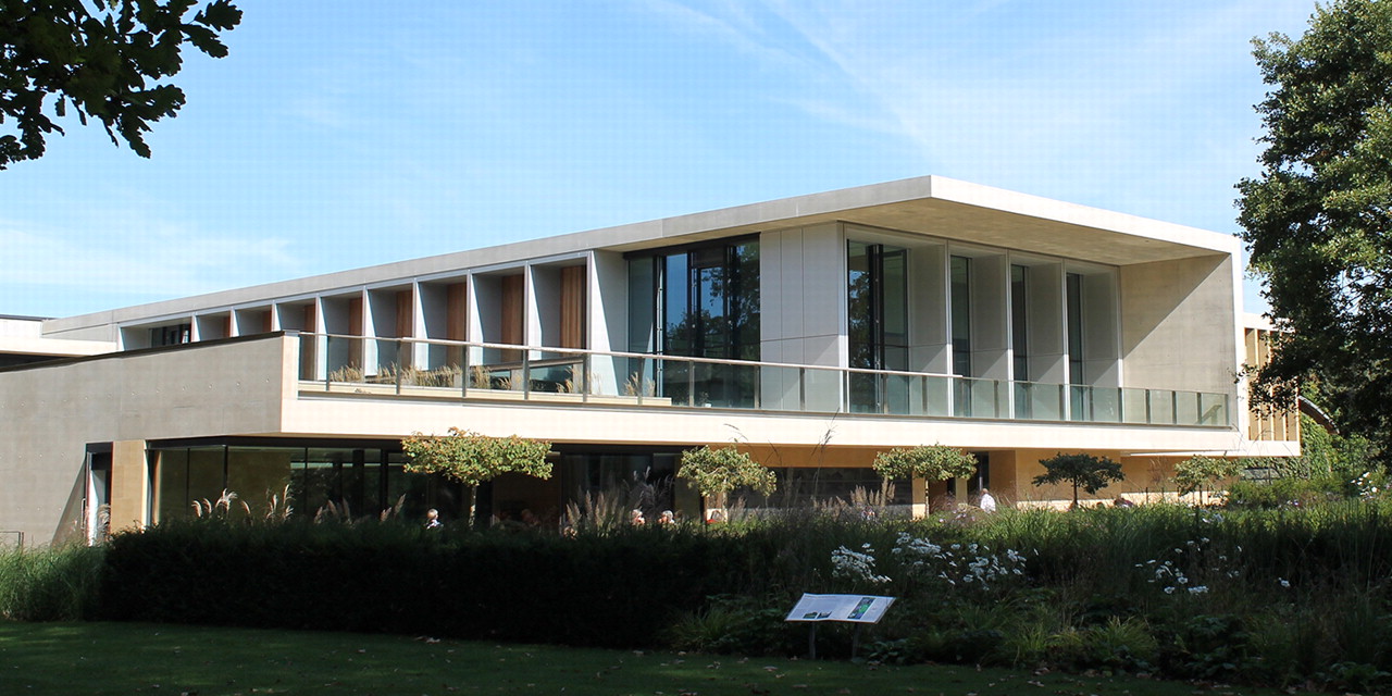
We’ll get together at 20:00 on Thursday April 28, at Mungos Bar (in Eliot College) Gulbenkian Cinema Cafe Bar.
We’ll be there for about an hour or so, and then we’ll head off to the poster session in the Woolf Building, so don’t worry about missing dinner or posters.
There will be a sign at The Company of Biologists’ stand in the exhibition space about this, and if anything changes (location, times) you can find the information there as well. If it’s not too cumbersome, I’ll try to bring the banner (pictured) to the bar for easy identification, otherwise there’ll be other ways to identify us.
If there is hope to fully understand stem cells, then the environment surrounding those stem cells must be understood too. A recent Development paper describes important results on niche establishment in Drosophila.
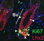
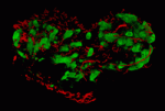
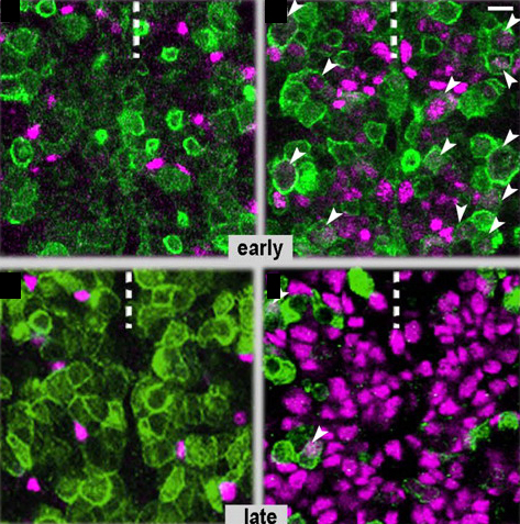
Stem cell niches play an important role in regulating stem cell self-renewal and differentiation. The Drosophila testis has two populations of stem cells that cluster around a niche called the “hub.” It was previously known where hub cells originated, but recent work describes how hub cells become specified. Notch and its two ligands, Delta and Serrate, are required for specifying hub cell fate during gonad development. Interestingly, endoderm tissue (posterior midgut cells, if you’re keeping score) provides the Delta signal, further emphasizing a well-known relationship between gut and germ cells. As seen in the above images of larval gonads, flies with mutations in Delta (left, bottom) or (right, bottom) had fewer hub cells (green) than control gonads (top).
For a more general description of this image, see my post at EuroStemCell, the European stem cell portal.
Okegbe, T., & DiNardo, S. (2011). The endoderm specifies the mesodermal niche for the germline in Drosophila via Delta-Notch signaling Development, 138 (7), 1259-1267 DOI: 10.1242/dev.056994
Here are the research highlights from the current issue of Development:
Cranial neural crest development: p53 faces up
The tumour suppressor p53 plays multiple roles in the prevention of cancer but its developmental functions are less clear. Here (see p. 1827), Eldad Tzahor and colleagues elucidate the key role that p53 plays in craniofacial development. During embryogenesis, cranial neural crest (CNC) cells give rise to the facial bones, cartilage and connective tissues. Neural crest development involves an epithelial-mesenchymal transition (EMT) that converts epithelial cells into migratory mesenchymal cells, which delaminate from the neural tube. Notably, EMT is an early step in tumour progression. The researchers report that craniofacial development is disrupted in p53 knockout mouse embryos. Then, they show that p53 is expressed in CNC progenitors in chick embryos but that its expression decreases as these cells delaminate from the neural tube. Moreover, p53 gain-of-function results in fewer migrating CNC cells, whereas p53 loss-of-function increases the EMT/delamination of CNC cells. These and other findings suggest that p53 coordinates CNC growth and EMT/delamination processes during craniofacial development.
Oiling the wheels of Hippo signalling
The Hippo signalling pathway, a conserved tumour suppressor pathway, is involved in other developmental processes in addition to proliferation control. For example, Hippo signalling in the posterior follicle cells (PFCs) of Drosophila ovaries is required for oocyte polarisation. Now, Trudi Schüpbach and colleagues report that a phospatidylinositol 4-kinase (PI4KIIIalpha), which catalyses the production of membrane phospholipids, is required in PFCs for oocyte polarisation and Hippo signalling (see p. 1697). The researchers isolated mutations in CG10260, which encodes PI4KIIIalpha, while screening for Drosophila genes required in follicle cells for oocyte polarisation. They show that PI4KIIIalpha loss in PFCs leads to oocyte polarisation defects similar to those caused by mutations in the Hippo signalling pathway, and that PI4KIIIalpha mutations cause misexpression of Hippo targets. Notably, the apical membrane localisation of Merlin, which is required for Hippo signalling, is lost in PI4KIIIalpha mutant PFCs, presumably because of changes in the cell membrane’s lipid composition. Together, these data reveal a new link in the Hippo signalling pathway.
Nu-age transposon silencing
The nuage, a perinuclear structure of unknown function, is present in the germline cells of many organisms. Now, on p. 1863, Haifan Lin and colleagues reveal a function for the Drosophila germline nuage. This structure contains Aubergine and Argonaute 3 (AGO3), two of the three PIWI proteins that are essential for germline development. PIWI proteins bind to PIWI-interacting RNAs (piRNAs) and function in transposon silencing. The researchers report that Partner of PIWIs (PAPI), a novel nuage component, is a TUDOR-domain protein that interacts with all three PIWI proteins through symmetrically dimethylated arginines in their N-terminal domains. This interaction is essential for AGO3 recruitment to the nuage and for transposon silencing. Importantly, the AGO3-PAPI complex associates with the P-body component TRAL/ME31B complex in the nuage and transposon activation occurs in tral mutant ovaries. Thus, the interaction in the nuage between the piRNA pathway and mRNA-degrading P-body components is involved in transposon silencing. The researchers suggest, therefore, that the nuage safeguards the germline genome against deleterious retrotransposition.
Invading heart morphogenesis with NFATC1
During cardiac morphogenesis, proepicardium cells envelop the myocardium to form the epicardium. Some epicardial cells subsequently undergo epithelial-to-mesenchymal transformation and invade the myocardium as epicardium-derived cells (EPDCs). This invasion step underlies the formation of the coronary vessels and fibrous matrix of the mature heart but how is it regulated? Michelle Combs, Katherine Yutzey and co-workers now reveal that NFATC1 promotes EPDC invasion into myocardium (see p. 1747). NFATC1, they report, is expressed in EPDCs in mouse and chick embryos and loss of its expression in EPDCs in mice decreases coronary vessel and fibrous matrix invasion into the myocardium. Other experiments in mouse embryos, chicken embryo hearts and isolated proepicardium cells indicate that NFATC1 activation by RANKL in EPDCs promotes expression of the extracellular matrix-degrading enzyme cathepsin K, promoting EPDC invasion into the myocardium. These new insights into heart morphogenesis, the authors suggest, could aid the development of EPDC-based therapies for cardiac diseases.
Timely neural identity decision making
The timing of cell identity decisions must be closely regulated during brain development. In Drosophila neuroblasts, the sequential expression of several transcription factors, including Hunchback (Hb), controls the temporal generation of diverse neural progeny. Because Hb is necessary and sufficient to specify early-born neurons, its expression has to be downregulated to allow specification of late-born progeny. Now, on p. 1727, Chris Doe and colleagues report that two pipsqueak-domain proteins – Distal antenna (Dan) and Distal antenna-related (Danr) – restrict Hb expression in neuroblasts and limit the numbers of early-born neurons. They show that Dan and Danr function independently of Seven-up (Svp), an orphan nuclear receptor that also regulates Hb expression in neuroblasts. Importantly, Hb misexpression can induce Dan and Svp expression in neuroblasts, which suggests that Hb can limit its own expression through a negative-feedback loop. The researchers conclude that Dan/Danr and Svp act in parallel pathways to limit Hb expression and allow neuroblasts to switch from making early-born to making late-born neurons at the proper time.
Longitudinal axon connections notched up
Development of the segmented central nerve cords of vertebrates and invertebrates involves the formation of longitudinal axon connections between successive segments. To establish these connections, a pathway must be marked for pioneer axons to follow and then the pioneers’ motility along that pathway must be promoted. But what are the molecular mechanisms that control these processes? On p. 1839, Edward Giniger and co-workers show how Notch signalling directs both processes in the developing Drosophila CNS. They show that canonical Notch signalling in specialised glial cells causes the extrusion of a mesh of fine filopodia by nearby differentiating neurons and shapes this mesh into a carpet that links adjacent segments. Simultaneously, non-canonical Notch signalling in the pioneer growth cones suppresses Abl tyrosine kinase signalling, which stimulates filopodial development and presumably also reduces substratum adhesion, thereby promoting the ability of pioneer axons to follow the carpet across segment borders. Thus, two parallel but separate Notch functions establish the first longitudinal connections in the fly CNS.
And…
Review: Small RNAs in early mammalian development: from gametes to gastrulation
Small non-coding RNAs, such as microRNAs, endo-siRNAs and piRNAs, are expressed throughout mammalian development and, here, Nayoung Suh and Robert Blelloch review emerging roles for these RNAs in the early stages of mammalian development, from gamete maturation through to gastrulation.
Last week, news that the Australian government was planning to slash the budget for medical research by more than half over the next three years leaked out and rocked the scientific community. Only one out of seven grants submitted to the National Health and Medical Research Council (NHMRC), Australia’s major funding body for medical research, is approved with at least another three deemed good enough for funding but ultimately rejected anyway due to lack of funds. With the potential upcoming cuts to medical funding, the NHMRC will only be able to fund one out of ten grants it reviews.
The gravity of this news cannot be understated. It is a sad disaster. Medical research is of prime importance especially when we take into account terrible disorders such as cancer, HIV/AIDS, cardiovascular diseases, diabetes and infectious diseases which our generation has to face. Cutting funding means saying no to progress against fighting those diseases and the many others. It means leaving people who can be saved to die. Yes, it is a murderous decision.
Developmental biology will not be spared from those cuts. Important and possibly life-changing research related to stem cells, for instance, will be halted, discontinued or rejected, plain and simple. Who will be affected? Well, it could be me, you, your family or friends… it could be anyone.
If the government (elected by the people, for the people) does not realize that it is going to spill its own people’s blood by cutting medical research funding, then it is up to the people to send them a message. And so it did. Rallies were organized today in several major Australian cities including Melbourne, Sydney, Canberra and Adelaide with more to follow in Perth, Darwin and Hobart later this week. A Twitter rally–that successfully trended #protectresearch, the Twitter hashtag adopted by the movement–was also carried out online.
I was at the Melbourne rally where an estimated 4000 demonstrators assembled in front of the State Library. Banners were everywhere, raised high up by outstretched arms, or rulers, meter rules or pipettes. Yes, scientists are an innovative bunch. “Medical research keeps my blood pumping,” “SOS: Save Our Science,” “Cut the nonsense, not the funding!” or “Gillard [the Australian prime minister], we’ll cure your dementia (no seriously… we will)!” were flung around.
Amidst the chorus of “Cures not cuts” and “research saves lives,” were a number of guest speakers. One of them was Nerissa Mapes, a 34-year-old woman who has been living with Parkinson’s disease for the past six years. She addressed the crowd with what was a poignant and deeply powerful speech. Her speech stressed on the important role scientists play in society. It was a wonderful way to remind us all that medical research is for the people. She concluded with this:
“There is no cure for Parkinson’s disease. But there is hope. And hope gives us courage. Don’t let them take this hope away from us.”
Medical research saves life. Cutting its funding is like handling death sentences at will.
The recipients of Canada’s most prestigious science awards, the Gairdner Awards, was recently announced. The Awards recognize researchers for their contributions to the field of medical research. The 2011 Gairdner Awards Recipients are:
2011 Canada Gairdner International Awards:
Adrian Peter Bird Ph.D., Howard Cedar M.D., Ph.D., and Aharon Razin Ph.D. for their discoveries on DNA methylation and its role in gene expression.
Shizou Akira M.D., Ph.D. and Jules A. Hoffman Ph.D. for their discoveries and definition of the family of Toll like receptors and the array of microbial compounds that they recognize to provide innate resistance to infection.
2011 Canada Gairdner Global Health Award:
Robert Black M.D., MPH for his contributions to improving child survival and for critical clinical and epidemiological studies to reduce childhood diarrheal deaths.
2011 Canada Gairdner Wightman Award:
Michael Hayden CM, OBC, M.B., Ch.B., Ph.D., FRCP (C), FRSC for his national and international leadership for medical genetics, entreprenuership and humanitarianism.
Awards winners will present lectures in October 2011 as part of the nation-wide celebration of the Gairdner Awards. A review of the 2010 Gairdner Award recipients and awards lectures can be found in a previous post. For more informatio about the Gairdners, visit the website.
You’ve seen the news: ES cells generate a 3D retinal structure. But what does this tell us about eye development?
In the developing embryo, the first step toward a functional eye is the formation of the optic vesicle from the neural tube. This optic vesicle then invaginates to form an optic cup, which in turn develops into the outer pigmented layer of the retina and the inner neurosensory layer.
Normally, this all takes place in the context of the developing organism, next to neighbouring tissues. But, in a paper published in Nature this week, the Sasai lab at the RIKEN institute in Japan describes how they generated an optic cup in culture, from mouse embryonic stem cells.
The lab had previously generated retinal precursors from mouse ES cells in culture, but those did not form three-dimensional structures. In this new study, they changed the cultured medium by adding Matrigel (containing basement-membrane components). This initiated the formation of small, polarized, spheres after six days in culture. These spheres then invaginated to form the optic cup structure, as shown in this video from the study:
The immediate relevance of this paper is the increased understanding it offers in the mechanisms behind eye development. The study suggests that formation of the retina occurs to a large extent via an intrinsic order that does not entirely depend on external forces. That does not mean that neighbouring tissues have no influence at all, but this influence appears smaller than previously believed.
While this does not mean that we can make custom eyes on demand just yet, the study does have some other clinical implications: If we can generate functional retina from induced pluripotent stem cells taken from patients’ tissues, these could be used in drug testing or disease modeling, and help increase our understanding of diseases that cause blindness.
We’re trying to gauge interest for an informal gathering of Node readers (in the form of drinks after dinner) at the upcoming BSDB meeting. We have a lot of readers among BSDB members, but don’t know if you’re all attending the meeting this year, and whether you’re interesting in meeting other readers and contributors. You can bring your non-Node-reading lab mates along, of course, and just take this as another opportunity to meet some people from other labs.
The meeting program is pretty full, but it looks like there’s a possibility to meet on Thursday April 28 between dinner and poster viewing, or that same night at the end of the poster viewing session. (The Thursday poster session for odd-numbered posters appears to be an hour longer than the time allotted for the even-numbered posters the next day, so this seemed the best moment.)
What do you think? Would you be interested in meeting other Node readers/writers? It will be very informal, and you don’t have to talk about the Node (but you can if you want to, and I’ll be happy to answer questions about the site).
Let us know via the poll if you’d be up for grabbing a drink on Thursday night. If we decide to go ahead with this, we’ll post a notice here, as well as at The Company of Biologists’ stand at the BSDB meeting with time and location details.
Congratulations to Stéphane Vincent of the IGBMC in Illkirch, France, who won the Node’s intersection image competition:
His image showing staining of a gut section of a E17.5 mouse embryo impressed the judges as well as the Node’s readers, receiving more than half of the votes.
Stéphane says: “I took this picture by chance: I was looking at the expression of Sox6 and slow Myosin Heavy Chain in the deep back muscles of a mutant mouse embryo and I saw this very nice “I” popping out in the gut tube… “
With this serendipitous image he has won a TipArt commission, and we hope we’ll get to see the final artwork he receives. In addition, Stéphane’s image will be used in a little project we’re working on to mark the Node’s upcoming first birthday in June.