The community site for and by
developmental and stem cell biologists

ReplicationDomain

Posted by , on 26 October 2010

ReplicationDomain is an online database resource for storing, sharing and visualizing DNA replication timing and transcription data, as well as other numerical epigenetic data types. Data is typically obtained from DNA microarrays or DNA sequencing. Our site has a user registration system that allows registered users to upload their own data sets. While non-registered users may freely view and download public data sets, registered users may upload their own data sets and view them privately, share them with other registered users, or make published data sets publicly available. In addition we have implemented additional mechanisms that allow users to restrict sharing of data sets to a user designated group of registered users. Further details on the database usage are in the User Guide Page, while data set details are in the Documentation Page.

Mus musculus_MM8_D3_ESC_Chr5_60200463-75644002
Thumbs up (3 votes)
Loading...

Categories: Research, Resources

Navigate the archive

Use our Advanced Search tool to search and filter posts by date, category, tags and authors.

In Development this week (Vol. 137, Issue 22)

Posted by , on 26 October 2010

Research highlights from the current issue of Development:

Novel Hh targets fly in

Hedgehog (Hh), a secreted morphogen, acts in a paracrine fashion to regulate tissue patterning during embryogenesis. Its tissue-specific effects are mediated by the transcription factor Cubitus interruptus (Ci), but how it exerts such effects is unclear. On p. 3887, Thomas Kornberg and colleagues address this question by identifying novel Drosophila Hh targets. Using chromatin-binding experiments to identify genes that are bound by Ci during Drosophila organogenesis, and by using expression data from wild-type embryos and Hh pathway mutants, they identified a set of Hhresponsive genes, many of which represent novel targets. Their validation of these targets in developing tissues, such as the dorsal ectoderm, showed that they are expressed in a tissue-specific manner, but, unexpectedly, that some targets are induced in an autocrine fashion. The authors also show that, in the tracheal primordium, some Hh target expression is subject to combinatorial control by Ci and an Hh-independent transcription factor. These unexpected features of Hh signalling provide new insights into our understanding of this pathway.

Modelling to get a head(fold)

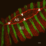
Formation of the head fold (HF), the first three-dimensional structure to form in the embryo, is a crucial event that initiates heart, foregut and brain development. Although the molecular factors involved in HF development are becoming known, the biophysical mechanisms governing this transformation have yet to be investigated. Now, on p. 3801, Larry Taber and colleagues combine experimental and modelling approaches to determine the forces that drive HF formation in chick embryos. They generated a computational model for HF formation, and by inducing three distinct morphogenetic mechanisms – convergent extension in the neural plate (NP), cell wedging along the anterior NP border, and cell shaping outside the NP – they were able to simulate HF formation in this model. By comparing the changes in tissue morphology, mechanical strains and regional tissue stresses observed in the model with those measured experimentally in ex ovo embryos, the authors confirm that these three modelled morphogenetic mechanisms can alone provide the forces that drive HF formation.

Embryonic variations on a histone theme

Numerous histone variants exist in eukaryotes, and the replacement of canonical histones with such variants probably contributes to chromatin remodelling. Chromatin remodelling occurs during fertilisation, as germ cells become totipotent zygotes, but the role of histone variants during this process is unknown. On p. 3785, Fugaku Aoki and colleagues assess the dynamics of histone H2A and its variants, H2A.X, H2A.Z and macroH2A, during mouse oogenesis and pre-implantation development. They report that all variants are present in oocytes; by contrast, only H2A.X is abundant in one-cell embryos. The authors confirmed this postfertilisation reduction in H2A, H2A.Z and macroH2A using transgenic mice that express tagged H2 variants, and by microinjecting embryos with mRNA for these variants. Domain-swapping experiments showed that the C-terminal 23 amino acids of H2A.X enable its incorporation into chromatin after fertilisation, and that the concomitant reduction of H2A.Z and macroH2A is required for normal development. The authors suggest that altered histone composition might therefore contribute to the genome remodelling, and hence reprogramming, that occurs postfertilisation.

Meristem homeostasis: it takes three

In plants, the shoot apical meristem (SAM) provides all the cells that are needed for post-embryonic growth and development of the leaves, stems and flowers. In Arabidopsis, a peptide ligand derived from CLAVATA3 (CLV3) regulates the SAM stem cell pool by signalling through two receptor complexes – a homodimer of the receptor-like kinase CLV1 and a heterodimer consisting of the receptor-like protein CLV2 and the protein kinase CRN/SOL2. Now, Shinichiro Sawa and colleagues report that the receptor-like kinase RPK2 also has a vital role in SAM maintenance (see p. 3911). They show that loss-of-function mutations in RPK2 result in SAM stem cell expansion and increased numbers of floral organs, as seen in clv1 and clv2 mutants. Notably, the RPK2 mutant phenotypes are additive with those of clv1 and clv2 mutations. Moreover, biochemical analyses in Nicotiana benthamiana reveal that RPK2 forms homodimers but does not associate with CLV1 or CLV2. The researchers propose, therefore, that three, rather than two, CLV3 signalling pathways regulate meristem homeostasis.

Lymphangiogenesis: macrophages show restraint

Lymphatic vessels play crucial roles during embryogenesis and in adult animals but the origin of their progenitor cells is controversial. Recent studies have suggested that during neo-lymphangiogenesis in inflammatory settings, macrophages transdifferentiate into lymphatic endothelial cells and/or release prolymphangiogenic growth factors, but are these mechanisms involved in the development of the normal lymphatic vasculature? On p. 3899, Natasha Harvey and co-workers suggest that the answer to this question is no. Using lineage tracing, the researchers show that lymphatic endothelial cells arise independently of the myeloid lineage during both embryogenesis and tumourstimulated lymphangiogenesis in the mouse. Thus, they suggest, macrophages are not the source of lymphatic endothelial progenitor cells in these settings. Furthermore, the dermal lymphatic vasculature in macrophage-deficient mice is hyperplastic because of increased lymphatic endothelial cell proliferation. So, rather than providing pro-lymphangiogenic growth factors, macrophages provide signals that restrain lymphatic endothelial cell proliferation. Given these results, any attempt to treat disease-stimulated lymphangiogenesis by targeting macrophages needs careful consideration, conclude the researchers.

Grainyhead heads up apical junction formation

Epithelial cell differentiation requires the formation of the apical junctional complex, a membrane-associated structure that includes adherens junctions (which mediate stable adhesion between epithelial cells) and tight junctions (which regulate the movement of water and solutes between epithelial cells). Now, on p. 3835, Kai Schmidt-Ott and colleagues report that the mammalian transcription factor grainyhead-like 2 (Grhl2), an epithelium-specific homologue of Drosophila Grainyhead, regulates the molecular composition of the apical junctional complex. Grhl2, they report, determines the expression levels of E-cadherin and claudin 4 (Cldn4) – key components of adherens junctions and tight junctions, respectively – in several types of epithelia. Other experiments reveal that Grhl2 regulates epithelial differentiation in vitro and in vivo, that Grhl2 deficiency in mice results in defective neural tube closure, and that Grhl2 associates with conserved cisregulatory elements in the Cldn4 and E-cadherin genes. Together, these data suggest that Grhl2 is a transcriptional activator of apical junctional complex components and is, therefore, a crucial participant in epithelial differentiation.

Also…


Since its discovery, FGF signalling has been implicated in numerous developmental processes and in disease. Now Karel Dorey and Enrique Amaya provide an update of the main developmental processes for which FGF signalling is vital during early vertebrate embryogenesis.

For more details, see the Primer article on p. 3731.

Thumbs up (No Ratings Yet)
Loading...

Tags: , , , , , , , , , , , , , , , , , ,
Categories: Research

Hello from Nairobi

Posted by , on 24 October 2010

My apologies for the lag in updates from field collections in China.  I got a little distracted with submitting a paper and writing a K99/R00 that seem to have consumed the last month of my life.  But what better reward than a 3 week trip to Kenya!

Yes, I am a roving postdoc.  Boston is starting to grow on me, but as one of my travel companions/colleagues said yesterday “the best thing to do in Boston is leave.” The current trip is an “ambassadorship” sponsored by the department of Genetics at HMS to represent and work for a non-profit organization called “Seeding Labs”.  Seeding Labs was started by a former HMS graduate student, Nina Dudnik, and aims to distribute research resources to developing countries to better equalize opportunities.  I got involved because in all the travels I’ve done, for work and for play, I’ve met people with that spark in the eye, the passion and drive, but not the essential things they need to do something with all of that energy.  By way of example, I met a graduate student at Xinjiang University who saved from her own salary for months to buy an antibody for the protein she was studying because the lab didn’t have the money.

The primary mission of Seeding Labs so far has been to collect surplus (working) laboratory equipment to distribute to universities all over the world, and a particularly close relationship has formed with Kenyatta University in Nairobi.  Phase 2 of the project is a personnel exchange.  Four fellows from KU spent 2 months this summer working at Novartis in Boston, and now the three of us from Harvard are spending two weeks at KU running workshops on teaching, grant writing, statistics, career development and graduate school applications, presentations skills, etc.  We’re also touring labs, meeting faculty and students, and doing everything we can to encourage growth and research development.  The first 48 hours has been invigorating and a lot of fun, and we’re squeezing in some trips to see wild animals amongst the work.

I’ll continue posting updates while I’m here, because I would love to bring awareness of this fabulous program to the Node community.  If any of you would like to learn more or get involved, check it out at www.seedinglabs.org

Thumbs up (3 votes)
Loading...

Categories: Resources

Postdoctoral position in Cell and Developmental Biology

Posted by , on 22 October 2010

Closing Date: 15 March 2021

St. Jude Children’s Research Hospital, Dept. of Genetics

POSTDOCTORAL POSITION in Cell and Developmental Biology is available to study the cellular and molecular mechanisms controlling the development of the lymphatic vasculature using available mouse models and its functional roles in health and disease. Highly motivated individuals who recently obtained a PhD. or MD degree and have a strong background in molecular and developmental biology are encouraged to apply. Interested individuals should send their curriculum vitae, a brief description of their research interests, and the names of three references to:

Guillermo Oliver, Ph.D (guillermo.oliver@stjude.org)

Department of Genetics

St. Jude Children’s Research Hospital

332 N. Lauderdale, Memphis, TN 38105

http://www.stjude.org/oliver

Thumbs up (No Ratings Yet)
Loading...

Tags:
Categories: Jobs

Biology lecture posters

Posted by , on 21 October 2010

One of my (many) geeky passions is the overlap between art and science: Science as art (think of the Nikon image competition) or art inspired by science. That last category includes these lecture announcement posters from UNC Chapel Hill.

Poster for a recent lecture by Peter Wilf

The posters are designed by developmental biologist Bob Goldstein, and printed by The Merch in Carrboro, who normally print posters for dance parties rather than biology lectures!

If you’ve seen the posters online before, I may have been responsible for that as well… I wrote about them on my old blog in early 2009, and the link got picked up by BoingBoing and subsequently by The Scientist, who interviewed Bob by phone about his posters (see video below).

In an additional small world connection, Bob’s former graduate student Erin started writing for the Node, featuring pretty images from papers. That brings us full circle from “art inspired by science” to “science as art”.

Thumbs up (9 votes)
Loading...

Tags: ,
Categories: Images, Research

Building blocks

Posted by , on 19 October 2010

Somites are the building blocks of the vertebrae, skeletal muscle and dermis…literally and figuratively.  Somites define the segmented features of vertebrate embryos, and are repeated blocks of epithelial cells formed sequentially, from anterior to posterior, and at regular intervals on either side of the neural tube.  A paper in the November 1 issue of Development helps our understanding of the signaling cascade that acts during somitogenesis.

Integrins are transmembrane proteins that function in cell adhesion, migration, signaling and proliferation, which are all important processes during development.  Previous research had demonstrated a role for integrins in the formation of somites in certain organisms, but the exact requirement or mechanism was not clear.  This month in Development, Rallis and colleagues show that β1-integrin is important for formation of all somites in chick embryos.  They also found that β1-integrin functions in “outside-inside” signaling, meaning that signals from the extracellular matrix bind to integrins and result in the activation of signaling within the cell.  Specifically, β1-integrin functions cell-autonomously to activate Wnt and Notch signaling, via ILK, which leads to compartmentalization and boundary formation of somites.

Images above show a schematic of a chick embryo (A) and β1-integrin (red) localization during somite formation (TOPRO3, in blue, stains nuclei and shows somite organization).  β1-integrin was found at the borders of both older, anterior somites (B) and newly formed somites (C).  There was also an anterior-posterior gradient of β1-integrin in the presomitic mesoderm prior to somite formation (white versus yellow arrow in D, and zoomed image in E).  Interestingly, β1-integrin was found in the core of somites (F, arrowhead), and showed a continued segmented pattern in much later embryos (G).

Reference:  Charalampos Rallis, Sheena M. Pinchin and David Ish-Horowicz (2010).  Cell-autonomous integrin control of Wnt and Notch signalling during somitogenesis.  Development 137, 3591-3601.  Paper can be found here.

Thumbs up (9 votes)
Loading...

Tags: ,
Categories: Images, Research

Tree of Life – biodiversity linked

Posted by , on 18 October 2010

From Arabidopsis to zebrafish, every species –living and extinct – is linked to every other species. Not just metaphorically, but also literally on the Tree of Life website, which ambitiously aims to create a linked database with information on every species and group of organisms.


(Image from Tree of Life, used under Attribution-NoDerivs 3.0 Unported license)

Launched in 1995 the site was originally developed for biologists who might need to find phylogenetic information, but got so many requests from students and educators that they expanded their reach and now Tree of Life also provide “treehouse” pages, featuring accessible information for a wider audience.

Incidentally, the educational aspect of Tree of Life reminded me of another project I recently heard about: Phylo is a trading card game of which the cards are produced online by volunteers. The cards can then be used to teach children about biodiversity, much like the treehouse pages on Tree of Life

Like Phylo, content on Tree of Life is also contributed by volunteers. Scientific content is peer reviewed, and contributed by scientists and science educators, but anyone can submit media (such as images) to the site.

There’s a lot to be discovered on the site, so have a look around. It’s a work in progress, as it will be a long time until they’ve filled it with complete pages for every organism!

Thumbs up (1 votes)
Loading...

Tags: , ,
Categories: Resources

Axon guidance, synaptic plasticity and regeneration meeting report

Posted by , on 13 October 2010

For anyone who has never been, Cold Spring Harbor Laboratories organize some really great conferences. The axon guidance, synaptic plasticity and regeneration conference, formerly the axon guidance synaptogenesis and plasticity conference, is biannually held in September and other than occasional heavy showers you can expect some fabulous weather. The campus, with the harbor that runs alongside it, is very beautiful especially with the first hint of autumn colour.

This year axon guidance and regeneration seemed to predominate over synaptic plasticity. The first day started with an evening session and whilst the jetlag can be a problem one of the other great things about these conferences is ‘The Leading Strand’, a password protected website that allows meeting participants to re-watch talks for a limited period of time after the conference. Sadly, but perhaps understandably, this appears to be becoming less and not more popular. Perhaps people feel that it’s unnecessary since there are no parallel sessions that force you to miss something.

The first session opened with some of our favourite axon guidance molecules Slit, Robo, Ephs and Ephrins. Adam Guy from the Neuronal Growth Mechanisms Lab, RIKEN Brain Science Institute, got the dubious honour of closing the session with a late night talk showing chemorepulsion of sensory axons in the chick spinal cord by a phospholipid, suggesting a specific axon guidance role and possibly a novel group of guidance molecules.

Wednesday opened with one of the few sessions that focused on synapses and circuits. This included a very interesting talk by Nicola Allen on an astrocyte secreted factor that can induce synapse formation in vitro. Nicola went on to describe the biochemical identification of the molecule and to show that hippocampal slice cultures from the knockout mouse do have an electrophysiological phenotype. The day finished with a plenary lecture which by Peter Devreotes on chemotaxis in which he sort to bring together the many different strands of research and show how they might fit together. It was a fascinating but rapid overview of the topic and there is clearly still much left to understand.

Thursday was easily the busiest day of the conference with talks until 3.30pm, a poster session until 5.30pm and then an evening session of talks from 7.30pm. You certainly get your money’s worth of science at Cold Spring Harbor!

Friday began with the second session on stem cells, regeneration and disease. In vivo laser axotomy appears to be the tool of choice for investigating regeneration at the moment and in combination with zebrafish and C.elegans, models which are so amenable to manipulation and live imaging, will certainly yield much useful and interesting data. After lunch we were treated to two more plenary lectures, the first given by Tom Jessell on ‘The nerves and networks of spinal motor control’ and the second by Eva Marder on ‘Compensation in robust network performance’, both were truly captivating and quite humbling.

The conference dinner on Friday evening was preceded by a very impressive performance by the up and coming violinist Hahn-Bin. After such a cultured beginning it was all downhill from then on…the less said about the drunken dancing the better!

Thumbs up (3 votes)
Loading...

Tags: , , ,
Categories: Events, Research

Nippon

Posted by , on 12 October 2010

Dear Reader,

My name is Dávid Molnár, I’m a third year Ph.D. student in the Department of Human Morphology and Developmental Biology at Semmelweis University (Budapest, Hungary). I’d like to share the story of my summer internship with You!

Thanks to the generous offer of Guojun Sheng, the team leader of the Laboratory for Early Embryogenesis in RIKEN Center for Developmental Biology (Kobe, Japan), I could spend three months in his lab in a Japanese world leading institute. It changed my working progress, my view on science and the way how I got there taught me a lot.

in the front: Guojun Sheng, in the background: Cantas Alev and Ruben Buys

in the front: Guojun Sheng
in the background: Cantas Alev and Ruben Buys

Almost a year ago in September of 2009, I lived the ordinary life of Hungarian Ph.D. students. The preparation for the ISDB 2009 conference brought a little excitement into my days and started a series of unpredicted events. Those days were also unique for me, because that was the first time in my life when I travelled by plane.

In the charming city of Edinburgh I met many people, who had just been well known names on the headers of articles before, but there I saw the persons themselves behind the papers. In the labyrinth of the exhibitors’ stands I found the desk of RIKEN Center for Developmental Biology. I had known before that it was a melting-pot of scientists from all over the world studying developmental biology on advanced level but at that time , due to my lack of foreign research experience, I couldn’t imagine how this kind of institute worked. I took a RIKEN brochure. At the end of the conference I realized that my baggage was heavy, so I started sorting out all the papers and tearing out the useful pages. That was the moment when I saw a report of Brendan McIntyre’s work who was a post doc in Dr. Sheng’s lab. The key words “blood, hematopoiesis, CD34, chicken embryo” aroused my interest immediately.

After the conference several weeks elapsed until I read those torn pages again. That time we just started introducing in situ hybridization to our techniques. We were interested in different hematopoietic markers e.g. CD34, but there was no commercial antibody against the chicken equivalent of this protein, and we were not that experienced in molecular biology methods. I sent an e-mail to Brendan McIntyre who was really helpful. He had already left Japan but answered my questions and forwarded my e-mail to team leader Guojun Sheng. From that time we exchanged plenty of letters with Dr. Sheng in which he shared all his remarks and advice about the ideas and experiments we were planning.

Surprisingly in late February I got an e-mail form Dr. Sheng: he offered me a short time internship in his laboratory in the RIKEN Center for Developmental Biology.

Then a hurrying organization started with visa application, obtaining the certificate of eligibility, exchanging letters, photos numerous documents. Because my financial sources were limited I had to look for external funding. Coincidentally one of my collegues after a lunch break mentioned an application which supports young researchers. We checked it out together – it was the Development Travelling Fellowship. First I estimated a low chance to receive the scholarship, but I convinced myself to try it. Getting closer and closer to the 1st of June, the determined date of departure, I got more and more excited. Nearly one week before my trip I got the result: I would get support from Development. It was unbelievable how lucky I was!

In less than a year I ended up with an invitation to Japan and a successful grant application, and soon after my first flight I got the chance to travel by plane again – immediately to the other half of the world. Since then I’ve been frequently thinking about what had happened if I hadn’t taken a brochure, if I hadn’t taken those torn pages, if I hadn’t been brave enough to write an e-mail to Dr. Sheng. The main conclusion of the previous year: always go with eyes wide open, because you never know what sort of chances will be served by life!

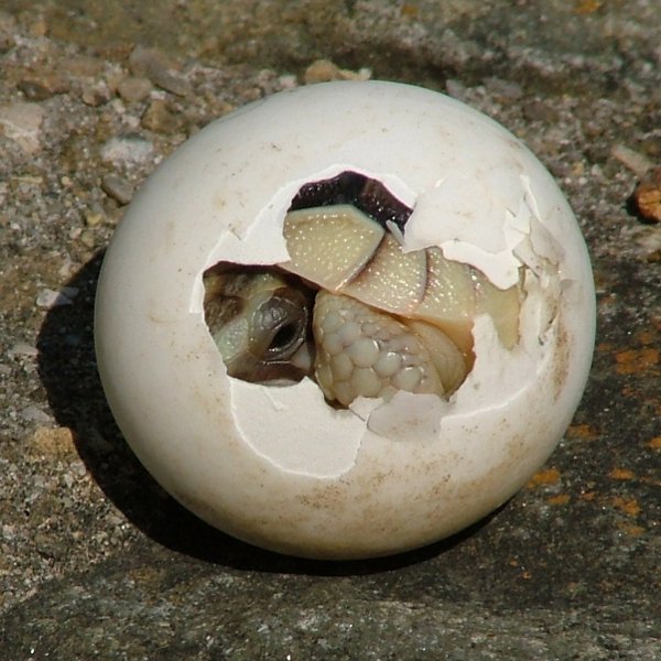
The turtle eggs have just arrived
The turtle eggs have just arrived!

My experiences in Japan absolutely fit into this series of successful events. In Guojun Sheng’s lab I spent three hard-working months under the supervison of Cantas Alev. During this time I tasted a little molecular biology in the fascinating environment of RIKEN CDB. I learned how to generate in situ probes and carry out whole mount in situs. I worked on No1 machines in the company of great scientists. Of course I traveled around the Kansai area and I managed to visit Okinawa too, but the main advantage of my stay – beside its scientific impact- is that I got to know how life goes on in Japan. At RIKEN CDB I experienced the atmosphere of high level science outside the institute I saw a peaceful and safe country with delicious food, inspiring history, beautiful geography and I was surrounded by kind and friendly people. I really enjoyed it and it’s easy to get used to it!

Unfortunately my internship finished just when I felt the people around me were pretty close to myself and work raised the possibilities of different projects. Nowadays I usually think about my friends who I met in Japan.

This year taught me to look for the chances and to be brave enough to use them, to realize the necessity of foreign experiences and how to fit to a changed environment.

I’m really thankful to Guojun Sheng and Cantas Alev. Their support brought me one step closer to molecular biology and extended my view on other fields of developmental biology. I truly believe that this three-month internship opened new doors which may result in a long-time collaboration between the two laboratories.

At last but not least I must say thank you to each and every member in the Laboratory for Early Embryogenesis (in alphabetical order): Cantas Alev, Manjula Brahmajosyula, MengChi Lin, Hiroki Nagai, Yukiko Nakaya, Fumie Nakazawa, Kanako Ota, Guojun Sheng, Erike Sukowati, Wei Weng, Yu Ping Wu. I’m also thankful to Ms. Naoko Yamaguchi who helped arrange the visit.

Finally I’d like to express my gratitude to all those at The Company of Biologists and Development who spent time on reading my application and supported my plan to come true!

Kobe - Harborland

Kobe – Harborland

Laboratory for Early Embryogenesis – RIKEN CDB – Kobe, Japan

Developmental Biology and Immunology Lab – Department of Human Morphology and Developmental Biology, Semmelweis University – Budapest, Hungary

Thumbs up (5 votes)
Loading...

Tags: , , , , , , ,
Categories: Funding, Lab Life, Research

In Development this week (Vol. 137, Issue 21)

Posted by , on 12 October 2010

Here are the research highlights from the current issue of Development:

Oct1: essential for trophoblast development

Most POU family transcription factors are temporally and spatially restricted during development and play pivotal roles in specific cell fate determination events. Oct1 (Pou2f1), however, is ubiquitously expressed in embryonic and adult mouse tissues; so, does Oct1 have a developmental role? On p. 3551, Fatima Cavaleri and colleagues report that Oct1 regulates trophoblast development during mouse embryogenesis. The researchers generate Oct1-null embryos and show that they fail to develop beyond the early primitive streak stage. Analysis of the mutant embryos reveals that they lack normal maternal-embryonic interfaces because of reduced extra-embryonic ectoderm (ExE) formation and the absence of the ectoplacental cone – two extra-embryonic tissues generated from the trophectoderm cells that overlie the inner cell mass (which forms the embryo). Other experiments indicate that Oct1 loss is incompatible with the derivation of trophoblast stem cells, which normally reside in the ExE. The researchers suggest, therefore, that Oct1 is primarily required for the maintenance and differentiation of the trophoblast stem cell compartment during early post-implantation development.

Schwann cells: more than just insulators

How neurons connect to their targets during embryogenesis has been intensively studied, but what maintains the position and connections of nerves during postembryonic growth? To investigate this, William Talbot and colleagues study the development of the posterior lateral line nerve (PLLn) in zebrafish embryos and larvae (see p. 3643). Using transmission electron microscopy, the researchers show that the PLLn – a peripheral nerve that innervates sensory organs in the epidermis – initially grows in the epidermis but that shortly after axon outgrowth, the epidermal basement membrane degrades and reforms on the nerve’s opposite side, thereby repositioning the nerve into the subepidermal space. Analysis of mutant and chimeric embryos shows that Schwann cells, which myelinate peripheral nervous system axons, are required for this process; without them, the PLLn becomes severely disorganised. Thus, by remodelling tissues in the vicinity of nerves, Schwann cells, which are traditionally regarded as static insulators, could play an important role in the proper organisation of nerves that innnervate other sensory organs during postembryonic growth.

WUSCHEL robustly plants stem cell homeostasis

Plant stem cell populations are maintained by the precise coordination of stem cell division and the rates of cell division and differentiation among stem cell progenitors. In the growing tips of higher plants (shoot apical meristems, SAMs), stem cell daughters produced by infrequent stem cell division in the central zone (CZ) are displaced towards the surrounding peripheral zone (PZ), where they divide faster and their progeny differentiate into leaves or flowers. Now, Venugopala Reddy and co-workers report that the homeodomain transcription factor WUSCHEL (WUS) mediates stem cell homeostasis in Arabidopsis (see p. 3581). The researchers use transient manipulation of WUS expression and live imaging to show that elevated WUS levels in the CZ induce CZ expansion and increase PZ cell division rates. Conversely, decreased WUS levels lead to a smaller CZ, reduced PZ cell division rates and increased responsiveness of PZ cells to the plant hormone auxin, which leads to enlarged organ primordia. Thus, by regulating stem cell numbers and growth and differentiation patterns, a single transcription factor robustly mediates plant stem cell homeostasis.

Haematopoietic cluster locations made transparent

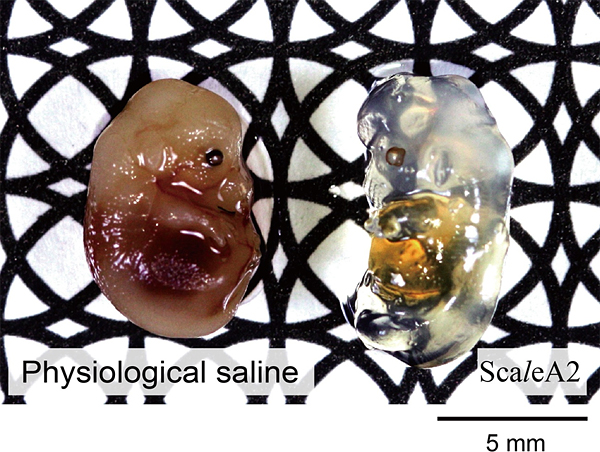
Haematopoietic clusters – cell aggregates that are associated with endothelium in the large blood vessels of midgestation vertebrate embryos – play a pivotal but poorly understood role in the formation of the adult blood system. To date, the opaqueness of whole embryos has prevented the systematic quantitation or mapping of all the haematopoietic clusters in mouse embryos but, on p. 3651, Tomomasa Yokomizo and Elaine Dzierzak remedy this situation. Using a technique to make whole mouse embryos transparent, combined with immunostaining and three-dimensional confocal microscopy, they show that the number of clusters peaks at embryonic day 10.5. Clusters are heterogeneous, they report, and localise to specific vascular subregions, such as the middle subregion of the aorta near to its junction with the vitelline artery. Finally, by combining flow cytometry and functional studies, the authors demonstrate that haematopoietic progenitor and stem cells are enriched within the cluster population. Together, these results provide novel insights into the spatial development of the adult blood system in mice.

Chordin downregulation waves on aortae fusion

During development, extensive remodelling of the embryonic vasculature, the first organ to develop, ensures that the embryo’s cells are constantly supplied with oxygen and nutrients. The first major vascular remodelling event in mammalian and avian embryos is fusion of the bilateral dorsal aortae at the midline to form the dorsal aorta. Takashi Mikawa and co-workers now show that a developmental switch in notochord activity signals this fusion in chick and quail embryos (see p. 3697). Prior to fusion, the researchers report, the notochord secretes positive and negative factors that together block the initiation of aortae fusion. Notably, whereas the expression of positive vascular regulators is maintained during fusion, the expression of negative regulators such as chordin, an antagonist of the positive regulator BMP, is downregulated along the anteroposterior axis. The discovery of this novel mechanism for modifying vascular pattern – modulation of vascular inhibitors without changes in positive vascular regulator levels – could aid the development of treatments for vascular injury.

Cut to fly airway remodelling

In insects that completely metamorphose, such as Drosophila, embryonically specified imaginal cells remain dormant until the larval stages when their coordinated proliferation and differentiation generates various adult organs. Now, on p. 3615, Chrysoula Pitsouli and Norbert Perrimon describe how embryonic cells – spiracular branch (SB) tracheoblasts – remodel the Drosophila abdominal airways during metamorphosis. The adult fly tracheal system consists of branched tracheal tubes (which transport air into the insect’s body) and spiracles (the external respiratory organs, which are surrounded by epidermal cells). The researchers show that embryonic SB tracheoblasts are multipotent cells that express the homeobox transcription factor Cut, which is necessary for their survival and normal development. SB tracheoblasts, they report, give rise to three distinct cell populations at the end of larval development, which generate the two components of the adult tracheal system and the surrounding epidermis. This dissection of the molecular events that underlie the formation of an adult tubular structure in Drosophila might shed light on mammalian tubular organ development, suggest the researchers.

Also…

In the first of our new Evolutionary crossroads in developmental biology series, Prigge and Bezanilla introduce Physcomitrella patens, a moss from this ancient, non-vascular plant lineage, studies of which are distinguishing ancestral developmental mechanisms from those that are novel innovations in flowering plants. See the Primer on page 3535.

This article is the first in a series of Primers on organisms and phyla that have been particularly informative for studying the evolution of developmental mechanisms and morphology. Other articles in this series will be published over the next 12 months.

Thumbs up (No Ratings Yet)
Loading...

Tags: , , , , , , , , , , ,
Categories: Research