The community site for and by
developmental and stem cell biologists

FASEB Excellence in Science Award for Gail Martin

Posted by , on 7 October 2010

The FASEB Excellence in Science Award is awarded annually to a woman whose research has made an exceptional contribution to the field of biological sciences. In 2011, this award will go to developmental biologist Gail Martin, of the University of California, San Francisco.

Martin’s current work focuses mainly on the role of FGF signalling in organ development, but in 1981 she was the first  to isolate embryonic stem cells from mouse blastocysts grown in vitro, and in fact coined the term “embryonic stem cell”.

The Society for Developmental Biology (SDB) has written a profile of Gail Martin that is currently in press at Developmental Biology. Martin was president of the SDB in 2006-2007, and in her interview she addresses her passion for mentoring students and postdocs, and her interest in sharing unpublished results. She will receive her award at the 2011 SDB meeting, to be held in Chicago in July.

Thumbs up (3 votes)
Loading...

Tags: , ,
Categories: News

Navigate the archive

Use our Advanced Search tool to search and filter posts by date, category, tags and authors.

American plant research gets a boost

Posted by , on 7 October 2010

In the US, basic plant research is relatively underfunded compared to other fields, with most of the available money going directly towards the development of practical agricultural applications.

Last year, the Howard Hughes Medical Institute (HHMI) organized a meeting called “Future Horizons in Plant Science”, where select scientists in the field concluded that there was a need for a funding boost in plant science. Now, the HHMI, together with the Gordon and Betty Moore Foundation (GBMF) is providing this boost in the form of $75 million for 15 researchers over the next five years.

This is a one-time competition to specifically target plant researchers at a time when they are not receiving as much funding as their colleagues working on animal organisms. To qualify, scientists need to currently be working as tenured or tenure-track researcher at a US institution. Applications for the new grants are due on November 9.

See HHMI press release for full details, including a link to the application website with even more information.

(Arabidopsis painting by Emmanuel Boutet through CC-BY-SA license on Wikimedia commons.)

Thumbs up (No Ratings Yet)
Loading...

Tags: , ,
Categories: Funding, News

Stunning cysts

Posted by , on 6 October 2010

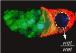
Hello to all of you Node readers!  My name is Erin Campbell and I’m the blogger behind HighMag Blog, a blog that features cell biology images a few times a week.  The great Eva Amsen contacted me about featuring some images on The Node, so I’m excited to be part of this growing community forum.  The first image I’m blogging about is from a paper in the October 1 issue of Development, and features the biologically complex and visually stunning Drosophila ovary.

Ovarian cyst development begins in the germarium, with a stem division that produces the cystoblast, which then divides 4 more times.  One cell in this 16-cell mass eventually becomes the oocyte, and the remaining cells serve as nurse cells to support the growth of the oocyte.  This 16-cell cyst then becomes a separate egg chamber after being surrounded by follicle cells and budding off of the germarium.  The hierarchy of signals and events that allow the differentiation of the cyst has been well studied, and a recent paper fills in the gaps in our understanding of this process.

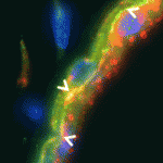
Tastan and colleagues report that the Drosophila homolog of the human ataxin 2-binding protein 1 (A2BP1) gene functions in the intermediate stages of cyst differentiation, bridging the expression of early and terminal differentiation markers.  Mutations in A2BP1 cause defects in cyst differentiation, as shown in the images above.  Germline cells are labeled in green using anti-VASA antibodies, membranes are labeled in red using an anti-1B1 antibody, and DNA is labeled in blue.  Compared with control ovaries, mutants exhibit a range of phenotypes:  A2BP1CC00511 cysts have extra nurse cells, A2BP1f02600 cysts exhibit a mildly tumorous phenotype, and A2BP1f01889 cysts have an extreme tumorous phenotype.

Reference: Ömür Y. Tastan, Jean Z. Maines, Yun Li, Dennis M. Mckearin and Michael Buszczak (2010). Drosophila Ataxin 2-binding protein 1 marks an intermediate step in the molecular differentiation of female germline cysts. Development 137, 3167-3176. Paper can be found here.

Thumbs up (10 votes)
Loading...

Tags: ,
Categories: Images, Research

Nobel Prize for Robert Edwards

Posted by , on 4 October 2010

Robert Edwards has just been announced as winner of the 2010 Nobel Prize in Physiology or Medicine, for his work on in vitro fertilization (IVF)

We speculated about the winners a few weeks ago, and he was not among anyone’s guesses, but this is a very exciting and timely choice. Just last Friday I mentioned that the Human Fertilisation and Embryology Authority will likely be cut. This is the body that regulates all IVF research in the UK, where Edwards work took place. Hopefully this award will bring the relevance of such regulatory bodies in the spotlight again.

Edwards worked together with gynecologist Patrick Steptoe, who died in 1988. Nobel Prizes are not awarded posthumously, which explains why he’s not included in the award.

The Nobel Prize website will be livestreaming all announcements of Nobel Prize winners this week. The Chemistry prize – which has predominantly been awarded to biology-related research the past decade – is announced on Wednesday.

Thumbs up (6 votes)
Loading...

Tags: ,
Categories: News

Who will regulate UK embryo research?

Posted by , on 1 October 2010

In an attempt to tighten the country’s budget, the UK government wants to cut a large number of arms-length non-governmental organisations. These “quangos” (quasi-autonomous non-governmental organizations) include regulatory bodies, advisory organs, and other committees.

Until last week, there were only vague speculations as to which funds would be cut, but the news has become more certain now that the BBC has received a leaked document specifically listing 177 non-governmental organizations that are to be cut. Among those is the Human Fertilisation and Embryology Authority (HFEA), which regulates legal and ethical aspects of anything involving IVF, but also sets standards and licensing requirements for any research done on human embryonic stem cells or human embryonic developmental studies in the UK.

Without the HFEA, who would oversee this area of research? This is a question the BBC asked HFEA chairwoman Lisa Jardine, who explained that the current functions of the HFEA would probably be split in three portions, with the research using human embryonic tissue ending up in a new regulatory body. Since that new body is not yet set up, she assumes that the HFEA will remain in charge of regulations until an appropriate alternative has been set up. (If you’re in the UK you can watch her response in a video, otherwise you can read a summary and partial transcript in this article on BioNews which gives a lot of background information).

On the HFEA website is a list of research projects that they have approved in the past. As you can see, it contains many fundamental developmental biology and stem cell research projects. Delays in approving such research in the UK would affect progress within the field, so hopefully Jardine’s prediction will hold true and cutting HFEA funding will not result in a gap in licensing.

The impending demise of the HFEA is not the only worry British stem cell and human embryo researchers have at the moment. The news comes at a time when UK-based researchers in all areas of science are threatened with cuts in research funding that would allow funding only for top and/or commercially viable projects. With scientists taking to the streets to protect funding for basic research, and with important regulatory bodies such as the HFEA under threat, interesting times are ahead for researchers in the UK.

Meanwhile, on the other side of the Atlantic, things could not be more different for hESC researchers: Not only is the US increasing basic science funding rather than reducing it, but it has just been announced that the federally imposed ban on stem cell research (previously on the Node) has been lifted, bringing US stem cell projects back on track. Quite the opposite story. Are any other countries currently awaiting regulations that affect stem cell or developmental biology research?

Thumbs up (3 votes)
Loading...

Tags: , , , , , , ,
Categories: Discussion, Funding, News

An interview with Irving Weissman at the 2010 ISSCR meeting

Posted by , on 30 September 2010

(This interview originally appeared in Development on September 28, 2010)

The International Society for Stem Cell Research (ISSCR) held their annual conference in San Francisco this June. At the time, the President of the society was Irving Weissman, who is currently on the board of directors of the ISSCR as past President. He is Professor of Pathology and Developmental Biology and also the Director of the Institute of Stem Cell Biology and Regenerative Medicine at Stanford University School of Medicine, where he works on the generation of myeloid and lymphoid lineages from haematopoietic stem cells. At the ISSCR meeting, we asked Professor Weissman about his role on the board of directors of the ISSCR, and also about the meeting and the field of stem cell research in general.

Thank you for taking the time for a quick interview. Are you enjoying the conference so far?

Of course, for those few moments I have without administrative duties.

What have been your highlights of the conference these first two days?

Well, there are different kinds of highlights. There is of course the highlight that just before the conference we launched our (the ISSCR’s) website on people in clinics who are practising for-profit, unproven therapies (www.closerlookatstemcells.org). This is our way of providing patients and their care-givers with the kind of advice they need in order to avoid being promised treatments that have neither the hope of treating or curing the disease nor of providing good medical practice.

How long have you been involved with the ISSCR; was it from the very start?

Yes, I was a member of the founding group, but I didn’t want the job of ISSCR President until it was more established.

How have you seen the ISSCR change over the past eight years?

I think we went from under a thousand people to nearly four thousand, both as members and as people who are participating in these meetings. We have also formed an alliance with the journal Cell Stem Cell.
(more…)

Thumbs up (No Ratings Yet)
Loading...

Tags: , ,
Categories: Interview

In Development this week (Vol 137, Issue 20)

Posted by , on 28 September 2010

A fateful look at early mouse lineage specification

The first cell lineages specified in the mouse embryo are the trophectoderm (TE), which generates the embryonic portion of the placenta, and the inner cell mass (ICM). The ICM subsequently forms the pluripotent epiblast (EPI, which produces the embryo) and the primitive endoderm (PrE, which generates other extraembryonic structures). Two papers shed new light on these crucially important early embryonic specification events.

On p. 3383, Janet Rossant and colleagues investigate the role that the intercellular adhesion molecule E-cadherin plays in the divergence of the TE and ICM. By embryonic day 3.5, the TE has formed a polarised epithelial layer that encloses the apolar ICM. The researchers show that the normal epithelial morphology of the TE is disrupted in mouse embryos lacking both maternal and zygotic E-cadherin function but that individual cells in the blastocyst still initiate TE- and ICM-like fates. Interestingly, most of the cells express the TE marker Cdx2, which suggests that organised epithelium formation is not necessary for TE-specific gene expression. Furthermore, individual cells in these embryos still generate an apical membrane domainthat correlates with Cdx2 expression. Thus, the epithelial integrity mediated by E-cadherin is not required for Cdx2 expression but is essential for setting normal TE/ICM ratios in mouse embryos.

On p. 3361, Anna-Katerina Hadjantonakis and colleagues identify a role for platelet-derived growth factor (PDGF) signalling in PrE expansion in mouse embryos. The PDGF receptor-α (PDGFRα) is an early marker of the PrE lineage and of extra-embryonic endoderm (XEN) cells, which can be isolated from mouse blastocysts as derivatives of the PrE lineage. By combining live imaging and lineage analysis, the researchers show that Pdgfra expression coincides with that of GATA6, the earliest expressed transcription factor in the PrE lineage. GATA6 expression, they report, is required for Pdgfra transcriptional activation, and PDGF signalling is essential for the establishment and proliferation of XEN cells in culture. Moreover, implantation-delayed Pdgfra-null mutant blastocysts contain reduced PrE cell numbers and, surprisingly, increased EPI cell numbers, indicating that reciprocal signalling between PrE and EPI tissues might regulate compartment size within peri-implantation mammalian embryos.

For more on early mouse lineage segregation, see also the review by Fredrik Lanner and Janet Rossant on p. 3351.

OMA-1/2: repressors of translation and transcription

Primordial germ cell specification requires global transcriptional repression. In C. elegans, the zygote (P0) undergoes four successive asymmetric divisions to generate the germline precursors P1, P2, P3 and finally P4, the germline founder. OMA-1 and OMA-2 (OMA1/2), cytoplasmic proteins degraded after the first mitotic cycle, repress global transcription in P0 and P1 by sequestering TAF-4, an RNA polymerase II pre-initiation complex component, while the maternal protein PIE-1 represses transcript elongation in P2-P4. Now, Rueyling Lin and colleagues report that OMA proteins repress transcription in P2-P4 indirectly by maintaining PIE-1 expression (see p. 3373). OMA-1/2, they show, repress zif-1 mRNA translation in oocytes; zif-1 encodes the substrate-binding subunit of the E3 ligase that marks PIE-1 for degradation. MBK-2, a kinase that is activated after fertilisation, controls OMA1/2 function, report the researchers. Thus, they suggest, MBK-2 phosphorylation of OMA1/2 acts as a key developmental switch in the oocyte-to-embryo transition by converting OMA proteins from specific translational repressors in oocytes to global transcriptional repressors in embryos.

Shh: new TWISTs to limb patterning

Sonic hedgehog (SHH) controls anterior-posterior (A-P) patterning in the mammalian limb. Its expression is normally restricted to the posterior limb bud but when expressed ectopically, it can change digit number and/or identity. Several transcriptional factors regulate Shh expression in the limb bud but how do they function together? On p. 3417, Xin Sun and co-workers report that interactions between two negative regulators of Shh expression (the ETS transcription factors ETV4/5 and the bHLH transcription factor TWIST1) and a positive regulator of Shh expression (the bHLH transcription factor HAND2) control A-P limb patterning in mice. By examining mutant limb buds, the researchers show that Twist1 is required to inhibit Shh expression in the anterior limb bud, and that it acts with the Etv genes to antagonise Hand2. Moreover, biochemical data indicate that the ETV proteins inhibit Shh expression by regulating the dimerisation of TWIST1/HAND2. Together, these findings highlight the importance of a precise balance between positive and negative regulators of Shh expression during limb A-P patterning.

Muscling in on motoneuron specification

Mammalian skeletal muscles contain several types of muscle fibres, each characterised by its contraction speed and molecular properties. Individual motor axons innervate a few dozen muscle fibres, usually all of the same type. How this striking ‘motor unit homogeneity’ is established is incompletely understood but, on p. 3489, Joshua Sanes and colleagues reveal that, in mice, signals from the muscle fibres influence the molecular properties of motoneurons that innervate them. The lack of markers for motoneuron types has impeded the study of motor unit homogeneity. Here, however, the researchers show that the motoneurons that innervate slow muscle fibres selectively express the synaptic vesicle protein SV2A and carry it to their nerve terminals. Notably, overexpression of the transcriptional co-regulator PGC1α in muscle fibres, which converts them to a slow phenotype, increases the number of SV2Apositive motoneurons. The researchers propose, therefore, that retrograde signals from muscles integrate with previously described anterograde influences of the nerve on the muscle fibre to match the properties of these synaptic partners to each other.

LIN-42-ing up development and stress

Environmental stresses, such as nutrient fluctuations, can affect developmental progression in animals. C. elegans larvae, for example, normally develop into adults through four larval stages under the control of heterochronic (developmental timing) genes such as lin-42, a homologue of the circadian rhythm gene period. But, when times are hard, C. elegans forms long-lived dauer larvae, an alternative third larval stage. Now, Ann Rougvie and co-workers report that lin-42 functions in dauer entry (see p. 3501). Loss of lin-42, they report, makes animals hypersensitive to dauer formation under stressful conditions, whereas misexpression of lin-42 in pre-dauer stages inhibits dauer formation. Other experiments suggest that LIN-42 acts in opposition to the ligand-free form of the nuclear receptor DAF-12, which integrates external cues and developmental decisions. Together, these results suggest that LIN-42 and DAF-12 are intimate partners in the decision to become a dauer larva and raise the possibility that Period-like proteins play a conserved role in coordinating intrinsic timing mechanisms with environmental conditions.

Thumbs up (No Ratings Yet)
Loading...

Tags: , , , , , , , , , , , , , , , , , ,
Categories: Research

Pleiades Promoter Project

Posted by , on 27 September 2010

ResearchBlogging.orgA recent paper in PNAS describes the development of MiniPromoters: human DNA promoters of less than 4 kb, designed to drive gene expression in specific areas of the brain. The initiative is called the Pleiades Promoter Project, and so far they have confirmed brain-region specific activity in knockin mice for 27 of their MiniPromoters. The final goal is to produce 160 fully characterized MiniPromoters to drive gene expression in defined brain regions.

The project is a spin-off of the Mouse Atlas Project (hence the name “Pleiades”: in Greek mythology, the Pleiades were the daughters of Atlas). To create the MiniPromoters, the researchers have computationally identified regulatory regions associated with brain-region or cell-type enriched gene expressions. Information about region-specific expression came from the Mouse Atlas Project as well as from the Allen Brain Atlas, which maps the human brain.


Several MiniPromoters driving LacZ in different areas of the mouse brain. (Part of Figure 5 from PNAS paper)

The MiniPromoters are designed to work in both mouse and human brains. While the Pleiades website emphasizes the ultimate goal of selective target delivery in gene therapy, the MiniPromoters can also be used to drive reporter expression in cell types of interest, for example to follow development of particular areas of the brain.
(more…)

Thumbs up (No Ratings Yet)
Loading...

Tags: , , ,
Categories: Research, Resources

Nobel Predictions

Posted by , on 23 September 2010

Thomson Reuters has predicted who they think will walk away with the Nobel Prizes in medicine, chemistry, physics, and economics. Their predictions for the prize for Physiology or Medicine include six names in four areas:
– Douglas Coleman and Jeffrey Friedman, for the discovery of leptin
– Ernest McCulloch and James Till, for the discovery of stem cells
– Ralph Steinman, for the discovery of dendritic cells
– Shinya Yamanaka, for the development of induced pluripotent stem cells

What do you think? Are any of these scientists going to be on the podium in Stockholm in a few months, or someone else?

I asked on Twitter, where Dean Griffiths guesses the prize will go to Shinya Yamanaka and John Gurdon, but is rooting for Till and McCulloch. Marios Stavridis thinks it’s too soon yet for Yamanaka, but that Till and McCulloch are long overdue a win.

Here’s my own guess: I think I agree with Marios that it’s too early for a win for Shinya Yamanaka. He’ll get it eventually, but probably not yet this year. And like Dean I’m also rooting for Till and McCulloch (I did my PhD in Toronto, so they’re “the home team” for me) but I also think Coleman and Friedman have a good chance. I watched the video for their Lasker Award (see previous post on the Node) and the whole story just screams “Nobel Prize” to me.

We’ll know soon enough, because the winners will be announced on October 4.

Thumbs up (2 votes)
Loading...

Categories: Discussion, News

Lasker Awards 2010

Posted by , on 21 September 2010

Today, the Lasker Foundation announced the winners of their 65th annual Lasker Awards, celebrating biomedical research. The winners all contributed to scientific research with strong implications for medical advancements:

Albert Lasker Basic Medical Research Award
Awarded to: Douglas Coleman & Jeffrey Friedman for the discovery of leptin, the “obesity gene”

Lasker-DeBakey Clinical Medical Research Award
Awarded to Napoleone Ferrara for the discovery of the role of VEGF in angiogenesis and the development of an anti-VEGF therapy for wet macular degeneration

Lasker-Koshland Special Achievement Award in Medical Science
Awarded to David Weatherall for lifetime research in the genetics of blood diseases, in particular for his work on thalassemia

The status of the awards becomes clear when you realize that in the past 65 years, as many as seventy-nine Lasker winners have also received a Nobel Prize. Winners of the Lasker receive $250,000 per award, and a statue, which you can see in the video below.

In Napoleone Ferrara’s award announcement video, he and others describe the role of VEGF in angiogenesis and wet macular degeneration:

Videos for the other winners are also available on the Lasker Foundation website.

Coleman, Friedman, Ferrara and Weatherall will receive their awards on Friday, October 1, in New York City.

And a little quiz: Aside from all winning a Lasker award this year, what connects these three research topics? (The simplest answer is only five letters long, but you can be more complicated. No prize, just for fun.)

Thumbs up (1 votes)
Loading...

Tags: , , , , , ,
Categories: News, Research, Video