Meeting Report: Young Embryologist Meeting, 2011
Posted by SophieP, on 23 May 2011
Sophie Pryor
Institute of Child Health
University College London


The third Young Embryologist Meeting, which brings together PhD students, Post-Doctoral and Young PI researchers, was held on Friday 6th May at King’s College, London. The meeting was initially established in 2008 by PhD students and Dr Yoshiyuki Yamamoto from the UCL Research Department of Cell and Developmental Biology (CDB), and is now organized and run by the Young Embryologist Network, a group of PhD students and Post-Docs from four London departments (UCL Dept. of CDB, Institute of Child Health, NIMR and Kings College London). The Young Embryologist Meeting is the main event organised by The Network, and aims to promote interaction and collaborations between early career researchers working on a variety of models and developmental systems. This year’s 130+ audience was comprised of researchers from 18 institutions (15 in the UK and 3 abroad).
The meeting opened with a keynote lecture by Prof. Sir John Gurdon, of the Gurdon Institute at the University of Cambridge, who is described on the Wellcome Trust’s website as ‘the man who made cloning possible, pioneering nuclear transfer and the ‘reprogramming’ of the fate of cells’. Prof. Gurdon spoke about the different methods of nuclear reprogramming, explaining the advantages of transplanting somatic nuclei into host oocytes (defined as those at the first meiotic prophase), rather than into eggs (those at second meiotic metaphase). He went on to discuss the mechanisms of nuclear reprogramming, namely: 1) the removal of differentiation markers, 2) chromatin de-condensation and protein:DNA exchange, 3) transcription activation and 4) the resistance of somatic cells to reprogramming as they become increasingly specialised. In conclusion, Prof. Gurdon described the emergence of a ‘time course’, saying that ‘the earlier you go back (towards pluripotency) the wider the reprogramming’.
The first session of the day, entitled ‘Organogenesis’, was made up of five talks on topics ranging from eye formation in the fruit fly to the genetics of mammalian cardiovascular development. The first speaker was Holger Apitz (NIMR), who presented his research into a novel mode of neurogenesis in the inner proliferation centre (IPC) of the Drosophila optic lobe. Holger discussed the identification of a new allele of Polycomblike (Pcl), which he has found to be involved in the specification and maintenance of outer (OPC) vs IPC neuroepithelial cell identity, regulation of EMT in subdomains of the IPC, and in preventing the premature differentiation of migrating progenitors into neuroblasts. Kenzo Ivanovitch (Dept. of CDB, UCL) showed fluorescent time lapse movies from his work on the division of the zebrafish eye field into two laterally located optic vesicles, and discussed the mechanism by which eye field cells acquire their polarity during this process. Lucy Freem (Institute of Child Health, UCL) spoke about the role of the Sox10 transcription factor in the formation of neural crest-derived intrinsic neurons during mammalian lung development, as well as describing the abnormal lung innervation in the Tbx1 mutant mouse. Divya Venkatesh (Institute of Genetic Medicine, Newcastle) discussed the development of the aortic arch arteries and her characterisation of the cardiovascular abnormalities in Pax9-null mice, which recapitulate human congenital heart defects. Closing the session was Andrew Economou (King’s College), who used mathematical simulations to describe his work on the role of Shh and Fgf signalling during formation of the regularly spaced ridges, or ‘rugae’ in the mammalian palate.
The second session consisted of four talks on different aspects of ‘Signalling and Epigenetics’. Ohad Shaham (Tel Aviv University, Israel) spoke about the function of Pax6 during eye development and his work to uncover novel roles of the transcription factor in cell cycle exit, lens epithelial cell survival and lens fibre cell differentiation in mouse embryos. He also discussed the results of his microarray analysis, which uncovered a high number of Notch pathway-related genes which are negatively regulated by Pax6. Characterisation of the Xenopus histone variant isoforms H2A.Z1 and H2A.Z2 was the subject of Jordan Price’s (University of Portsmouth) talk, where he presented the morphological and paralysis phenotypes produced by knockdown of each of these isoforms, respectively. Richard Wells (Sheffield University) discussed his research into the role of the JAK/STAT cytokine pathway in the Drosophila embryonic hindgut, which provides a valuable model to study the acquisition of left-right asymmetry during organogenesis. He showed that asymmetrical JAK/STAT signalling regulates gut curvature by controlling tissue stability via the localisation of Fascillin III. Last to speak was Paula Alexandre (King’s College), who reported on neuronal differentiation in the zebrafish brain and proposed a model, involving the dynamic remodelling of cellular protrusions, to explain neuronal spacing in the spinal cord.
The lunchtime poster session was followed by three talks on the subject of ‘Evo-Devo’. Adrien Demilly (Institut Jacques Monod, Paris) addressed the uncertainty surrounding the timing of CNS and neurogenesis evolution by describing the neurectoderm architecture in the annelid marine worm Platynereis dumerilii. In particular, he discussed the possible involvement of Wnt signalling in an organising centre at the ventral midline. The evolution of cerebellar development was discussed by Thomas Butts (King’s College), who described how species variation in cerebellar structure is largely due to variations in granule neuron progenitor (GNP) proliferation patterns. He presented a model whereby the successive evolution of GNP transit amplification explains the complexity of the amniote cerebellum. The final speaker, Dorit Hockman (Cambridge University), spoke about the different embryonic origins of oxygen-sensing cells in the carotid body and lung airway epithelia, before describing her work to identify the signalling pathways required for the specification of O2-sensing cell fate.
The day concluded with a lively careers question and answer session by Prof. Claudio Stern (Dept. of CDB, UCL), Prof. Jon Clarke (King’s college) and Dr Claudia Linker (King’s College). One audience member asked the three PIs what qualities they would look for when hiring a post-doc. Prof. Clarke said he tries to find those with a ‘genuine interest’ who have the ability to ‘present their research well, ask sensible questions and are capable of generating their own ideas’. He also spoke of his amazement at how many people come to visit his lab without doing any preparatory reading about the research that goes on there! Another question was: ‘How important is it to do a post-doc abroad?’ Prof. Stern stressed that the important thing is to find a good lab and spoke of the benefits of change and increasing the breadth of your research. Dr Linker described her own experience of working with different model systems, saying that ‘moving around is good for you and your cv’. The panel members were also asked what had been their hardest career moment, to which Prof. Clarke answered ‘knowing when to go for your first PI position’. Prof. Stern described the challenge of being successful at highly competitive interviews, and also voiced concerns about the effect that current government cuts to research funding would have on those looking for jobs in the future. He advised the audience that ‘if you’re convinced you want to do science, just go for it!’
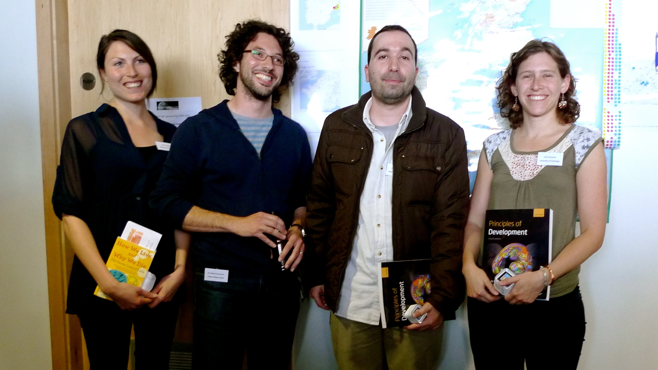
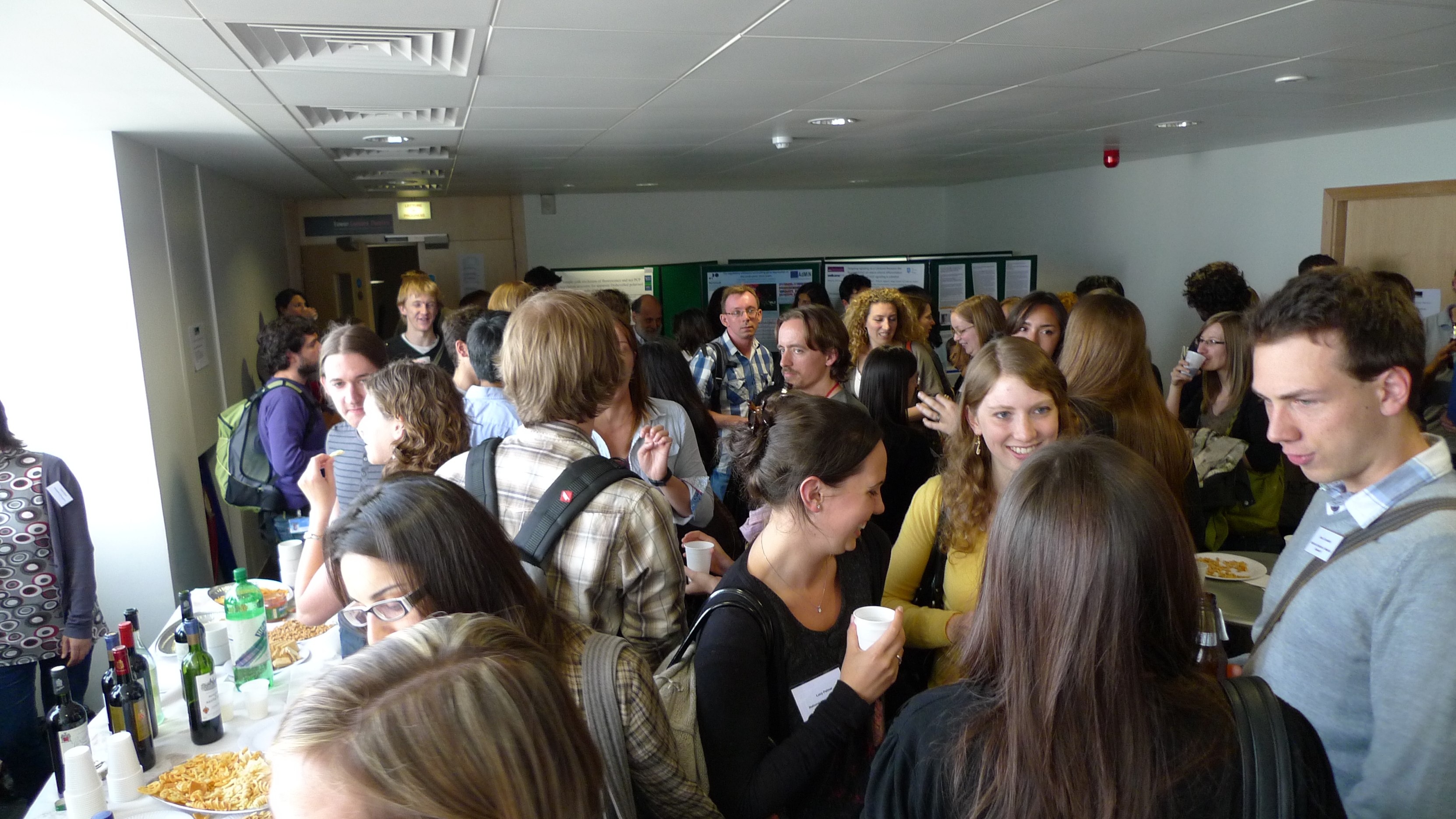
The day was a great success and allowed young embryologists to present their research in a relaxed environment. The lunchtime poster session and evening wine reception provided the opportunity for attendees to discuss other people’s work and exchange ideas about many aspects of developmental biology. Congratulations to Dorit Hockman (Cambridge University) and Andrew Economou (Kings College London), who were awarded first and second prize respectively for best talks of the day and to Ricardo Laranjeiro (University College London) and Emily Hoggar (University of Sheffield) who were awarded first and second poster prizes, respectively.
For more information about the Young Embryologist Network, please see the website (http://www.ucl.ac.uk/cdb/yen) or find us on Facebook.
 (7 votes)


 (No Ratings Yet)