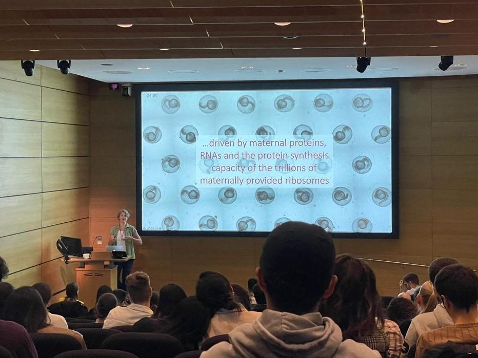
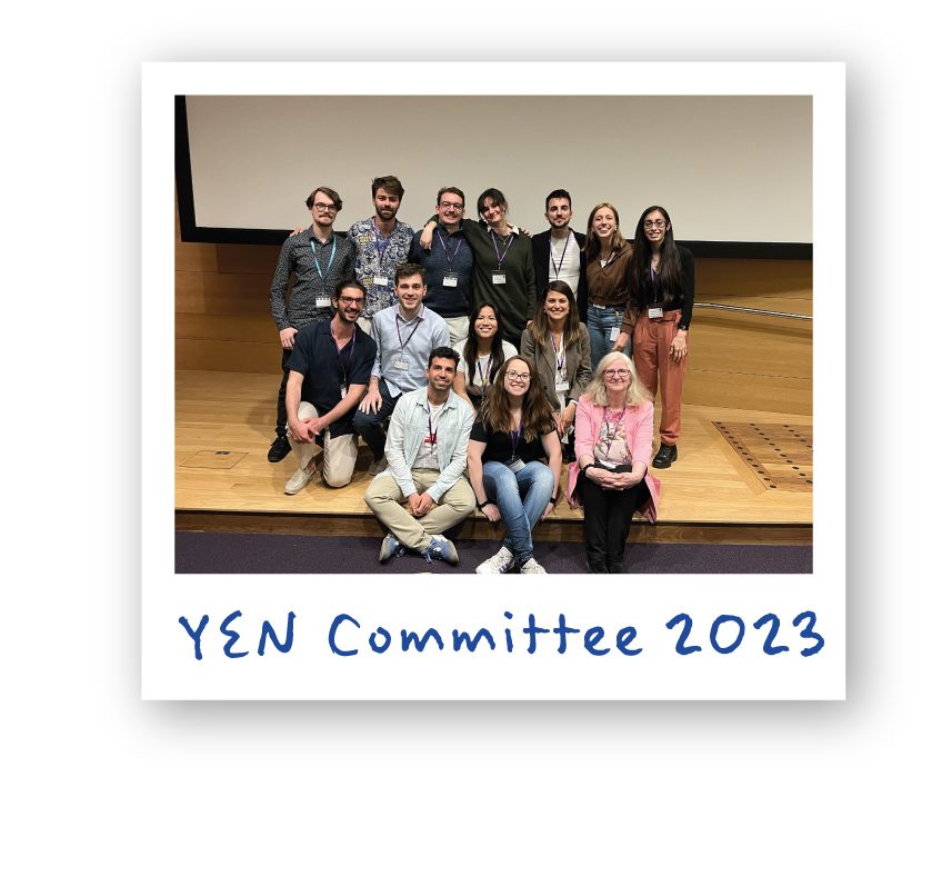
The Young Embryologist Network (YEN) Conference was held in May at the Francis Crick Institute. This year, apart from the scientific talks, the programme included a panel discussion on ‘Equality, diversity and inclusion in science’. We caught up with the two panelists, Alison Forbes (Head of EDI, the Francis Crick Institute), and Rafael Galupa (Scientist & social entrepreneur), to talk more in-depth about this important topic.
How did you find the YEN2023 meeting?
Alison: I loved it, great fun to speak to a large audience of early career scientists. I enjoy speaking to scientists as an ‘outsider’ (I studied humanities, I’m in HR, I am not a researcher). It means I can bring a fresh eye to science as an industry, and I think the audience enjoyed that.
Rafael: As a scientist, I really enjoyed myself and was very impressed and excited by all the science presented! This is a great meeting for me to attend regularly in the future and where to send my future lab members. As a scientist concerned with EDI issues, I thought it was great that the organisers included three different moments in the scientific programme to discuss such topics.
Could you tell us more about your background? What made you decide to be involved with EDI initiatives?
Alison: I grew up in Hackney, London and went to a north London grammar school, then a Russell Group university.
I am from a middle-class, academic family. But I always found snobbery, elitism and other ‘-isms’ irritating, which is why I am drawn to this work. I remember my grammar school teachers repeatedly telling us that we were ‘the best of the best’. That attitude never sat right with me.
I worked at UCL for 12 years in widening participation, working on social class and diversity in undergraduate admissions. I ran long term programmes for highly able teenagers from London state schools. They were mostly from low-income backgrounds, lots of refugee or immigrant families, usually parents had not been to university. These students were intelligent, sharp and witty, asked challenging questions, and were tough and resilient.
They were highly ambitious, as were their parents. I learned a lot from these young people, and got to see ‘my kids’ progress to UCL or other excellent universities. I also worked with UCL undergraduates, and saw how working class or minoritised students struggled with imposter syndrome. I remember young people telling me they had nowhere quiet to study, that they had had 4 teachers for Maths A level in one school year, or how their parents pressured them to apply for medicine and would not let them consider any other subjects.
It gave me a better understanding of how class, social mobility and privilege affect careers, and how oblivious we can be of own privilege (‘Well, I worked hard to get here!’).
I moved into EDI through a secondment at UCL. It is a fascinating and evolving field. There is much to be done, and you can have a real impact. I’ve been at the Crick since November 2021, and I love it.
Rafael: I grew up in the suburbs of Lisbon, surrounded by a big extended family. Because my dad went to university and became a power engineer, my household lived comfortably, but this wasn’t the case for the others in the family, or for the households of my school friends. I grew up side by side with these inequalities, and perhaps that’s why I’ve always felt a responsibility to address them.
I started volunteering when I was sixteen, in an orphanage – those kids had a completely different social structure around them (compared to mine), but not necessarily less supportive; this made me understand the importance of a support network, whichever nature it has, and how it is possible to build one if we don’t have the privilege to have one more “naturally”.
Later, in the University of Lisbon, while studying Molecular Biology and Genetics, I was part of a support group for students with disabilities. I had a weekly studying session with a younger undergraduate who was losing his vision. This was a major challenge for him, aggravated by how the classes, the studying materials, basically everything!, were very poorly accessible.
During my PhD, I came across Native Scientists, a nonprofit organisation that connects students with underserved children via interactive workshops, and to this day I have been involved in such activities. During my postdoc I participated in the project Letters to a Prescientist, which promotes letter exchanges between scientists worldwide and students at US-based schools where at least 60% of students qualify for “Free or Reduced Price Lunch”.
This experience inspired me and a friend to start a similar organisation, Cartas com Ciência, targeting students from low-income communities in Portuguese-speaking countries. Many studies show how students from low-income communities are less likely to pursue university studies and choose STEM careers, which promotes the perpetuation of the low-income cycle. The reasons for such stats are many, but include the lack of science role models around those students and often not knowing what a scientific career is. This is something that we as scientists can help eradicate! And there are many other levels at which we can act. So how not to get involved with initiatives that promote such fundamental values as equality, diversity and inclusion?
What do you think is the current biggest challenge in achieving an equal, diverse, and inclusive research culture?
Alison: There isn’t one single challenge, and there isn’t one single solution. It is complex.
Until we have urgency on these issues within government, and within science leadership, progress may well remain slow.
When something is urgent, we make it happen. For example, the sudden pivot by many employers to remote working during COVID. Home working was something disabled employees had asked for before COVID to help manage their conditions, but because allowing remote working was not the norm and was not urgent, it wasn’t seen as possible.
Many organisations are risk averse. They don’t want to be ‘the first’ to do something– they want to their peers to go first, so they can get assurance that an intervention works, and will not cause reputational damage. For example, after some Russell Group universities piloted contextual undergraduate admissions, it became common practise. Similar for interventions like reverse mentoring or various positive action schemes. So, the organisations that do ‘go first’ and take that risk should be credited.
Rafael: As Alison stated, there isn’t one biggest challenge. We all need to change our mindset, but especially people at the higher management of our research institutions. We need a more inclusive, healthier, people-centric work culture. I am sure that things will change – how fast, it remains to be seen. Planck’s principle basically says that change does not occur because individuals change their minds but rather because successive generations have different views (or in more crude terms, “one funeral at a time”). This will certainly be the case, but I also believe in the power of education, and so training in inclusive leadership, for instance, might accelerate things, as a good friend working in EDI, Roshni Mooneeram, taught me. We also need funding agencies and governments to take the lead and set the “tone” – change at those levels is the most likely to have a domino effect. Also, something that is at the root of many of the problems in research institutes (and in society in general) is the frenetic pace at which we live and work – complex problems, such as EDI issues but also most of the scientific questions we address, need us to pause more frequently, to think and reflect.
Why is it important to raise awareness on EDI in scientific meetings?
Alison: Scientists need to understand equity, diversity and inclusion for several reasons:
- Equity principles should inform research design. Scientists should be considering sex as a biological variable, the ethnic diversity of patients in clinical trials, and how to design equitable collaborations with scientist colleagues in lower income countries.
- Scientists should be recruited by an evidence-based, equitable, transparent process. When a PI recruits a post doc, do they use a process designed to minimise bias and to attract diverse candidates? For example, do they name blind applications, use standard interview questions that are scored, and do they have gender and if possible ethnicity mix on interview panels? There is a lot of evidence that diverse teams are more high performing, but often common recruitment practises in science penalise minoritized applicants.
- Most scientists agree public communication is important. But if scientists do not have a nuanced understanding of ‘how they come across’ when speaking to diverse audiences, it can harm public health messages. In the pandemic we saw high vaccine hesitancy in communities of colour in the UK and USA, and among some patient communities with chronic conditions. This was partly because of low trust due to prior experiences of racism, or histories of medical trauma. So, scientists have to understand those contexts when they speak to different audiences.
Rafael: Scientific meetings provide a great platform to discuss EDI because on the one hand, we can reach more people at once; on the other hand, everyone paused their everyday work and is in a new environment, which is more conducive for reflexion and thinking about issues that most of us do not normally think about. Moreover, such complex issues need collective thinking and collective actions, and scientific meetings definitely help with those.
What can early-career researchers do, both individually and as a community?
Alison: As an individual, start with self-education. Read some books and listen to podcasts; I’ve recommended some below.
Then interrogate your small daily actions and decisions. Do you ask questions about diversity in committee? For example, do you ask to see gender or ethnicity pay gap data, or recruitment data, or ask about what inclusion training your institute provides? Who do you defend, advocate for, or invite to join you at conferences? And who do you interrupt or ignore? Who do you seek to build relationships with?
As a wider community, you have power in numbers. Networks like the Node, the EDIS Group or grassroots organisations like Black in Cancer or BBSTEM have a big collective impact and should be nurtured.
Rafael: As Alison stated, join ongoing initiatives – they assume many different forms and flavours, so I’m sure that everyone can find a way to contribute that is aligned with their personality and availability. It’s also important to remember that we all have a certain degree of control over what happens around us (and to us as well) and certainly over how we do things. As we often say at Cartas com Ciência, EDI is about what we do but also how we do it and with whom we do it. Alison’s questions illustrate how we can incorporate such thinking in whichever actions or initiatives we think of doing, from setting a collective journal club, to organising a meeting or a public engagement activity.
Where can people find out more on this topic?
Alison: Nature ran a special issue about racism in science in October 2022. I also recommend their Nature careers: Working scientist podcast series. They often have episodes about inclusion, for example their mini-series Science diversified.
The EDIS Group (equality diversity and inclusion in science) have excellent resources and toolkits.
The book Invisible Women: exposing data bias in a world designed for men by journalist and campaigner Caroline Criado-Perez has some excellent chapters about research design. Female cells, animals, and patients are often under-studied in discovery research and clinical trials. This leads to outcomes like women being much more likely to be misdiagnosed for heart attacks, because they present with ‘atypical’ symptoms. Her book emphasises the importance of studying sex as a biological variable.
Another book I recommend often is The Class Ceiling: why it pays to be privileged by LSE academics Sam Friedman and Daniel Lauriston. They studied labour market data and found clear evidence of a pay gap between middle- and working-class employees in similar roles. They conducted anonymous interviews about experiences of class at work, with staff in a large TV company, an architecture firm and a financial firm. They don’t look specifically at science as an industry, but these themes are common across all sectors. A really insightful and entertaining read.
Rafael: The major scientific journals in life sciences have been regularly publishing special issues on EDI topics, for instance, Cell Press has collections on “Building inclusivity in science”, “Black in science”, “Women in science”, “LGBTQ+ in science” (here). Or the “Diversity, equity and inclusion in science” collection from Nature Human Behaviour (here). Hopefully all those articles are open access! Science published a short and comprehensive article on “How to begin building a culture of diversity, equity, and inclusion in your research group” (open access).
Two amazing books on how science has actually contributed to sexism and racism, I recommend Angela Saini’s books Inferior and Superior (respectively). They present and digest lots of interesting facts and studies, and Angela does a great job in always trying to compare and contrast different perspectives. Plus, it’s fun to read. On improving scientific culture in general, I recommend Uri Alon’s initiatives and available materials (here).
Want to find out what else happened at the YEN Conference? Read the detailed meeting report written by Ioakeim (Makis) Ampartzidis, Courtney Lancaster, Danielle Liptrot, and Rosie Marshall.
To learn more about YEN, follow them on twitter @YEN_community.
 (4 votes)
 Loading...
 (No Ratings Yet)