Behind the Paper: the discovery of a novel cell type involved in fly touch sensing
Posted by Federica Mangione, on 23 May 2023
A recent paper in Nature Cell Biology entitled ‘Co-option of epidermal cells enables touch sensing’ reports a new type of specialised epidermal cells involved in touch sensing in Drosophila. We caught up with Dr Federica Mangione, the first author of this paper, to find out more about the story behind the paper.
How did you get started on this project?
I joined the Tapon lab for my postdoc, aiming to study how cell differentiation impacts the structure and function of the somatosensory system, using my beloved Drosophila as an in vivo model. I initially wanted to understand how the tactile bristles, hair follicle-like structures decorating the adult epidermis, develop to allow the adult fruit fly to sense tactile stimuli. Given that the terminal differentiation of these touch-sensitive organs was largely unexplored, what I did first was to visualize this developmental stage, combining bristle-specific genetic labelling with live imaging. While analysing the cellular dynamics underlying the differentiation of the four lineage-related cell types that make up each bristle within the epidermis, I identified the epidermal F-Cell as a novel cell type in the assembly of the mature tactile organ. The temporal and spatial precision underlying the acquisition of the F-Cell fate led us to pursue a detailed study on the structure and function of the tactile bristle and its association with the F-Cell.
What was known about the role of epidermal cells in the function of touch-sensitive organs before your work?
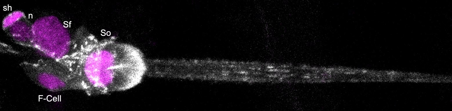
The epidermis is the outermost layer of animals’ body, and it integrates various specialized cells and cellular structures that associate with sensory neurons to shape the sense of touch. In mammals, specialized cells of epidermal origin include the Merkel cells and a subset of the cells making up the hair follicles. While both Merkel cells and hair follicles associate with specific subsets of sensory neurons for touch sensing, the specific role of the epidermal cells of the hair follicles in touch sensation is not well understood. Our work shows that the tactile bristles, hair follicle-like structure in the Drosophila epidermis, associate with specialized epidermal cells, the F-Cell, to sense touch and reveal that the insect epidermis also contains specialized epidermal cells involved in sensory detection.
Can you summarise your key findings?
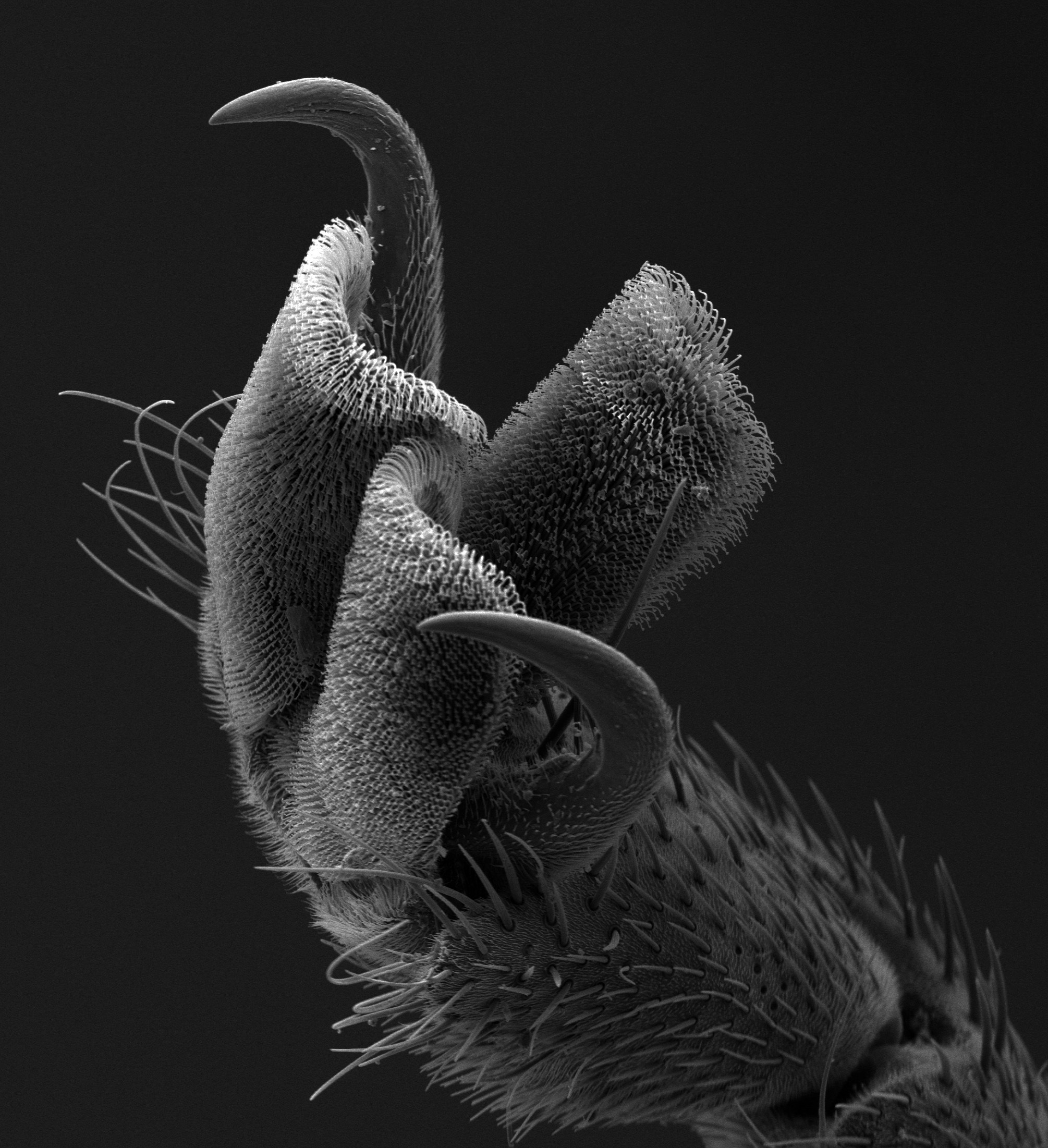
One key finding is that F-Cell fate specification occurs post-mitotically: selective elimination of the F-Cell through laser microsurgery induced de novo specification of the F-Cell fate in the remaining epidermal cell adjoining each tactile bristle. The precise dynamic of this event led us to perform a series of genetic and optical experiments which, together, indicated that the shaft cell of the bristle is orchestrating a short-range signalling to select F-Cell fate within the epidermis. Another key finding is that this short-range signalling is dependent on the conserved epidermal growth factor receptor (EGFR) signalling. While corroborating these findings, we asked what happens after F-Cell fate acquisition. Through temporal volume electron microscopy (vEM) and light microscopy during the terminal differentiation of the epidermis, we established that the F-Cell is the only epidermal cell that changes shape to wrap around the tactile bristle. The close association between the F-Cell and the tactile bristle suggested a functional requirement for this cell in touch sensing. Through in vivo electrophysiological recordings and behavioural assays, we found that the F-Cell is indeed essential for touch sensing. Altogether, our work established that the F-Cell is a specialized epidermal cell which shapes the functional assembly of the tactile bristles.
This interdisciplinary work involved diverse expertise, can you talk a bit about your experience collaborating with people in other fields?
Collaborating with people with different known-how and points of view has been incredibly beneficial, both professionally and personally. By working closely with Catherine Maclachlan in the team of Lucy Collinson (The Francis Crick Institute, London, UK), I have learned the many steps behind the beauty of an EM image and the power of vEM in gaining information on cellular morphologies in 3D. I am looking forward to discovering more about the cells of the tactile bristles through EM as I carry on working together with this great team of experts! Working with Joshua Titlow in the team of Ilan Davis (University of Oxford, UK) has been a fantastic experience too! He guided me through the complex process of generating a successful recording of neuronal activity and I am so grateful for his dedication and patience during my many visits to Oxford. This collaboration definitely stoked my passion for neuroscience, which will stay with me for the rest of my career. I feel fortunate to have met Michel Gho (Sorbonne University, Paris, FR) while I was characterizing the genetic basis of F-Cell fate specification. Collaborating with a leading expert in bristle development and genetics such as him has been an absolute honour and of great help to focus on EGFR as a key signal for F-Cell fate specification. All the support from our collaborators have been crucial to shape this paper the way it is!
Did you have any eureka moment that has stuck with you?
One in particular, indeed! While performing live imaging, I observed that, at some point during their differentiation, the cells expressing a bristle-specific cell marker changed from four to five. I could not detect any cell division within the bristle lineage or outside in the epidermis at that time, so how did this switch from four to five occurs? And why? This observation had me puzzled for a while! Three decades of studies established that each bristle is composed of four cell types, all derived from a single precursor cell, so why was I counting 5 instead? How is it that 2+2=5? The eureka moment arrived within the first 2 years of studies, when I found that specific manipulations of bristle cells were affecting the appearance of the F(ifth)-Cell. This told me that the F-Cell is co-opted by the tactile bristle form the neighbouring epidermis, and this makes the impossible possible: 2+2 really equals 5! The 2 pairs of sister cells in the bristle progenitor lineage require “a fifth element” for the assembly of a functional tactile organ. Eureka!
And the flipside: were there any moments of frustration or despair?
Finding a new cell type is very exciting! A flipside, however, is that gaining insights into what makes an uncharacterized cell type unique is quite challenging experimentally. For example, after first observing the F-Cell, I didn’t have any genetic tools to manipulate gene expression in a restricted manner for quite a long time. Also, many of the genetic manipulations I was initially testing affected the fate of bristle cells too, preventing me from cleanly disentangling bristle and F-Cell specification. These obstacles were successfully overcome by combining laser microsurgery with live imaging to target individual cells and in a temporally controlled manner (PMID: 36685184). Performing light and electron microscopy imaging was also technically challenging, especially at later stages of development, when the epidermis become stiffer to form the adult exoskeleton of the fruit fly. We dedicated a lot of effort in optimizing sample preparations and imaging set ups, which definitely worthwhile as we were able to gain access to these developmental stages too! The limited/intermittent access to the lab during the pandemic was also very frustrating of course. Support from the lab helped me in keeping a positive attitude and I saved ‘positive energy stores’ for optimizing experimental designs and close gaps in the project once I was back in the lab.
Why do you think the role of the F-Cell was not characterized before?
I have been asked this question many times, and yet I am not sure that there is a ‘right’ answer. The beauty of Drosophila bristles has attracted scientistic for three decades, as they are a powerful cellular context to address fundamental questions about cell fate determination and asymmetric cell divisions. Surprisingly, however, their differentiation dynamics (how the mature tactile organ is built from its constituent cells) was understudied. The reason behind this lack of knowledge remained mysterious to me until I started performing live imaging during bristle differentiation, which revealed various technical challenges. Thus, I would say that the F-Cell remained uncharacterized as accessing to late developmental stages is not straightforward. My love for microscopy and support from the lab and collaborators helped me in overcoming some of these limitations and gaining accessibility to these developmental stages.
What is next for you after this paper?
I discovered the F-Cell shortly after starting my postdoc in the Tapon lab and collecting the data for this paper has been a great journey. This journey is still going on! There are many aspects of the development of this cell and its association with the tactile bristle that I wish to explore further. I am now completing my postdoctoral training and I am very excited about the prospect of leading a research team in the future and gain more insight into the cell biology of touch!
If you are interested in learning more about the volume electron microscopy (vEM) technique used in this paper, check out the FocalPlane post ‘Inputs and Outputs of vEM in a Sensory System‘.
 (2 votes)
 (No Ratings Yet)