Synthetic embryos have dominated the discussion on #devbio Twitter over the past few weeks, following ground-breaking publications from the Hanna and Zernicka-Goetz labs. The labs have produced synthetic mouse embryos with astonishing similarity to the ‘real thing’, which has prompted discussions on where this technology can go next, the distinction between embryo-like models and real embryos, ethical implications and responsible reporting to the general public. With fantastic timing, next Monday (12 September) at 16.00 BST, we will be livestreaming our panel discussion on technical, ethical and legal challenges of studying early human development from our Journal meeting on human development, where we expect to cover some of the topics discussed on Twitter in more depth. The stream of our panel discussion, as well as talks from Sarah Teichmann and Sergiu Pasca, will be hosted on the Node. For now, we have picked out a few of our favourite Tweets and replies for you to read (to check out the discussion, simple click on the tweets below).
– they do not have organismal potential – there's lots of work to do in the mouse system to boost efficiency (now 0.1 – 2%), which should imo be prioritized before going human
We are of course excited about our embryo-like model but wish to stress that it is important not to think of our embryo-model as being a real embryo – even if it is getting close to the real thing
Half-baked idea alert: if people are gonna take in vitro wombs to their logical conclusion, they should start with marsupials! Less important imprint regulation, and later implantation. And you only need to get this far. MAKE A KOALA DAMMIT pic.twitter.com/AJrg6uXck7
If you’ve seen something that we should include in our ‘Developing news’ report or you would like to help us put the blog together, please get in touch at thenode@biologists.com
Metabolic enhancement of mammalian developmental pausing Vera A. van der Weijden, Maximilian Stoetzel, Beatrix Fauler, Dhanur P. Iyer, Mohammed Shahraz, David Meierhofer, Steffen Rulands, Theodore Alexandrov, Thorsten Mielke, Aydan Bulut-Karslioglu
wnt16 regulates spine and muscle morphogenesis through parallel signals from notochord and dermomyotome Claire J. Watson, W. Joyce Tang, Maria F. Rojas, Imke A.K. Fiedler, Ernesto Morfin Montes de Oca, Andrea R. Cronrath, Lulu K. Callies, Avery Angell Swearer, Ali R. Ahmed, Visali Sethuraman, Sumaya Addish, Gist H. Farr III, Arianna E. Gomez, Jyoti Rai, Adrian T. Monstad-Rios, Edith M. Gardiner, David Karasik, Lisa Maves, Bjorn Busse, Yi-Hsiang Hsu, Ronald Young Kwon
β1 integrin regulates alveolar epithelial cell differentiation following injury Jennifer M.S. Sucre, Fabian Bock, Nicholas M. Negretti, John T. Benjamin, Peter M. Gulleman, Xinyu Dong, Kimberly T. Ferguson, Christopher S. Jetter, Wei Han, Yang Liu, Seunghyi Kook, Jason Gokey, Susan H. Guttentag, Jonathan A. Kropski, Timothy S. Blackwell, Roy Zent, Erin J. Plosa
Single cell, whole embryo phenotyping of pleiotropic disorders of mammalian development Xingfan Huang, Jana Henck, Chengxiang Qiu, Varun K. A. Sreenivasan, Saranya Balachandran, Rose Behncke, Wing-Lee Chan, Alexandra Despang, Diane E. Dickel, Natja Haag, Rene Hägerling, Nils Hansmeier, Friederike Hennig, Cooper Marshall, Sudha Rajderkar, Alessa Ringel, Michael Robson, Lauren Saunders, Sanjay R. Srivatsan, Sascha Ulferts, Lars Wittler, Yiwen Zhu, Vera M. Kalscheuer, Daniel Ibrahim, Ingo Kurth, Uwe Kornak, David R. Beier, Axel Visel, Len A. Pennacchio, Cole Trapnell, Junyue Cao, Jay Shendure, Malte Spielmann
Multi-organ functions of yolk sac during human early development Rachel A Botting, Issac Goh, Antony Rose, Simone Webb, Justin Engelbert, Yorick Gitton, Emily Stephenson, Mariana Quiroga Londoño, Michael Mather, Nicole Mende, Ivan Imaz-Rosshandler, Dave Horsfall, Daniela Basurto-Lozada, Nana-Jane Chipampe, Victoria Rook, Pavel Mazin, MS Vijayabaskar, Rebecca Hannah, Laure Gambardella, Kile Green, Stephane Ballereau, Megumi Inoue, Liz Tuck, Valentina Lorenzi, Kwasi Kwakwa, Clara Alsinet, Bayanne Olabi, Mohi Miah, Chloe Admane, Dorin-Mirel Popescu, Meghan Acres, David Dixon, Rowen Coulthard, Steven Lisgo, Deborah J Henderson, Emma Dann, Chenqu Suo, Sarah J Kinston, Jong-eun Park, Krzysztof Polanski, Stijn Van Dongen, Kerstin B Meyer, Marella de Bruijn, James Palis, Sam Behjati, Elisa Laurenti, Nicola K Wilson, Roser Vento-Tormo, Alain Chédotal, Omer Bayraktar, Irene Roberts, Laura Jardine, Berthold Göttgens, Sarah A Teichmann, Muzlifah Haniffa
Tissue-Like 3D Standard and Protocols for Microscope Quality Management Benjamin Abrams, Thomas Pengo, Rebecca C. Deagle, Nelly Vuillemin, Tse-Luen Wee, Linda M. Callahan, Megan A. Smith, Kristopher E. Kubow, Anne-Marie Girard, Joshua Z. Rappoport, Carol J. Bayles, Lisa A. Cameron, Richard Cole, Claire M. Brown
Junior scientists spotlight social bonds in seminars for diversity, equity, and inclusion in STEM Evan A. Boyle, Gabriela Goldberg, Jonathan C. Schmok, Jillybeth Burgado, Fabiana Izidro Layng, Hannah A. Grunwald, Kylie M. Balotin, Michael S. Cuoco, Keng-Chi Chang, Gertrude Ecklu-Mensah, Aleena K. S. Arakaki, Noorsher Ahmed, Ximena Garcia Arceo, Pratibha Jagannatha, Jonathan Pekar, Mallika Iyer, DASL Alliance, Gene W. Yeo
What an exciting time to be studying embryonic development! Emerging experimental systems, methods and analyses allow addressing a whole new set of fascinating questions, as well as revisit older ones. In particular, the field of patterning and morphogenesis, which investigates how embryos establish their body plan with the correct cell types and shape, currently experiences rapid technological and conceptual transformations. For example, microfluidics and microfabrication now permit manipulating the culture conditions of developing embryos with extended flexibility; organoid systems allow the reconstitution of tissues and structures entirely from stem cells; microscopy methods such as light sheet microscopy, live sensor of signalling pathways, optogenetic tools allow dissecting developmental mechanisms over unprecedented temporal and spatial scales. Together, these new possibilities will be key to structure the field of patterning and morphogenesis over the next decades. This research will be carried out, in part, by a new generation of emerging leaders. From July 25th to 29th of 2022, scientists who recently established their labs in Europe gathered in Les Treilles to discuss the emerging quantitative approaches to study embryonic development. From nematodes, flies, sea urchin, frogs, zebrafish, cow and mouse to human, a broad variety of developmental processes were covered from fertilization to organogenesis.
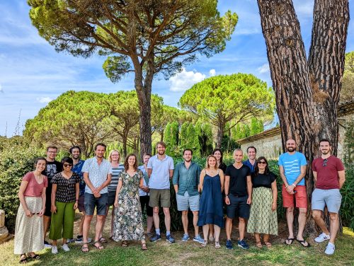
From left to right: Diana, Kuba, Rashmi, Tommaso, Romain, Ina, Rita, Kate, Tim, Elias, Anne-Cécile, Verena, Patrick, Nicolas, Mariaceleste, Jean-Léon and Renaud.
Observing embryonic development is key to understand it.
Technological developments in light sheet microscopy have allowed imaging of large-scale morphogenetic events while maintaining cellular resolution. Kate McDole, who locked herself in a room during the pandemic to build a new light sheet microscope, presented never-seen-before cell movements during mouse gastrulation, shedding light on the origin of cells constituting the different germ layers. Rita Mateus also used light sheet microscopy to image the patterns involved in the formation of the zebrafish pectoral fins and better characterise how those patterns scale during growth and regeneration. As the fins emerge, other organs, like the heart, also form complex structures such as the muscular trabecular ridges that enable proper heart contraction. Using quantitative live imaging and clonal analysis, Rashmi Priya explained how complex tissue architecture is built during trabecular morphogenesis. Zebrafish was also at the heart of Diana Pinheiro’s work, as she reported on a mechanism explaining the coordination of mesoderm progenitors migration and specification by Nodal signalling during gastrulation. When missing, Nodal signaling causes characteristic phenotypes that expert zebrafish embryologists can easily recognize. Patrick Müller leveraged deep learning analyses to develop algorithms that outperform embryologists at identifying the phenotypes of zebrafish gastrulation mutants.
Together, these studies show how imaging large-scale shape changes and quantitatively tracking them can help understand the global patterning and morphogenesis of embryos.
Down to the sub-cellular scale, high resolution microscopy reveals the mechanisms employed by cells to shape embryos. In particular, Anne-Cécile Reymann looked into the influence and distribution of maternally deposited regulators of the actin cytoskeleton during the early cleavages of c elegans embryos. In contrast, Tommaso Cavazza reported on how microtubules help coordinate chromosome segregation in cow zygotes before the nuclear envelope of the parental pronuclei breaks down. Later during mammalian development, Jean-Léon Maître described how protrusions help cells control the formation and positioning of a fluid-filled lumen that helps setting the first axis of symmetry of the embryo. A different set of protrusions was introduced by Jakub Sedzinski to explain how cells can insert themselves within epithelia and increase the complexity of tissues. Finally, peering into fly embryos, Timothy Saunders quantified the scaling of Bcd gradients by combining fluorescence correlation spectroscopy and mutants affecting egg size.
Altogether, bridging the sub-cellular scale to embryonic changes is key to understand the molecular and physical regulation of embryonic development.
Probing and challenging embryos in novel ways
Exploring how cells change their chemical composition and physical properties during development will help understand their behaviour. Mariaceleste Aragona quantitatively analysed the effect of tissue stretching in vivo using clonal analysis and single cell sequencing to understand how cells change composition and fate after a long-term physical perturbation. To understand how cells sense mechanical perturbations,Verena Ruprecht investigated the short-term response of cells to compression by combining high resolution microscopy, microfabrication and optical tweezers. Tweezers were also on the menu for Nicolas Mincwho developed magnetic tweezers strong and precise enough to move the entire mitotic spindle within cells to induce division asymmetries and explore the mechanics of the cytoplasm.
Understanding how embryos develop requires the need to finely perturb this process in controlled ways. Romain Levayer leveraged the powerful genetics of Drosophila and optogenetics to induce the death of a precise number of cells and determine the range of robustness of epithelia to cell delamination. Another way to control in space and time the molecular regulation of embryonic development is microfluidics: using precise oscillations of chemical compound, Ina Sonnen could explore the coupling of biological clocks during somitogenesis. Finally, Elias Barriga measured endogenous electric fields and applied electric fields of similar magnitude to steer the migration of neural crest cells in Xenopus embryos.
Together, discussing the current research and future projects of young European scientists revealed the promises and challenges of applying quantitative methods to developmental biology. On the one hand, quantitative methods allow dissecting developmental mechanisms with unprecedented precision. On the other hand, the integration of multiple spatial and temporal scales, and different source of biological information (such as chemical, mechanical and electrical signals) remain extremely challenging. Moreover, tackling these problems when most biologists lack the appropriate training in mathematics and coding can be challenging. A consensus appeared regarding the need to include more biology-oriented maths in university programmes, as this will be key to anchor biology to the realm of modern science. Importantly, Renaud Pourpre, who carried out several initiatives to bring microscopy data to the public, reminded us how microscopy images constitute a straightforward avenue to communicate around science to a lay audience (see CellWorlds documentary). By the same token, there is no doubt that the beauty of developing embryos will continue motivating biologists and other scientists to expand their toolbox to decipher the mechanisms of development.
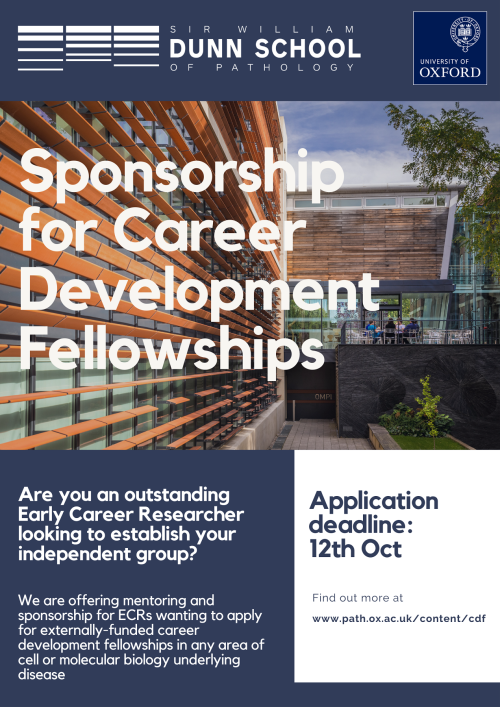
Interested in the cell or molecular mechanisms underlying disease? Do you have an outstanding record and an innovative research plan?
The Dunn School of Pathology at the University of Oxford is looking for outstanding early career researchers seeking a stimulating and supportive environment in which to establish their research group as externally-funded fellows. We are specifically looking for researchers seeking mentoring and sponsorship to apply for career development fellowships (e.g. Wellcome Trust Career Development Award, MRC Career Development Award, CRUK Career Development Fellowship, UKRI Future Leaders, etc). Researchers who succeed in securing a fellowship will then be invited to establish their independent group in the department, benefiting from a generous support package, comprehensive mentorship, career development training and opportunities to recruit Oxford undergraduate and postgraduate students.
Successful candidates will have an outstanding track record in any area of biomedical research, with a particular focus on the fundamental cell and molecular biology underlying disease. The Department celebrates diversity and we welcome applicants from diverse backgrounds that are currently underrepresented at the University of Oxford.
More information and contact for informal queries can be found on our website: https://www.path.ox.ac.uk/content/cdf The deadline for applications is the 12th of October 2022.
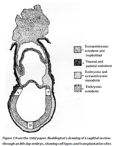
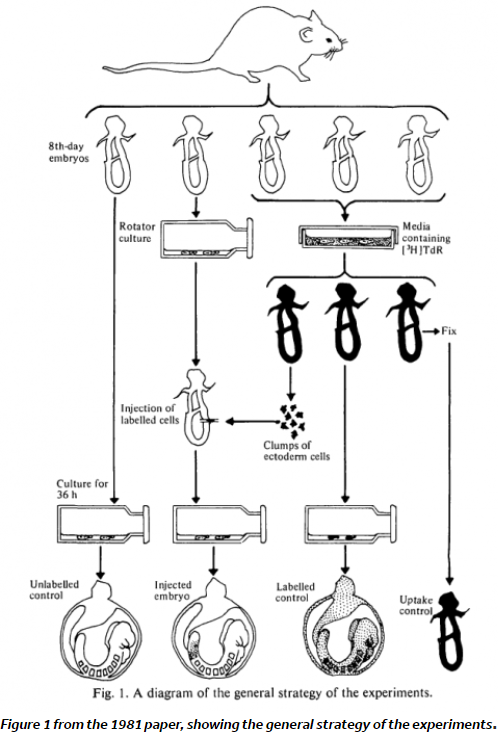
Are you just starting out in your career as a developmental biologist (or feeling nostalgic for those times)? The vast amount of literature can feel a bit daunting when you are tackling a new topic. To help you navigate the field, and to focus on some of the lesser known classics, we asked prominent researchers to recommend their hidden gems from history. Their selection includes Rosa Beddington’s chimeras, Morgan’s planarian experiments and principles of morphogenesis from Gustafson and Wolpert. Let us know if you come across any forgotten classics that we should include in our series.
“Imagine a society where female reproductive rights are a matter of state concern. Where working class females are actively suppressed from having their own children, and only the nobility are allowed to reproduce. I am, of course, talking about honey bees.”
Dr Sally Le Page
In the latest episode of the Genetics Unzipped podcast, Dr Sally Le Page looks at the genetics of societies, exploring how genes underpin the rigid social structures and roles in bees, and how they can rise up the ranks to become queen bee.
If you enjoy the show, please do rate and review on Apple podcasts and help to spread the word on social media. And you can always send feedback and suggestions for future episodes and guests to podcast@geneticsunzipped.com Follow us on Twitter – @geneticsunzip
Read on for our news roundup of the past few weeks, with an emphasis on what has caught our eyes on twitter. This post is focussed on career advice, conferences and related conversations.
— Dr Lisa Nivison-Smith (@LNivisonSmith) June 29, 2022
Can posting a preprint help advance your career in science, especially if you are a student? Learn more in the video below, where Sónia Gomes Pereira discusses her experience with posting a #preprint. https://t.co/9uBhnwWVzA
Numerous faculty job ads are starting to appear, application materials are being prepared – wanted to share advice for applicants on past & future research statements based on my experience as a faculty search chair last year. First, a few thoughts…🧵
— American Society for Cell Biology (@ASCBiology) August 11, 2022
Earlier this year, we heard from Dhruv Raina about his move to industry following his PhD in Dr Christian Schröter’s lab at the Max Planck Institute of Molecular Physiology (MPI).
For the latest job listings at all levels, including an opportunity for a new Features and Reviews Editor at Journal of Cell Science, check out our activejobs board.
Conferences
The return to in-person conferences has been wonderful, but what have we learnt due to the restrictions imposed during lockdowns, and how can we improve future conferencing?
I love going to conferences but i must find a way to bring my family with me otherwise no matter how fun or important it is the guilt is killing me. Perhaps half of a conference day should be free time? (to allow you to be with family)
Today I spoke up at a #FENS2022 discussion event about pandemic effects on mentoring. I spoke about my frustration regarding transmission mitigation at the meeting. I sorta highjacked the discussion and I'm sorry about that, but I just couldn't not say anything and 1/n
— Dr. Maria Veldhuizen 😷💉 (@margaveldhuizen) July 12, 2022
We are delighted to be live–streaming our session on recent recent progress and ethical challenges in studying early human development from our upcoming meeting, From Stem cells to Human Development on Monday 12 September 16:00 BST. More details will follow on the Node shortly.
In vitro models of development have been a talking point on Twitter in the last few weeks. The thread from Dr Martinez Arias sums up some of the key points.
The recent reports on the generation of embryo-like structures from mouse pluripotent stem cells by the @jacob_hanna and @MZG_Lab groups are an important landmark in developmental and reproductive biology. Thread
— Alfonso Martinez Arias (@AMartinezArias) August 7, 2022
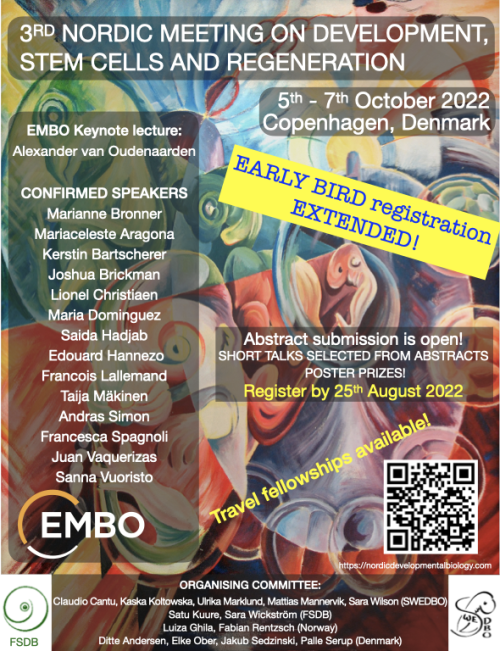
We are arranging a 2-day conference together with the Swedish Society for Developmental Biology (SWEDBO), Finnish Society for Developmental Biologists, and Danish & Norwegian Developmental Biologists the 3rd Nordic Meeting on Development, Stem Cells and Regeneration in Copenhagen in October 5-7th, 2022
The line up of invited speakers is outstanding and brings together experts in developmental and stem cell biology and regeneration! Attending the meeting is a great opportunity to meet with developmental biologists, from the Nordic countries and beyond. All speakers will be there ‘in person’ allowing for lots of networking.
There are many opportunities for short talks, poster prizes, and student and postdoc activities are planned in addition. The registration is now open and it includes membership in one of the Nordic Developmental Biology associations! Follow the link to register: https://nordicdevelopmentalbiology.com
Dr. Denise Allen and Dr. Tomasz Nowakowski at the University of California, San Francisco recently published an article in Science where they revealed a dual origin for astrocytes in the human cortex. Using a combination of fate mapping and single cell analysis, they revealed that the two stem cell niches in the developing cortex give rise to spatially, morphologically, and molecularly distinct populations of astrocytes. The Node asked them to give us a behind the scenes look at how the story came together:
How did you get started on this project?
TJN: For a very long time, we have been interested in the question of why the brains of primates and humans are so much larger and complex than the brains of mice, which we study frequently in the laboratory. Differences in brain size can be found very early on during development, and therefore it was plausible to hypothesize that differences in the way radial glia, which act as neural stem cells, develop could contribute to these differences. In our prior work, we found that animals with large brains, such as primates or humans, contain a greater diversity of radial glia subtypes compared to mice. In particular, we found that based on gene expression profiles, radial glia could be divided into truncated radial glia and outer radial glia, which are located in two anatomically distinct niches of the developing cortex.
DA: During my undergraduate neuroscience classes I was always struck by the deep knowledge we have about the development of neurons in the cerebral cortex, but astrocytes and other glia seemed to be so often overlooked. During my rotation in the Nowakowski lab, I became fascinated with Tom’s preliminary data that suggested distinct subtypes of radial glia could give rise to distinct astrocyte populations. I was really excited by the fact that large brain mammals, including primates and humans, seem to have a different repertoire of radial glia compared to rodents, as well as much more complex astrocytes. So the possibility to study the unique features of human development with a focus on astrocytes was a dream come true.
What was already known about the developmental trajectories of radial glia in the developing brain prior to your work?
A lot of work has been done probing the differentiation of outer radial glia (also known as basal radial glia). Numerous papers have shown that they give rise to neurons, oligodendrocytes and supposedly the majority of astrocytes, but the role of truncated radial glia has not been studied in great detail. Previous studies have suggested that because few mitotic cells can be found in the ventricular zone stem cell niche during midgestation in primates and humans, that the truncated radial glia that reside in this zone are unlikely to serve as a major source of new cells. We decided to challenge this assumption by labeling progenitors in the ventricular zone and determining the fates of the resulting cells. To our surprise, we found that neurons, oligodendrocytes, and astrocytes continue to be produced by ventricular zone progenitors.
Can you summarize your findings? What was the key experiment?
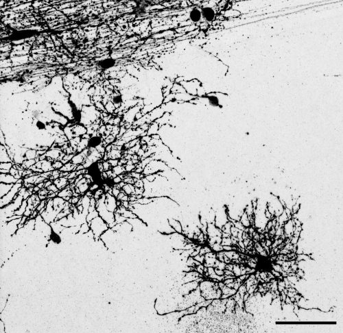
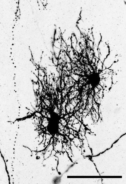
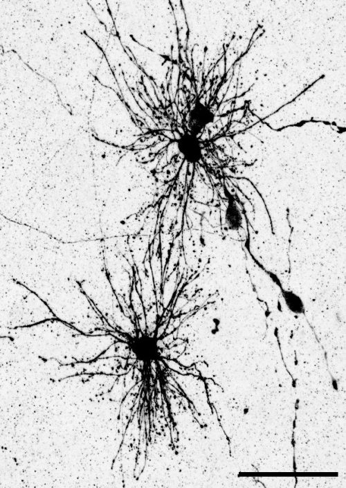
The key experiment involved labeling progenitor cells that occupy anatomically distinct niches that truncated and outer radial glia cells, and tracing the fates of cells that they produce. We found that while both populations broadly produce similar cell types (neurons, astrocytes and oligodendrocytes), they produce very distinct subtypes of astrocytes. In a series of very striking results, we found that truncated radial glia give rise to astrocytes that migrate to the cortical plate, while outer radial glia give rise to astrocytes that do not migrate, and instead differentiate locally in the outer subventricular zone.
We often speak about the diversity of neurons, but classical studies have also shown that astrocytes can be remarkably diverse, even in early development. I wanted to further explore the diversity of the astrocyte subtypes we identified, but it is challenging to connect these classical descriptions of astrocyte subtypes to modern-day descriptions such as those derived from single cell sequencing. To solve this problem, I took advantage of a method called Patch-seq which gave us the ability to aspirate the contents of a cell that was previously defined based on its morphology and position, and then performing sequencing of that cellular contents to determine a molecular identity. This analysis was key for bridging our cellular definitions based on morphology and developmental cell lineage, and linking them to molecular markers. This allowed me to bring the story in full circle.
When doing the research, did you have any particular result or eureka moment that has stuck with you?
The very first experiment I performed that involved labeling these two different stem cell niches resulted in a distribution of cell types that could not have been more different. Many comparisons in developmental biology rest upon small differences between conditions. To see such a stark difference in the distribution of cells–especially of glia–was an exciting moment that defined the course of the project very early on in my PhD.
Another surprising finding was when we started closely comparing classical drawings by Ramon y Cajal and Retzius and to images of our astrocyte subtypes. Remarkably, we found that our “dense bulbous” astrocytes were clearly depicted in those early records, but these cells have rarely been mentioned in modern literature. This realization gave us a lot of confidence that the cells we were observing were a real phenomenon and not an experimental artifact. These cells had just been lying in wait, waiting for someone to put the spotlight on them.
And what about the flipside? Any frustrations or despair?
The Universe really conspired against us when we were trying to finish experiments for the revision of the paper. I set up the last three revision experiments in late December, when we suddenly found out that it was time to move our lab to a new building at UCSF. We came up with an elaborate system to keep the cultures going while we moved and they seemed to have survived, until someone suddenly noticed that the incubator had failed and the alarm hadn’t gone off. What followed was two months of issue after issue trying to repeat these last two experiments, but we finally got there in the end!
Where will this story take the lab?
This work has inspired several new projects in the lab. We are excited to examine if similar findings can be replicated in other models of brain development such as cerebral organoids, what these unique subpopulations look like in the adult brain, and what role they might play in disease states. I’m also hoping this work will also attract more trainees interested in glial development to the lab!
What is next for you/the lab after this paper? Let us know if you are continuing this research, or starting/looking for a new position.
Denise has graduated and is currently interviewing for computational biologist roles in biotech. She is looking forward to delving into the “big data” side of biology, and working towards making a significant impact on patients’ lives.
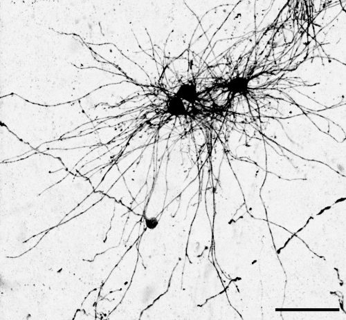
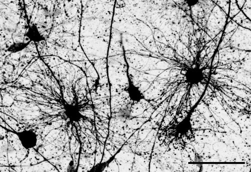
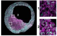
“Dense bulbous” astrocytes derived from the VZ“Dense bulbous” astrocytes derived from the VZ“Dense bulbous” astrocytes derived from the VZ “Dense smooth” astrocytes derived from the OSVZ“Dense smooth” astrocytes derived from the OSVZ (1 votes) Loading...
Development’s biennial meeting, From Stem Cells to Human Development, will be taking place in mid-September (11-14) at beautiful Wotton House in Surrey, UK. After a very successful virtual meeting in 2020, we’re excited to be meeting in person again, but we wanted to explore ways of making part of the meeting accessible to the broader community. We’re therefore delighted to announce that we’ll be livestreaming one session of this meeting, and the recording is available below.
Session details (all times GMT+1): 16:00 Sarah Teichmann (Wellcome Sanger Institute, UK): Human development: one cell at a time 16:30 Sergiu Pasca (Stanford University, USA): From stem cells to assembloids: constructing and deconstructing human nervous system development and disease 17:00 Panel discussion: Technical, ethical and legal challenges of studying early human development Chair: Patrick Tam (University of Sydney, Australia) Panellists: Amander Clark (University of California Los Angeles), Robin Lovell-Badge (The Francis Crick Institute, UK), Sergiu Pasca, Sarah Teichmann, Magdalena Zernicka-Goetz (University of Cambridge, UK and CalTech, USA) 18:00 Close
 (No Ratings Yet)
 (6 votes)