May in preprints
Posted by the Node, on 15 June 2022
Welcome to our monthly trawl for developmental biology (and related) preprints.
The preprints this month are hosted on bioRxiv, arXiv and preprints.org – use these links to get to the section you want.
- Patterning & signalling
- Morphogenesis & mechanics
- Genes & genomes
- Stem cells, regeneration & disease modelling
- Plant development
- Evo-devo
Developmental biology
| Patterning & signalling
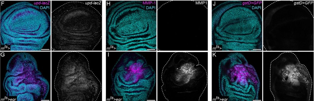
Mutual repression between JNK/AP-1 and JAK/STAT stratifies cell behaviors during tissue regeneration
Janhvi Jaiswal, Raphael Engesser, Andrea Armengol Peyroton, Vanessa Weichselberger, Carlo Crucianelli, Isabelle Grass, Jens Timmer, Anne-Kathrin Classen
Variable Shh and Fgf8 positioning in regenerating axolotl limb guarantees consistent limb morphogenesis in different limb sizes
Saya Furukawa, Sakiya Yamamoto, Rena Kashimoto, Yoshihiro Morishita, Akira Satoh
Par3/Bazooka promotes Notch pathway target gene activation
Jun Wu, Neeta Bala Tannan, Linh T. Vuong, Yildiz Koca, Giovanna M. Collu, Marek Mlodzik
Drosophila embryos spatially sort their nutrient stores to facilitate their utilization
Marcus D. Kilwein, Matthew R. Johnson, Jonathon M. Thomalla, Anthony Mahowald, Michael A. Welte
Timed Notch Inhibition drives Photoreceptor fate specification in Human Retinal Organoids
Shereen H. Chew, Cassandra Martinez, Kathleen R. Chirco, Sangeetha Kandoi, Deepak A. Lamba
Wnt signalling, cell fate determination and anteroposterior polarity of the skate gill arch skeleton
Jenaid M. Rees, Victoria A. Sleight, Stephen J. Clark, Tetsuya Nakamura, J. Andrew Gillis
The Wnt/TCF7L1 transcriptional repressor axis drives primitive endoderm formation by antagonizing naive and formative pluripotency
Paraskevi Athanasouli, Martina Balli, Anchel De Jaime-Soguero, Annekatrien Boel, Sofia Papanikolaou, Bernard K. van der Veer, Adrian Janiszewski, Annick Francis, Youssef El Laithy, Antonio Lo Nigro, Francesco Aulicino, Kian Peng Koh, Vincent Pasque, Maria Pia Cosma, Catherine Verfaille, An Zwijsen, Bjorn Heindryckx, Christoforos Nikolaou, Frederic Lluis
Cachd1 is a novel Frizzled- and LRP6-interacting protein required for neurons to acquire left-right asymmetric character
Gareth T. Powell, Yuguang Zhao, Ana Faro, Heather Stickney, Laura Novellasdemunt, Pedro Henriques, Gaia Gestri, Esther Redhouse White, Jingshan Ren, Weixian Lu, Rodrigo M. Young, Thomas A. Hawkins, Florencia Cavodeassi, Quenten Schwarz, Elena Dreosti, David W. Raible, Vivian S. W. Li, Gavin J. Wright, E. Yvonne Jones, Stephen W. Wilson
Morphogen gradient orchestrates pattern-preserving tissue morphogenesis via motility-driven (un)jamming
Diana Pinheiro, Roland Kardos, Édouard Hannezo, Carl-Philipp Heisenberg
Multifunctional role of GPCR signaling in epithelial tube formation
Vishakha Vishwakarma, Thao Phuong Le, SeYeon Chung
Dandy Walker-like malformations in mutant mice demonstrate a role for PDGF-C/PDGFRα signalling in cerebellar development
Sara Gillnäs, Radiosa Gallini, Liqun He, Christer Betsholtz, Johanna Andrae
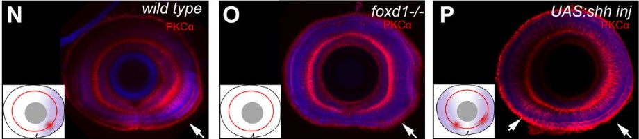
Foxd1 dependent induction of temporal retinal character is required for visual function
María Hernández-Bejarano, Gaia Gestri, Clinton Monfries, Lisa Tucker, Elena I. Dragomir, Isaac H. Bianco, Paola Bovolenta, Stephen W. Wilson, Florencia Cavodeassi
Stromal Hippo-YAP signaling in stem cell niche controls intestinal homeostasis
Kyvan Dang, Alka Singh, Jennifer L. Cotton, Zhipeng Tao, Haibo Liu, Lihua J. Zhu, Xu Wu, Junhao Mao
Suppressor of Fused regulation of Hedgehog Signaling is Required for Proper Astrocyte Differentiation
Danielle M. Spice, Joshua Dierolf, Gregory M. Kelly
Retinoic acid signaling in mouse retina endothelial cells is required for early angiogenic growth
Christina N. Como, Cesar Cervantes, Brad Pawlikowski, Julie Siegenthaler
Functional substitutions of amino acids that differ between GDF11 and GDF8 impact skeletal development and skeletal muscle
John Lian, Ryan G. Walker, Andrea D’Amico, Ana Vujic, Melanie J. Mills, Kathleen A. Messemer, Kourtney R. Mendello, Jill M. Goldstein, Krystynne A. Leacock, Soraya Epp, Emma V. Stimpfl, Thomas B. Thompson, Amy J. Wagers, Richard T. Lee
Fin ray branching is defined by TRAP+ osteolytic tubules
João Cardeira-da-Silva, Anabela Bensimon-Brito, Marco Tarasco, Ana S. Brandão, Joana Rosa, Paulo J. Almeida, António Jacinto, M. Leonor Cancela, Paulo J. Gavaia, Didier Y. R. Stainier, Vincent Laizé
Opportunistic binding of EcR to open chromatin drives tissue-specific developmental responses
Christopher M. Uyehara, Mary Leatham-Jensen, Daniel J. McKay
RanBP1 plays an essential role in directed migration of neural crest cells during development
Elias H Barriga, Delan N Alasaadi, Chiara Mencarelli, Roberto Mayor, Franck Pichaud
Feedback between a retinoid-related nuclear receptor and the let-7 microRNAs controls the pace and number of molting cycles in C. elegans
Ruhi Patel, Himani Galagali, John K. Kim, Alison R. Frand
Maintenance of neurotransmitter identity by Hox proteins through a homeostatic mechanism
Weidong Feng, Honorine Destain, Jayson J Smith, Paschalis Kratsios
Early patterning followed by tissue growth establishes proximo-distal identity in Drosophila Malpighian tubules
Robin Beaven, Barry Denholm
Development of dim-light vision in the nocturnal coral reef fish family, Holocentridae
Lily G. Fogg, Fabio Cortesi, David Lecchini, Camille Gache, N. Justin Marshall, Fanny de Busserolles
Gradients in cellular metabolism regulate development of tonotopy along the cochlea
Thomas S Blacker, James DB O’Sullivan, Val Yianni, Mohi Ahmed, Zoe F Mann
Patterning of the female reproductive tract along antero-posterior and dorso-ventral axes is dependent on Amhr2+ mesenchyme in mice
Shuai Jia, Jillian Wilbourne, McKenna J Crossen, Fei Zhao
Repulsive Sema3E-Plexin-D1 signaling coordinates both axonal extension and steering via activating an autoregulatory factor, Mtss1
Namsuk Kim, Yan Li, Ri Yu, Anji Song, Hyo-Shin Kwon, Mi-Hee Jun, Jin-Young Jeong, Mi-Jin Kim, Jung-Woong Kim, Won-Jong Oh
Inter-organ Wingless/Ror/Akt signaling regulates nutrient-dependent hyperarborization of somatosensory neurons
Yasutetsu Kanaoka, Koun Onodera, Kaori Watanabe, Tadao Usui, Tadashi Uemura, Yukako Hattori
A CXCL12 morphogen gradient uncovers lung endothelial heterogeneity and promotes distal vascular growth
Prashant Chandrasekaran, Nicholas M. Negretti, Aravind Sivakumar, Maureen Peers de Nieuburgh, Joanna Wang, Nigel S. Michki, Fatima N. Chaudhry, Hongbo Wen, Sukhmani Kaur, MinQi Lu, Jarod A. Zepp, Lisa R. Young, Jennifer M.S. Sucre, David B. Frank
A new model of Notch signaling: Control of Notch receptor cis-inhibition via Notch ligand dimers
Daipeng Chen, Zary Forghany, Xinxin Liu, Haijiang Wang, Roeland M.H. Merks, David A. Baker
Morphogen Directed Coordination of GPCR Activity Promotes Primary Cilium Function for Downstream Signaling
Shariq S. Ansari, Miriam E. Dillard, Yan Zhang, Mary Ashley Austria, Naoko Boatwright, Elaine L. Shelton, Amanda Johnson, Brandon M. Young, Zoran Rankovic, Camenzind G. Robinson, John D. Schuetz, Stacey K. Ogden
Notch signaling functions in non-canonical juxtacrine manner in platelets to amplify thrombogenicity
Susheel N. Chaurasia, Mohammad Ekhlak, Geeta Kushwaha, Vipin Singh, Ram L. Mallick, Debabrata Dash
Sequential functional differentiation of luminal epithelial cells regulated by maternal and embryonic signaling in the mouse endometrium
Hai-Quan Wang, Dong Li, Jingyu Liu, Yue Jiang, Ji-Dong Zhou, Zhi-Long Wang, Xin-Yi Tang, Yang Zhang, Xin Zhen, Zhi-Wen Cao, Xiao-Qiang Sheng, Chao-Fan Yang, Qiu-ling Yue, Li-jun Ding, Ya-li Hu, Zhi-Bin Hu, Chao-Jun Li, Gui-Jun Yan, Hai-Xiang Sun
pop-1/TCF, ref-2/ZIC and T-box factors regulate the development of anterior cells in the C. elegans embryo
Jonathan D. Rumley, Elicia A. Preston, Dylan Cook, Felicia L. Peng, Amanda L. Zacharias, Lucy Wu, Ilona Jileaeva, John Isaac Murray
| Morphogenesis & mechanics
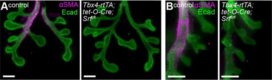
Plasticity in airway smooth muscle differentiation during mouse lung development
Katharine Goodwin, Bezia Lemma, Adam Boukind, Celeste M. Nelson
TGF-β signaling and Creb5 cooperatively regulate Fgf18 to control pharyngeal muscle development
Jifan Feng, Xia Han, Yuan Yuan, Courtney Kyeong Cho, Eva Janečková, Tingwei Guo, Siddhika Pareek, Jing Bi, Junjun Jing, Mingyi Zhang, Thach-Vu Ho, Yang Chai
Vertebrate extracellular matrix protein hemicentin-1 interacts physically and genetically with basement membrane protein nidogen-2
Jin-Li Zhang, Stefania Richetti, Thomas Ramezani, Daniela Welcker, Steffen Lütke, Hans-Martin Pogoda, Julia Hatzold, Frank Zaucke, Douglas R. Keene, Wilhelm Bloch, Gerhard Sengle, Matthias Hammerschmidt
Quantitative analysis of three-dimensional cell organisation and concentration profiles within curved epithelial tissues
Chaitra Prabhakara, Krishnan Swaminathan Iyer, Madan Rao, Timothy E Saunders, Satyajit Mayor
Primary cilia regulate Meibomian glands development and dimensions without impairing lipid composition of the meibum
Céline Portal, Yvonne Lin, Varuni Rastogi, Cornelia Peterson, James Foster, Amber Wilkerson, Igor Butovich, Carlo Iomini
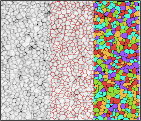
A quantitative biophysical principle to explain the 3D cellular connectivity in curved epithelia
Pedro Gómez-Gálvez, Pablo Vicente-Munuera, Samira Anbari, Antonio Tagua, Carmen Gordillo-Vázquez, Jesús A. Andrés-San Román, Daniel Franco-Barranco, Ana M. Palacios, Antonio Velasco, Carlos Capitán-Agudo, Clara Grima, Valentina Annese, Ignacio Arganda-Carreras, Rafael Robles, Alberto Márquez, Javier Buceta, Luis M. Escudero
Coupled organoids reveal that signaling gradients drive traveling segmentation clock waves during human axial morphogenesis
Yusuf Ilker Yaman, Roya Huang, Sharad Ramanathan
Machine learning directed organoid morphogenesis uncovers an excitable system driving human axial elongation
Giridhar M. Anand, Heitor C. Megale, Sean H. Murphy, Theresa Weis, Zuwan Lin, Yichun He, Xiao Wang, Jia Liu, Sharad Ramanathan
Testicular macrophages are recruited during a narrow time window by fetal Sertoli cells to promote organ-specific developmental functions
Xiaowei Gu, Anna Heinrich, Tony DeFalco
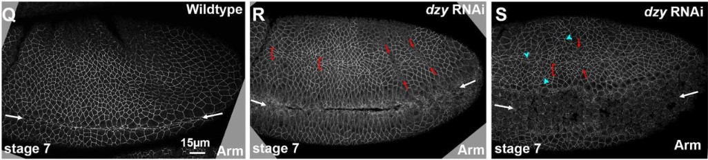
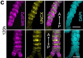
A ratchet-like apical constriction drives cell ingression during the mouse gastrulation EMT
Alexandre Francou, Kathryn V. Anderson, Anna-Katerina Hadjantonakis
Rap1 uses Canoe-dependent and Canoe-independent mechanisms to regulate apical contractility and allow embryonic morphogenesis without tissue disruption
Kia Z. Perez-Vale, Kristi D. Yow, Melissa Greene, Noah J. Gurley, Mark Peifer
Morphogenetic forces planar polarize LGN/Pins in the embryonic head during Drosophila gastrulation
Jaclyn Camuglia, Soline Chanet, Adam C Martin
| Genes & genomes
Transcriptome profiling of histone acetylation and methylation writers and erasers families across male germ cell development and pre-cleavage zygote
Candela R. González, Camila Perez Lujan, Alfredo D. Vitullo, Betina González
Developmental heterogeneity of embryonic neuroendocrine chromaffin cells and their maturation dynamics
Natalia Akkuratova, Louis Faure, Polina Kameneva, Maria Eleni Kastriti, Igor Adameyko
The ELT-3 GATA Factor Specifies Endoderm in Caenorhabditis angaria in an ancestral gene network
Gina Broitman-Maduro, Morris F. Maduro
Lineage-Specific Differences and Regulatory Networks Governing Human Chondrocyte Development
Daniel Richard, Steven Pregizer, Divya Venkatasubramanian, Pushpanathan Muthuirulan, Zun Liu, Terence D. Capellini, April M. Craft
CRISPR loss of function screening to identify genes involved in human primordial germ cell-like cells development
Young Sun Hwang, M. Andrés Blanco, Kotaro Sasaki
Western diet-induced shifts in the maternal microbiome are associated with altered microRNA expression in baboon placenta and fetal liver
Kameron Y. Sugino, Ashok Mandala, Rachel C. Janssen, Sunam Gurung, MaJoi Trammell, Michael W. Day, Richard S. Brush, James F. Papin, David W. Dyer, Martin-Paul Agbaga, Jacob E. Friedman, Marisol Castillo-Castrejon, Karen R. Jonscher, Dean A. Myers
Direct specification of lymphatic endothelium from non-venous angioblasts
Irina-Elena Lupu, Nils Kirschnick, Sarah Weischer, Ines Martinez-Corral, Aden Forrow, Ines Lahmann, Paul R. Riley, Thomas Zobel, Taija Makinen, Friedemann Kiefer, Oliver A. Stone
Zygotic genome activation by the totipotency pioneer factor Nr5a2
Johanna Gassler, Wataru Kobayashi, Imre Gáspár, Siwat Ruangroengkulrith, Maximilian Kümmecke, Pavel Kravchenko, Maciej Zaczek, Antoine Vallot, Laura Gomez Hernandez, Laura Cuenca Rico, Sabrina Ladstätter, Kikuë Tachibana
Transcription factors that specify cell fate activate cell cycle regulator genes to determine cell numbers in ascidian larval tissues
Kenji Kobayashi, Miki Tokuoka, Hiroaki Sato, Manami Ariyoshi, Shiori Kawahara, Shigeki Fujiwara, Takeo Kishimoto, Yutaka Satou
Identifying novel regulators of placental development using time series transcriptomic data and network analyses
Ha T. H. Vu, Haninder Kaur, Kelby R. Kies, Rebekah R. Starks, Geetu Tuteja
Foxe1 orchestrates thyroid and lung cell lineage divergence in mouse stem cell-derived organoids
Barbara F. Fonseca, Cindy Barbée, Mirian Romitti, Sema Elif Eski, Pierre Gillotay, Daniel Monteyne, David Perez Morga, Samuel Refetoff, Sumeet Pal Singh, Sabine Costagliola
Recapitulation of the embryonic transcriptional program in insect pupae
Alexandra M Ozerova, Mikhail S. Gelfand
Site pleiotropy of a stickleback tooth and fin enhancer
Alyssa J. Rowley, Tyler A. Square, Craig T. Miller
Polycomb proteins translate histone methylation to chromatin folding
Ludvig Lizana, Negar Nahali, Yuri B. Schwartz
Centrosome Loss And Cell Proliferation Defects Underlie Developmental Failure In Haploid Zebrafish Larvae
Kan Yaguchi, Daiki Saito, Triveni Menon, Akira Matsura, Takeomi Mizutani, Tomoya Kotani, Sreelaja Nair, Ryota Uehara
Differential integration of activation and repression signals in a multi-enhancer system
Peter H. Whitney, Bikhyat Shrestha, Jiahan Xiong, Tom Zhang, Christine A. Rushlow
The H3K27me3 epigenetic mark is crucial for callus cell identity and for the acquisition of new fate during root and shoot regeneration
Tali Mandel, Udi Landau, Tommy Kaplan, Leor Eshed Williams
Context-dependent transcriptional regulation by Drosophila Polycomb Response Elements
Rory T. Coleman, Gary Struhl
Single cell transcriptomics identifies conserved regulators of neurosecretory lineages
Julia Steger, Alison G. Cole, Andreas Denner, Tatiana Lebedeva, Grigory Genikhovich, Alexander Ries, Robert Reischl, Elisabeth Taudes, Mark Lassnig, Ulrich Technau
Single-cell profiling coupled with lineage analysis reveals distinct sacral neural crest contributions to the developing enteric nervous system
Weiyi Tang, Jessica Jacobs-Li, Can Li, Marianne E. Bronner
Developmentally regulated alternate 3’ end cleavage of nascent transcripts controls dynamic changes in protein expression in an adult stem cell lineage
Cameron W. Berry, Gonzalo H. Olivares, Lorenzo Gallicchio, Gokul Ramaswami, Alvaro Glavic, Patricio Olguín, Jin Billy Li, Margaret T. Fuller
m6A epitranscriptomic modification regulates neural progenitor-to-glial cell transition in the retina
Yanling Xin, Qinghai He, Huilin Liang, Jingyi Guo, Qi Zhong, Ke Zhang, Jinyan Li, Yizhi Liu, Shuyi Chen
DNA methylation Dependent Restriction of Tyrosine Hydroxylase Contributes to Pancreatic β-cell Heterogeneity
Nazia Parveen, Jean Kimi Wang, Supriyo Bhattacharya, Janielle Cuala, Mohan Singh Rajkumar, Xiwei Wu, Hung-Ping Shih, Senta K. Georgia, Sangeeta Dhawan
Single cell transcriptomics of ferrets reveal a common temporal pattern of progenitors in brain development of gyrencephalic mammals
Merve Bilgic, Quan Wu, Taeko Suetsugu, Yuji Tsunekawa, Atsunori Sitamukai, Mitsutaka Kadota, Osamu Nishimura, Shigehiro Kuraku, Fumio Matsuzaki
Gene expression analysis of the Xenopus laevis early limb bud proximodistal axis
D.T. Hudson, J. S Bromell, R.C. Day, T McInnes, J.M. Ward, C.W. Beck
RNA exosome ribonuclease DIS3 degrades Pou6f1 to promote mouse pre-implantation cell differentiation
Di Wu, Jurrien Dean
Single-cell RNA sequencing identifies phenotypically, functionally, and anatomically distinct stromal niche populations in human bone marrow
Hongzhe Li, Sandro Bräunig, Parashar Dhapolar, Göran Karlsson, Stefan Lang, Stefan Scheding
Loss of transcriptional heterogeneity in aged human muscle stem cells
Emilie Barruet, Katharine Striedinger, Pauline Marangoni, Jason H. Pomerantz
Deeply conserved super-enhancers maintain stem cell pluripotency in placental mammals
Juqing Zhang, Yaqi Zhou, Wei Yue, Zhenshuo Zhu, Xiaolong Wu, Shuai Yu, Qiaoyan Shen, Qin Pan, Wenjing Xu, Rui Zhang, Xiaojie Wu, Xinmei Li, Yayu Li, Yunxiang Li, Yu Wang, Sha Peng, Shiqiang Zhang, Anmin Lei, Xinbao Ding, Fan Yang, Xingqi Chen, Na Li, Mingzhi Liao, Wei Wang, Jinlian Hua
Early-life nutrition interacts with developmental genes to shape the brain and sleep behavior in Drosophila melanogaster
Gonzalo H. Olivares, Franco Núñez-Villegas, Noemi Candia, Karen Oróstica, Franco Vega-Macaya, Nolberto Zúñiga, Cristian Molina, Trudy F. C. Mackay, Ricardo A. Verdugo, Patricio Olguín
Resetting of H3K4me2 during mammalian parental-to-zygote transition
Chong Wang, Yong Shi, Jia Guo, Senlin Shi, Shiqi Yi, Kaiyue Hu, Xiqiao Xu, Yaqian Wang, Yang Li, Jiawei Xu
Affinity-optimizing variants within cardiac enhancers disrupt heart development and contribute to cardiac traits
Granton A Jindal, Alexis T Bantle, Joe J Solvason, Jessica L Grudzien, Agnieszka D’Antonio-Chronowska, Fabian Lim, Sophia H Le, Reid O Larsen, Adam Klie, Kelly A Frazer, Emma K Farley
Affinity-optimizing variants within the ZRS enhancer disrupt limb development
Fabian Lim, Genevieve E Ryan, Sophia H Le, Joe J Solvason, Paige Steffen, Emma K Farley
Sex-specific transcript diversity is regulated by a maternal pioneer factor in early Drosophila embryos
Mukulika Ray, Ashley Mae Conard, Jennifer Urban, Joseph Aguilera, Annie Huang, Erica Larschan
The epigenomic landscape of single vascular cells reflects developmental origin and identifies disease risk loci
Chad S. Weldy, Paul P. Cheng, Albert J. Pedroza, Alex R. Dalal, Disha Sharma, Hyun-Jung Kim, Huitong Shi, Trieu Nguyen, Ramendra Kundu, Michael P. Fischbein, Thomas Quertermous
| Stem cells, regeneration & disease modelling
Chromatin remodeler Chd7 regulates photoreceptor development and outer segment length
Laura A. Krueger, Jessica D. Bills, Zun Yi Lim, Jennifer M. Skidmore, Donna M. Martin, Ann C. Morris
Adgrg6/Gpr126 is required for myocardial Notch activity and N-cadherin localization to attain trabecular identity
Swati Srivastava, Felix Gunawan, Alessandra Gentile, Sarah C. Petersen, Didier Y.R. Stainier, Felix B. Engel
Modeling Down syndrome neurodevelopment with isogenic cerebral organoids
Jan T. Czerminski, Oliver D. King, Jeanne B. Lawrence
Endothelial Brg1 fine-tunes Notch signaling during zebrafish heart regeneration
Chenglu Xiao, Junjie Hou, Fang Wang, Yabing Song, Jiyuan Zheng, Lingfei Luo, Jianbin Wang, Wanqiu Ding, Xiaojun Zhu, Jing-Wei Xiong
PKN2 deficiency leads both to prenatal ‘congenital’ cardiomyopathy and defective angiotensin II stress responses
Jacqueline J T Marshall, Joshua J Cull, Hajed O Alharbi, May Zaw Thin, Susanna TE Cooper, Christopher Barrington, Hannah Vanyai, Thomas Snoeks, Bernard Siow, Alejandro Suáarez-Bonnet, Eleanor Herbert, Daniel J Stuckey, Angus Cameron, Fabrice Prin, Andrew C. Cook, Simon L Priestnall, Sonia Chotani, Owen J L Rackham, Daniel N Meijles, Tim Mohun, Angela Clerk, Peter J Parker
Human primed and naïve PSCs are both competent in differentiating into bona fide trophoblast stem cells
Sergey Viukov, Tom Shani, Jonathan Bayerl, Daoud Sheban, Yonatan Stelzer, Noa Novershtern, Jacob Hanna

Single cell analysis reveals the molecular signaling and cellular composition of the regenerating Hydra head
Aide Macias-Muñoz, Lisa Y. Mesrop, Heidi Yahan Liang, Ali Mortazavi
Expanding EMC Foldopathies: Topogenesis Deficits Alter the Neural Crest
Jonathan Marquez, Mustafa K. Khokha
PAX4 loss of function alters human endocrine cell development and influences diabetes risk
Hwee Hui Lau, Nicole A. J. Krentz, Fernando Abaitua, Marta Perez-Alcantara, Jun-Wei Chan, Jila Ajeian, Soumita Ghosh, Benoite Champon, Han Sun, Alokkumar Jha, Shawn Hoon, Nguan Soon Tan, Daphne Gardner, Shih Ling Kao, E Shyong Tai, Anna L Gloyn, Adrian Kee Keong Teo
Delineation and birth of a multi-layer intestinal stem cell niche
Neil McCarthy, Guodong Tie, Shariq Madha, Judith Kraiczy, Adrianna Maglieri, Ramesh A. Shivdasani
Replication stress tolerance and management differs between naïve and primed pluripotent cells
Georgia R. Kafer, Aisling O’Connor, Samuel Rogers, Pierre Osteil, Christopher B. Nelson, Hilda A. Pickett, Patrick P.L. Tam, Anthony J. Cesare
“A dynamic in vitro model of Down Syndrome neurogenesis with Trisomy 21 gene dosage correction”
Prakhar Bansal, Erin. C Banda, Heather R. Glatt-Deeley, Christopher E. Stoddard, Darcy T. Ahern, Yuvabharath Kondaveeti, Michael Nicouleau, Stefan F. Pinter
The autism-associated gene SYNGAP1 regulates human cortical neurogenesis
Marcella Birtele, Ashley Del Dosso, Tiantian Xu, Tuan Nguyen, Brent Wilkinson, Jean-Paul Urenda, Gavin Knight, Roger Moore, Ritin Sharma, Patrick Pirrotte, Randolph S. Ashton, Eric J. Huang, Marcelo P. Coba, Giorgia Quadrato
Splashed E-box and AP-1 motifs cooperatively drive regeneration-response and shape regeneration abilities
Teruhisa Tamaki, Takafumi Yoshida, Eri Shibata, Hidenori Nishihara, Haruki Ochi, Atsushi Kawakami
Musashi1 and its short C-terminal variants regulate pluripotency states in embryonic stem cells
Youwei Chen, Hailin Zhang, Jiazhen Han, Qianyan Li, Ying Chen, Gufa Lin
Airway Basal Cells Show Regionally Distinct Potential to Undergo Metaplastic Differentiation
Yizhuo Zhou, Ying Yang, Jun Qian, Jian Ge, Debora Sinner, Hongxu Ding, Andrea Califano, Wellington V. Cardoso
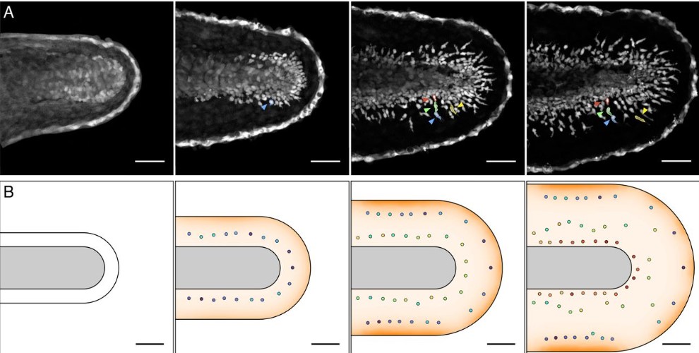
Tbx2 is essential for cochlear inner hair cell development and regeneration
Zhenghong Bi, Xiang Li, Minhui Ren, Yunpeng Gu, Tong Zhu, Shuting Li, Guangqin Wang, Suhong Sun, Yuwei Sun, Zhiyong Liu
Failure of digit tip regeneration in the absence of Lmx1b suggests Lmx1b functions disparate from dorsoventral polarity
Alejandro Castilla-Ibeas, Sofía Zdral, Laura Galán, Endika Haro, Lila Allou, Víctor M. Campa, Jose M. Icardo, Stefan Mundlos, Kerby C. Oberg, Marian A. Ros
Oxytocin promotes epicardial cell activation and heart regeneration after cardiac injury
Aaron H. Wasserman, Amanda R. Huang, Yonatan R. Lewis-Israeli, McKenna D. Dooley, Allison L. Mitchell, Manigandan Venkatesan, Aitor Aguirre
Cellular reprogramming with ATOH1, GFI1, and POU4F3 implicate epigenetic changes and cell-cell signaling as obstacles to hair cell regeneration in mature mammals
Amrita A. Iyer, Ishwar Hosamani, John D. Nguyen, Tiantian Cai, Sunita Singh, Lisa Beyer, Hongyuan Zhang, Hsin-I Jen, Rizwan Yousaf, Onur Birol, Jenny J. Sun, Russell S. Ray, Yehoash Raphael, Neil Segil, Andrew K. Groves
Loss of mitochondrial Chchd10 or Chchd2 in zebrafish leads to an ALS-like phenotype and Complex I deficiency independent of the mt-ISR
Virginie Petel Légaré, Christian J. Rampal, Mari J. Aaltonen, Alexandre Janer, Lorne Zinman, Eric A. Shoubridge, Gary A.B. Armstrong
Mitochondrial pyruvate metabolism regulates the activation of quiescent adult neural stem cells
Francesco Petrelli, Valentina Scandella, Sylvie Montessuit, Nicola Zamboni, Jean-Claude Martinou, Marlen Knobloch
Orchestration of alternative splicing regulates bone marrow mesenchymal stem cells fate during aging
Ye Xiao, Guang-Ping Cai, Xu Feng, Qi Guo, Yan Huang, Tian Su, Chang-Jun Li, Xiang-Hang Luo, Yong-Jun Zheng, Mi Yang
Prenatal inflammation perturbs fetal hematopoietic development and causes persistent changes to postnatal immunity
April C. Apostol, Diego A. López, Eric J. Lebish, Clint H. Valencia, Mari Carmen Romero-Mulero, Polina Pavlovich, Gloria E. Hernandez, E. Camilla Forsberg, Nina Cabezas-Wallscheid, Anna E. Beaudin
Biomechanical property of limbal niche maintains stemness through YAP
Swarnabh Bhattacharya, Abhishek Mukherjee, Sabrina Pisano, Shalini Dimri, Eman Knaane, Anna Altshuler, Waseem Nasser, Sunanda Dey, Lidan Shi, Ido Mizrahi, Ophir Jokel, Aya Amitai-Lange, Anna Kaganovsky, Michael Mimouni, Sergiu Socea, Peleg Hasson, Chloe Feral, Haguy Wolfenson, Ruby Shalom-Feuerstein
Developmental Morphogens Direct Human Induced Pluripotent Stem Cells Towards an Annulus Fibrosus-Like Cell Phenotype
Ana P. Peredo, Tonia K. Tsinman, Edward D. Bonnevie, Xi Jiang, Harvey E. Smith, Sarah E. Gullbrand, Nathaniel A. Dyment, Robert L. Mauck
SOX15 regulates stem cell pluripotency and promotes neural fate during differentiation by activating Hes5
Eun-Bee Choi, Munender Vodnala, Prince Saini, Madeleine Zerbato, Jaclyn J. Ho, Sharath Anugula, Jianing Wang, Shannan J. Ho Sui, Joon Yoon, Carla Inouye, Yick W. Fong
| Plant development
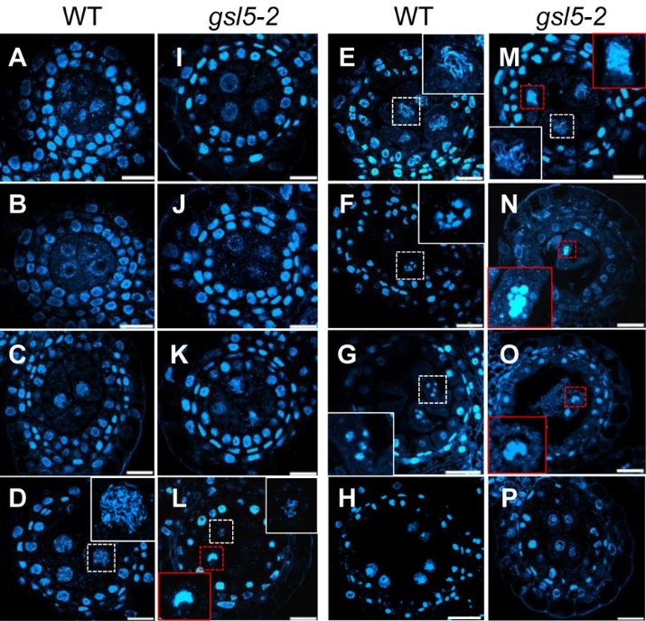
Rice GLUCAN SYNTHASE-LIKE5 promotes Callose deposition in Anthers to maintain proper Male Meiosis Initiation and Progression
Harsha Somashekar, Manaki Mimura, Katsutoshi Tsuda, Ken-Ichi Nonomura
Comparative Transcriptome Analysis of Hypocotyls During the Developmental Transition of C3 cotyledons to C4 Leaves in Halimocnemis mollissima: Exploring Root-Shoot Signalings Involved in the Establishing of C4 traits
Mahdis Zolfaghar, Mohammad Reza Ghaffari, Ali Mohammad Banaei-Moghaddam
LDL1 and LDL2 histone demethylases interact with FVE to regulate flowering in Arabidopsis
Mahima, Sourav Chatterjee, Sharmila Singh, Ananda K. Sarkar
Phosphorylation Status of B Beta Subunit Controls PP2A activity in Ethylene-mediated Root Growth Inhibition
Zhengyao Shao, Bo Zhao, Prashanth Kotla, Jackson G. Burns, Jaclyn Tran, Meiyu Ke, Xu Chen, Karen S. Browning, Hong Qiao
A molecular phenology scale of fruit development
Giovanni Battista Tornielli, Marco Sandri, Marianna Fasoli, Alessandra Amato, Mario Pezzotti, Paola Zuccolotto, Sara Zenoni
The DC1 domain protein BINUCLEATE POLLEN is required for pollen development in Arabidopsis thaliana
Leonardo A. Arias, Sebastián D’Ippolito, Jésica Frik, Natalia L. Amigo, Claudia A. Casalongué, Gabriela C. Pagnussat, Diego F. Fiol
The peptide GOLVEN10 controls nodule and lateral root organogenesis and positioning along the longitudinal root axis
Sonali Roy, Ivone Torres-Jerez, Shulan Zhang, Wei Liu, Katharina Schiessl, Clarissa Boschiero, Hee-Kyung Lee, Patrick X. Zhao, Jeremy D. Murray, Giles E. D. Oldroyd, Wolf-Rüdiger Scheible, Michael Udvardi
Sucrose rather than GA transported by AtSWEET13 and AtSWEET14 supports pollen fitness at late anther development stages
Jiang Wang, Xueyi Xue, Houqing Zeng, Jiankun Li, Li-Qing Chen
Ethylene signaling increases reactive oxygen species accumulation to drive root hair initiation in Arabidopsis
R. Emily Martin, Eliana Marzol, Jose M. Estevez, Gloria K. Muday
A network of CLAVATA receptors buffers auxin-dependent meristem maintenance
Amala John, Elizabeth Sarkel Smith, Daniel S. Jones, Cara L. Soyars, Zachary L. Nimchuk
TOR kinase controls Arabidopsis shoot development by translational repression of cytokinin catabolic enzymes
Denis Janocha, Anne Pfeiffer, Yihan Dong, Ondřej Novák, Miroslav Strnad, Lyuba A Ryabova, Jan U. Lohmann
A very long chain fatty acid responsive transcription factor, MYB93, regulates lateral root development in Arabidopsis
Yuta Uemura, Saori Kimura, Tomomichi Ohta, Takamasa Suzuki, Kosuke Mase, Hiroyuki Kato, Satomi Sakaoka, Yuki Komine, Kazuhiro Hotta, Motoyuki Shimizu, Atsushi Morikami, Hironaka Tsukagoshi
The pentatricopeptide repeat protein MTSF3 is required for nad2 mRNA stability and embryogenesis in Arabidopsis
Chuande Wang, Lisa Blondel, Martine Quadrado, Céline Dargel-Graffin, Hakim Mireau
| Evo-devo

Conserved meiotic mechanisms in the cnidarian Clytia hemisphaerica revealed by Spo11 knockout
Catriona Munro, Hugo Cadis, Sophie Pagnotta, Evelyn Houliston, Jean-René Huynh
Variant Polycomb complexes in Drosophila consistent with ancient functional diversity
Hyuckjoon Kang, Janel R. Cabrera, Barry M. Zee, Heather A. Kang, Jenny Marie Jobe, Maeve B. Hegarty, Aurelie E. Barry, Alexander Glotov, Yuri B. Schwartz, Mitzi I. Kuroda
Evolution and development of male-specific leg brushes in Drosophilidae
Kohtaro Tanaka, Olga Barmina, Ammon Thompson, Jonathan H. Massey, Bernard Y. Kim, Anton Suvorov, Artyom Kopp
Developmental evidence for parental conflict in driving Mimulus species barriers
Gabrielle D. Sandstedt, Andrea L. Sweigart
Machine-learning dissection of Human Accelerated Regions in primate neurodevelopment
Sean Whalen, Fumitaka Inoue, Hane Ryu, Tyler Fair, Eirene Markenscoff-Papadimitriou, Kathleen Keough, Martin Kircher, Beth Martin, Beatriz Alvarado, Orry Elor, Dianne Laboy Cintron, Alex Williams, Md. Abul Hassan Samee, Sean Thomas, Robert Krencik, Erik M. Ullian, Arnold Kriegstein, Jay Shendure, Alex A. Pollen, Nadav Ahituv, Katherine S. Pollard
Molecular and developmental signatures of genital size macro-evolution in bugs
Bruno C. Genevcius, Denis C. Callandrielo, Tatiana T. Torres
Cell Biology
A Van Gogh/Vangl tyrosine phosphorylation switch regulates its interaction with core Planar Cell Polarity factors Prickle and Dishevelled
Ashley C. Humphries, C. Clayton Hazelett, Claudia Molina-Pelayo, Danelle Devenport, Marek Mlodzik
Multiple roles of Pax6 in corneal limbal epithelial cells and maturing epithelial cell adhesion
Sweetu Susan Sunny, Jitka Lachova, Naoko Dupacova, Zbynek Kozmik
Extrinsic regulation of interneuron specification and migration
Fabrizia Pipicelli, Natalia Baumann, Rossella Di Giaimo, Christina Kyrousi, Rebecca Bonrath, Denis Jabaudon, Silvia Cappello
AGS3 antagonizes LGN to balance oriented cell divisions and cell fate choices in mammalian epidermis
Carlos Patiño Descovich, Kendall J. Lough, Akankshya Jena, Jessica J Wu, Jina Yom, Danielle C. Spitzer, Manuela Uppalapati, Katarzyna M. Kedziora, Scott E. Williams
Translation of the ERM-1 membrane-binding domain directs erm-1 mRNA localization to the plasma membrane in the C. elegans embryo
Lindsay P. Winkenbach, Dylan M. Parker, Robert T. P. Williams, Erin Osborne Nishimura
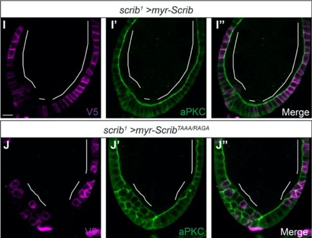
Minimal functional domains of the core polarity regulator Dlg
Mark J. Khoury, David Bilder
Nucleoli and the nucleoli-centromere association are dynamic during normal development and in cancer
Aaron Rodrigues, Kyle L. MacQuarrie, Emma Freeman, Kurt Leano, Alexander B Willis, Zhaofa Xu, Angel A Alvarez, Steven Kosak, Yongchao Ma, Bethany E Perez White, Daniel R Foltz, Sui Huang
Modulation of Spindle Assembly Checkpoint efficiency by cell fate in a chordate embryo
Marianne Roca, Lydia Besnardeau, Elisabeth Christians, Alex McDougall, Janet Chenevert, Stefania Castagnetti
Actin limits egg aneuploidies associated with female reproductive aging
Sam Dunkley, Binyam Mogessie
The circadian clock mediates daily bursts of cell differentiation by periodically restricting cell differentiation commitment
Zhi-Bo Zhang, Joydeb Sinha, Zahra Bahrami-Nejad, Mary N. Teruel
A CRISPR-screen in intestinal epithelial cells identifies novel factors for polarity and apical transport
Katharina MC Klee, Michael W Hess, Michael Lohmüller, Sebastian Herzog, Kristian Pfaller, Thomas Müller, Georg F Vogel, Lukas A Huber
Dynamic Features of Chromosomal Instability during Culture of Induced Pluripotent Stem Cells
Casey O. Dubose, John R. Daum, Christopher L. Sansam, Gary J. Gorbsky
Capacitation promotes a shift in the energy metabolism in murine sperm
Maximiliano Tourmente, Ester Sansegundo, Eduardo Rial, Eduardo R. S. Roldan
An actomyosin-polarized membrane reservoir mediates the unequal divisions of Drosophila neural stem cells
Bryce LaFoya, Kenneth E. Prehoda
Modelling
Echolocation-like model of directed cell migration within growing tissues
Tricia Y. J. Loo, Harsha Mahabaleshwar, Tom Carney, Timothy E. Saunders
Cellular compartmentalisation and receptor promiscuity as a strategy for accurate and robust inference of position during morphogenesis
Krishnan S Iyer, Chaitra Prabhakara, Satyajit Mayor, Madan Rao
Computing Minimal Boolean Models of Gene Regulatory Networks
Guy Karlebach, Peter N Robinson
Modelling realistic 3D deformations of simple epithelia in dynamic homeostasis
Domenic P.J. Germano, Stuart T. Johnston, Edmund J. Crampin, James M. Osborne
Tools & Resources
Morphodynamic Atlas for Drosophila Development
Noah P Mitchell, Matthew F Lefebvre, Vishank Jain-Sharma, Nikolas Claussen, Marion K Raich, Hannah J Gustafson, Andreas R Bausch, Sebastian J Streichan
Newly raised anti-c-Kit antibody detects interstitial cells of Cajal in the gut of chicken embryos
Rei Yagasaki, Yuuki Shikaya, Teruaki Kawachi, Masafumi Inaba, Yuta Takase, Yoshiko Takahashi
Chimeric 3D-gastruloids – a versatile tool for studies of mammalian peri-gastrulation development
Alexandra E. Wehmeyer, Katrin M. Schüle, Alexandra Conrad, Chiara M. Schröder, Simone Probst, Sebastian J. Arnold
Sex-specific topological differences in germline cell lineage trees
Rocky Diegmiller, Jasmin Imran Alsous, Duojia Li, Yukiko M. Yamashita, Stanislav Y. Shvartsman
Generation of a human ovarian granulosa cell model from induced pluripotent stem cells
Dirk Hart, Daniel Rodríguez Gutiérrez, Anna Biason-Lauber
Combined Lineage Tracing and scRNA-seq Reveals Unexpected First Heart Field Predominance of Human iPSC Differentiation
Francisco X. Galdos, Carissa Lee, Soah Lee, William Goodyer, Sharon Paige, Gabriela V. Escobar, Adrija Darsha, Aimee Beck, Sidra Xu, Rasmus O. Bak, Matthew Porteus, Sean M. Wu
Modelling the structure of Short Gastrulation and generation of a toolkit for studying its function in Drosophila
Sophie L Frampton, Catherine Sutcliffe, Clair Baldock, Hilary L Ashe
Pax6 mutant cerebral organoids partially recapitulate phenotypes of Pax6 mutant mouse strains
Nurfarhana Ferdaos, Sally Lowell, John O. Mason
MorphoFeatures: unsupervised exploration of cell types, tissues and organs in volume electron microscopy
Valentyna Zinchenko, Johannes Hugger, Virginie Uhlmann, Detlev Arendt, Anna Kreshuk
Reconstruction and deconstruction of human somitogenesis in vitro
Yuchuan Miao, Yannis Djeffal, Alessandro De Simone, Kongju Zhu, Andrew Silberfeld, Jong Gwan Lee, Jyoti Rao, Oscar A. Tarazona, Alessandro Mongera, Pietro Rigoni, Margarete Diaz-Cuadros, Laura Min Sook Song, Stefano Di Talia, Olivier Pourquié
Single-cell atlas of mouse limb development reveals a complex spatiotemporal dynamics of skeleton formation
Svetlana Markman, Mor Zada, Eyal David, Amir Giladi, Ido Amit, Elazar Zelzer
GliaMorph: A modular image analysis toolkit to quantify Müller glial cell morphology
Elisabeth Kugler, Isabel Bravo, Xhuljana Durmishi, Stefania Marcotti, Sara Beqiri, Alicia Carrington, Brian M. Stramer, Pierre Mattar, Ryan B. MacDonald
LOMAR: LOcalization Microscopy Analysis in R
Maria Theiss, Alvis Brazma, Virginie Uhlmann, Jean-Karim Hériché
A CRISPR-del-based pipeline for complete gene knockout in human diploid cells
Takuma Komori, Shoji Hata, Akira Mabuchi, Mariya Genova, Tomoki Harada, Masamitsu Fukuyama, Takumi Chinen, Daiju Kitagawa
Tissue libraries enable rapid determination of conditions that preserve antibody labeling in cleared mouse and human tissue
Theodore J. Zwang, Rachel E. Bennett, Maria Lysandrou, Benjamin Woost, Anqi Zhang, Charles M. Lieber, Douglas S. Richardson, Bradley T. Hyman
Parallel clonal and molecular profiling of hematopoietic stem cells using RNA barcoding
Edyta E. Wojtowicz, Jayna Mistry, Vladimir Uzun, Anita Scoones, Desmond W. Chin, Laura Kettyle, Francesca Grasso, Allegra M. Lord, Graham Etherington, Charlotte Hellmich, Petter S. Woll, Mirjam E. Belderbos, Kristian M. Bowles, Claus Nerlov, Wilfried Haerty, Leonid V. Bystrykh, Sten Eirik W. Jacobsen, Stuart A. Rushworth, Iain C. Macaulay
Induced pluripotent stem cells and cerebral organoids from the critically endangered Sumatran rhinoceros
Vera Zywitza, Silke Frahm, Norman Krüger, Anja Weise, Frank Göritz, Robert Hermes, Susanne Holtze, Silvia Colleoni, Cesare Galli, Micha Drukker, Thomas B. Hildebrandt, Sebastian Diecke
Introducing CELLBLOKS®: a novel organ-on-a-chip platform allowing a plug-and-play approach towards building organotypic models
Valon Llabjani, M.R. Siddique, Anaïs Makos, Afaf Abozoid, Valmira Hoti, Francis L Martin, Imran I. Patel, Ahtasham Raza
MuSiC2: cell type deconvolution for multi-condition bulk RNA-seq data
Jiaxin Fan, Yafei Lyu, Qihuang Zhang, Xuran Wang, Mingyao Li, Rui Xiao
New hypotheses of cell type diversity and novelty from comparative single cell and nuclei transcriptomics in echinoderms
Anne Meyer, Carolyn Ku, William Hatleberg, Cheryl A. Telmer, Veronica Hinman
The Network Zoo: a multilingual package for the inference and analysis of biological networks
Marouen Ben Guebila, Tian Wang, Camila M. Lopes-Ramos, Viola Fanfani, Deborah Weighill, Rebekka Burkholz, Daniel Schlauch, Joseph N. Paulson, Michael Altenbuchinger, Abhijeet Sonanwane, James Lim, Genis Calderer, David van Ijzendoorn, Daniel Morgan, Alessandro Marin, Cho-Yi Chen, Alex Song, Kate Shutta, Dawn DeMeo, Megha Padi, John Platig, Marieke L. Kuijjer, Kimberly Glass, John Quackenbush
Single-Cell Transcriptome Analyses Reveal the Cell Diversity and Developmental Features of Human Gastric and Metaplastic Mucosa
Ayumu Tsubosaka, Daisuke Komura, Hiroto Katoh, Miwako Kakiuchi, Takumi Onoyama, Asami Yamamoto, Hiroyuki Abe, Yasuyuki Seto, Tetsuo Ushiku, Shumpei Ishikawa
Machine Learning Reveals The Effect of Maternal Age on The Mouse Pre-Implantation Embryo Developmental Timing
Nati Daniel, Tanya Wasserman, Zohar Adler, Tomer Czyzewski, Yonatan Savir
Slim: interoperable web viewer and annotation tool for quantitative microscopy tissue imaging
Chris Gorman, Davide Punzo, Igor Octaviano, Steve Pieper, William J.R. Longabaugh, David A. Clunie, Ron Kikinis, Andrey Y. Fedorov, Markus D. Herrmann
SODA: a TypeScript/JavaScript Library for Visualizing Biological Sequence Annotation
Jack W. Roddy, George T. Lesica, Travis J. Wheeler
Single-Cell Atlas of the Drosophila Leg Disc Identifies a Long Non-Coding RNA Associated with Distal Leg Development
Joyce Tse, Tsz Ho Li, Jizhou Zhang, Alan Lee, Ivy Lee, Zhe Qu, Xiao Lin, Jerome Hui, Ting-Fung Chan
Research practice & education
PhD and postdoc training outcomes at EMBL: changing career paths for life scientists in Europe
Junyan Lu, Britta Velten, Bernd Klaus, Mauricio Ramm, Wolfgang Huber, Rachel Coulthard-Graf
Expansion of information in scientific research papers
Malika Abdullaeva, John J. Bromfield, I. Martin Sheldon
Gender-based disparities and biases in science: an observational study of a virtual conference
Junhanlu Zhang, Rachel Torchet, Hanna Julienne
The U.S. academic job market survives the SARS-CoV-2 global pandemic
Ariangela J Kozik, Ada Hagan, Nafisa M Jadavji, Christopher T Smith, Amanda Haage
Improving Interdisciplinary Teaching through a Complexity Lens
Sarah Neitzel, Yuhao Zhao, Carrie Diaz Eaton
When Citizen Science meets Teaching: A storified citizen science computer game to teach and contribute to genomic data analysis
Chrisostomos Drogaris, Alexander Butyaev, Elena Nazarova, Harsh Patel, Akash Singh, Brenden Kadota, Jérôme Waldispühl
Twitter as a community communication tool for international neuroscience conferences
Niall W. Duncan, Russell Shean
Community review: a robust and scalable selection system for resource allocation within open science and innovation communities
Chris L. B. Graham, Thomas E. Landrain, Amber Vjestica, Camille Masselot, Elliot Lawton, Leo Blondel, Luca Haenel, Bastian Greshake Tzovoras, Marc Santolini
 (No Ratings Yet)
 (1 votes)