In the latest episode of Genetics Unzipped, Kat Arney takes a look at the genetics of giants and the science of small. Why do some species grow so large? What’s the genetic legacy behind Charles Byrne and the Giants of Ireland? And what was it about life on a Mediterranean island that miniaturised a mammoth?
If you enjoy the show, please do rate and review on Apple podcasts and help to spread the word on social media. And you can always send feedback and suggestions for future episodes and guests to podcast@geneticsunzipped.com Follow us on Twitter – @geneticsunzip
CSF1R-dependent macrophages control postnatal somatic growth and organ maturation Sahar Keshvari, Melanie Caruso, Ngari Teakle, Lena Batoon, Anuj Sehgal, Omkar L. Patkar, Michelle Ferrari-Cestari, Cameron E. Snell, Chen Chen, Alex Stevenson, Felicity M. Davis, Stephen J. Bush, Clare Pridans, Kim M. Summers, Allison R. Pettit, Katharine M. Irvine, David A. Hume
Cells of the human intestinal tract mapped across space and time R Elmentaite, N Kumasaka, HW King, K Roberts, M Dabrowska, S Pritchard, L Bolt, SF Vieira, L Mamanova, N Huang, I Goh Kai’En, E Stephenson, J Engelbert, RA Botting, A Fleming, E Dann, SN Lisgo, M Katan, S Leonard, TRW Oliver, CE Hook, K Nayak, F Perrone, LS Campos, C Dominguez-Conde, K Polanski, S Van Dongen, M Patel, MD Morgan, JC Marioni, OA Bayraktar, KB Meyer, M Zilbauer, H Uhlig, MR Clatworthy, KT Mahbubani, K Saeb Parsy, M Haniffa, KR James, SA Teichmann
Parallel functional testing identifies enhancers active in early postnatal mouse brain Jason T. Lambert, Linda Su-Feher, Karol Cichewicz, Tracy L. Warren, Iva Zdilar, Yurong Wang, Kenneth J. Lim, Jessica Haigh, Sarah J. Morse, Cesar P. Canales, Tyler W. Stradleigh, Erika Castillo, Viktoria Haghani, Spencer Moss, Hannah Parolini, Diana Quintero, Diwash Shrestha, Daniel Vogt, Leah C. Byrne, Alex S. Nord
tiRNA signaling via stress-regulated vesicle transfer in the hematopoietic niche Youmna S. Kfoury, Fei Ji, Michael Mazzola, David B. Sykes, Allison K. Scherer, Anthony Anselmo, Yasutoshi Akiyama, Francois Mercier, Nicolas Severe, Konstantinos D. Kokkaliaris, Thomas Brouse, Borja Saez, Jefferson Seidl, Ani Papazian, Pavel Ivanov, Michael K. Mansour, Ruslan I. Sadreyev, David T. Scadden
Functional, metabolic and transcriptional maturation of stem cell derived beta cells Diego Balboa, Tom Barsby, Väinö Lithovius, Jonna Saarimäki-Vire, Muhmmad Omar-Hmeadi, Oleg Dyachok, Hossam Montaser, Per-Eric Lund, Mingyu Yang, Hazem Ibrahim, Anna Näätänen, Vikash Chandra, Helena Vihinen, Eija Jokitalo, Jouni Kvist, Jarkko Ustinov, Anni I. Nieminen, Emilia Kuuluvainen, Ville Hietakangas, Pekka Katajisto, Joey Lau, Per-Ola Carlsson, Sebastian Barg, Anders Tengholm, Timo Otonkoski
Sequential defects in cardiac lineage commitment and maturation cause hypoplastic left heart syndrome Markus Krane, Martina Dreßen, Gianluca Santamaria, Ilaria My, Christine M. Schneider, Tatjana Dorn, Svenja Laue, Elisa Mastantuono, Riccardo Berutti, Hilansi Rawat, Ralf Gilsbach, Pedro Schneider, Harald Lahm, Sascha Schwarz, Stefanie A. Doppler, Sharon Paige, Nazan Puluca, Sophia Doll, Irina Neb, Thomas Brade, Zhong Zhang, Claudia Abou-Ajram, Bernd Northoff, Lesca M. Holdt, Stefanie Sudhop, Makoto Sahara, Alexander Goedel, Andreas Dendorfer, Fleur V.Y. Tjong, Maria E. Rijlaarsdam, Julie Cleuziou, Nora Lang, Christian Kupatt, Connie Bezzina, Rüdiger Lange, Neil E. Bowles, Matthias Mann, Bruce Gelb, Lia Crotti, Lutz Hein, Thomas Meitinger, Sean Wu, Daniel Sinnecker, Peter J. Gruber, Karl-Ludwig Laugwitz, Alessandra Moretti
Multiple 9-1-1 complexes promote homolog synapsis, DSB repair, and ATR signaling during mammalian meiosis Catalina Pereira, Gerardo A. Arroyo-Martinez, Matthew Z. Guo, Michael S. Downey, Emma R. Kelly, Kathryn J. Grive, Shantha K. Mahadevaiah, Jennie Sims, Vitor Marcel Faça, Charlton Tsai, Carl J. Schiltz, Niek Wit, Heinz Jacobs, Nathan L. Clark, Raimundo Freire, James M. A. Turner, Amy M. Lyndaker, Miguel A. Brieño-Enríquez, Paula E. Cohen, Marcus B. Smolka, Robert S. Weiss
Measuring nonapoptotic caspase activity with a transgenic reporter in mice P. J. Nicholls, Thomas F. Pack, Nikhil M. Urs, Sunil Kumar, Yang Zhou, Gabor Turu, Evan Calabrese, Wendy L. Roberts, Ping Fan, Valeriy G. Ostapchenko, Monica S. Guzman, Flavio Beraldo, Vania F. Prado, Marco A. M. Prado, Ivan Spasojevic, Joshua C. Snyder, Kafui Dzirasa, G. Allan Johnson, Marc G. Caron
A field guide to cultivating computational biology Anne E Carpenter, Casey S Greene, Piero Carnici, Benilton S Carvalho, Michiel de Hoon, Stacey Finley, Kim-Anh Le Cao, Jerry SH Lee, Luigi Marchionni, Suzanne Sindi, Fabian J Theis, Gregory P Way, Jean YH Yang, Elana J Fertig
Sarah Jacquelyn Smith, Lance Davidson and Mark Rebeiz
One of the biggest mysteries in the developmental evolution field is the puzzle of how new morphological structures come about. If you think about it, every anatomical structure in the multicellular world was new at some point in time. And yet, we currently only have a rough picture of how insect wings, beetle horns, or turtle shells initially evolved. There are multiple ways that this question can be answered, and it is an exciting time to be studying how genetic programs of gene expression translate into the physical manifestations of development that form new tissue configurations. In studying one such novelty in Drosophila, we recently learned that there is more than meets the eye to generating extreme deformations in cellular shape1. We also learned to listen more carefully to our collaborators…
Novelty in Drosophila genital traits
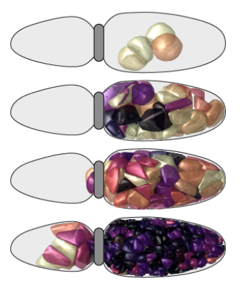
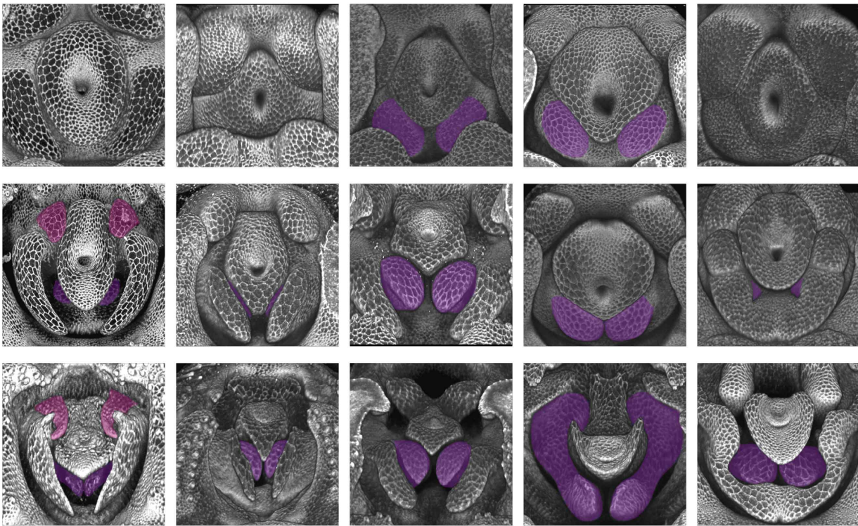
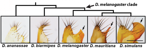
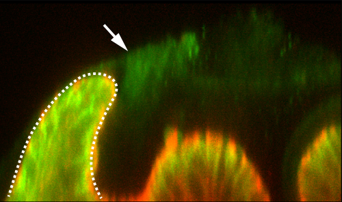
Reproductive structures are notorious for their rapid evolution among internally fertilizing species2. The chitinous cuticle shapes in the genitalia of Drosophila melanogaster are indeed quite different from its nearest relatives. In fact, these differences in genital morphology are used to discriminate this model organism from its closest relatives3. Namely, the posterior lobe is a recently evolved hook-shaped structure which is attached to a cuticular plate known as the lateral plate (Fig. 1). The posterior lobe is highly divergent in size and shape in species that possess this structure and is absent in more distantly related Drosophila species (Fig. 1), which offer a useful reference for understanding how the genitalia likely developed before the evolution of this structure. Because the posterior lobe is present in the highly tractable Drosophila melanogaster, we can exploit the bountiful genetic tools of this model organism to examine and dissect its development. Our previous work had suggested that many genes expressed in the posterior lobe were re-deployed, or ‘co-opted’ from another organ system, the posterior spiracle4, which is an extension of the larval tracheal system. Identifying genes expressed in a structure however doesn’t tell you much about how it is built, and thus studies of genetic control mechanisms must be complemented by studies at the cellular level (and vice versa)5.
Figure 1. The posterior lobe is a highly divergent structure unique to the D melanogaster clade. The lobe is the protrusion on the right-hand side (arrow), which is attached to the structure known as lateral plate which is also shown. D. ananassae and D. biarmipes are two species which express the ancestral state of lacking this structure. Scale bar = 100µM (bottom right)
The major discriminating feature of Drosophila melanogaster is a single cell tall!
When first examining the morphogenesis of the posterior lobe, we had no idea where to start. We sought the advice of a colleague across campus, Lance Davidson, who studies the biomechanics of development at the University of Pittsburgh Swanson School of Engineering. Lance was very excited to see our preliminary movies of posterior lobe development in which we monitored apical cell junctions (Movie 1). Even with this new perspective on lobe development, we had no idea exactly what was going on – we could see the apical surfaces of cells changing size and shape as the posterior lobe forms, but of course seeing these changes and knowing what processes cause these alterations are two very different things.
Movie 1. Movie of posterior lobe morphogenesis. Apical junctions are labeled by GFP-tagged armadillo protein.
However, apical cell shape changes cover only 2D of the 3D story and examining the entire shape of the cell using single cell fluorescent labeling (Movie 2), indicated that cells of the posterior lobe drastically change their shape along the apical-basal axis, and become extremely tall and thin throughout development, allowing the posterior lobe to project off of the lateral plate. Understanding how this extreme cell shape is controlled and spatially patterned would provide a context in which to connect the activity of gene regulatory networks to the patterning of cellular traits. Initially, we became quite interested in the role that cytoskeletal regulators might play in this process: patterning and stabilizing filaments of both actin and microtubules. However, our investigation uncovered an unexpected perspective on this cellular behavior.
Movie 2. Single cell labeling of the posterior lobe reveals that it is a singe cell tall. Clonal expression of mTFP1 was induced via the heat shock promoter and associated temperature shift.
Listen to your collaborators!
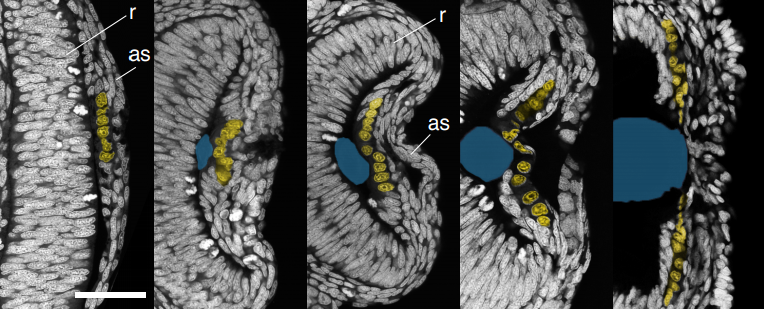
One day over our frequent lunchtime conversations, Lance raised the point that the posterior lobe has a cantilevered structure: it leans to one side, and he mentioned that such configuration is rare in epithelial structures. This led him to speculate that maybe we should be looking for some kind of “tether” that would pull the tissue into this arrangement. I remember quite vividly discussing this interesting model with members of the lab, and quickly dismissing it because we had never seen such a tether with our own eyes. Of course, we had only been looking at epithelial and nuclear markers at that point, and so any material that could serve as a tether might go unnoticed if it is translucent and doesn’t express the markers we used. This idea lingered in the back of our minds with little notice before we realized that perhaps Lance was onto something. Performing certain antibody stains, in particular for a septate junction protein Macroglobulin (a kind gift from Dr. Rob Ward, U. Kansas), we noticed that there was a mysterious pattern of background staining in our samples when visualized on the confocal (Figure 2). This caused us to consider the potential role of the apical extracellular matrix in building a posterior lobe.
Figure. 2. Non-specific staining of extracellular material in a stain for septate junctions (green, Macroglobulin). A cross-section of the posterior lobe (dashed lines) leans towards the detected extracellular material.
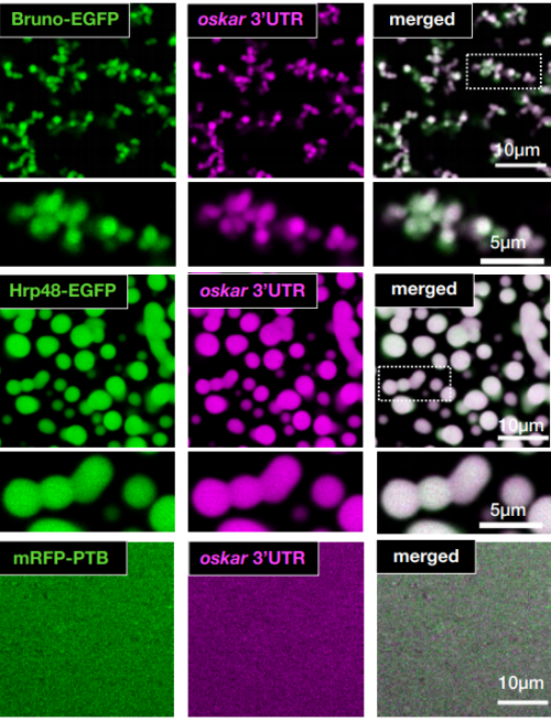
Looking beyond what the eye can see
Although basal extracellular matrices are well known to play critical roles in epithelial sheets6, recent work has shown that apical matrices are also important. Several recent papers had shown that the zona pellucida domain-encoding protein Dumpy is important for shaping the wing by attaching its distal tip to an overlying cuticle7,8. We examined a fluorescently tagged fusion protein for Dumpy, and this revealed a surprisingly intricate web of apical matrix throughout the genitalia that showed prominent connections to the posterior lobe (Movie 3). We developed fluorescent lectin staining protocols to show that this matrix exists in species that lack lobes. However, we found that strong aECM connections to where the lobe would otherwise form are much less pronounced in non-lobed species. Finally, what made this narrative compelling from an evolutionary sense is that RNAi experiments showed that Dumpy expression is required for cells of the lobe to achieve their height. Together, these results demonstrated how making prominent aECM connections is important to the formation of a new structure, and was likely subject to evolutionary changes which alter how the epithelium deposits and interacts with the matrix.
Movie 3. A GFP-tagged Dumpy expression during posterior lobe development reveals a complex and dynamic network of apical ECM.
Evolution of novelties: more than meets the eye
What makes this finding surprising is that it reveals the layers of complexity even in such a simple morphological novelty. We had not anticipated that such a complex matrix would exist outside the cells that form a morphologically novel structure. As we generally don’t look to matrices when studying cellular processes, it may be that other epithelial structures have equally elaborate and uncharted apical matrices. More broadly, the story highlights how studying novelties can unveil processes previously unknown and shows how we can zero in on proximal cellular mechanisms that assemble the elaborate structures we see in the multicellular world. Comparing cellular behaviors between species thus offers a unique window into how genetic programs drive physical processes in developing tissues. We suspect that studying the regulatory sequences and networks controlling the expression patterns of Dumpy and other apical ECM components will allow us to go beyond simplistic models of network co-option and the evolution of novelty.
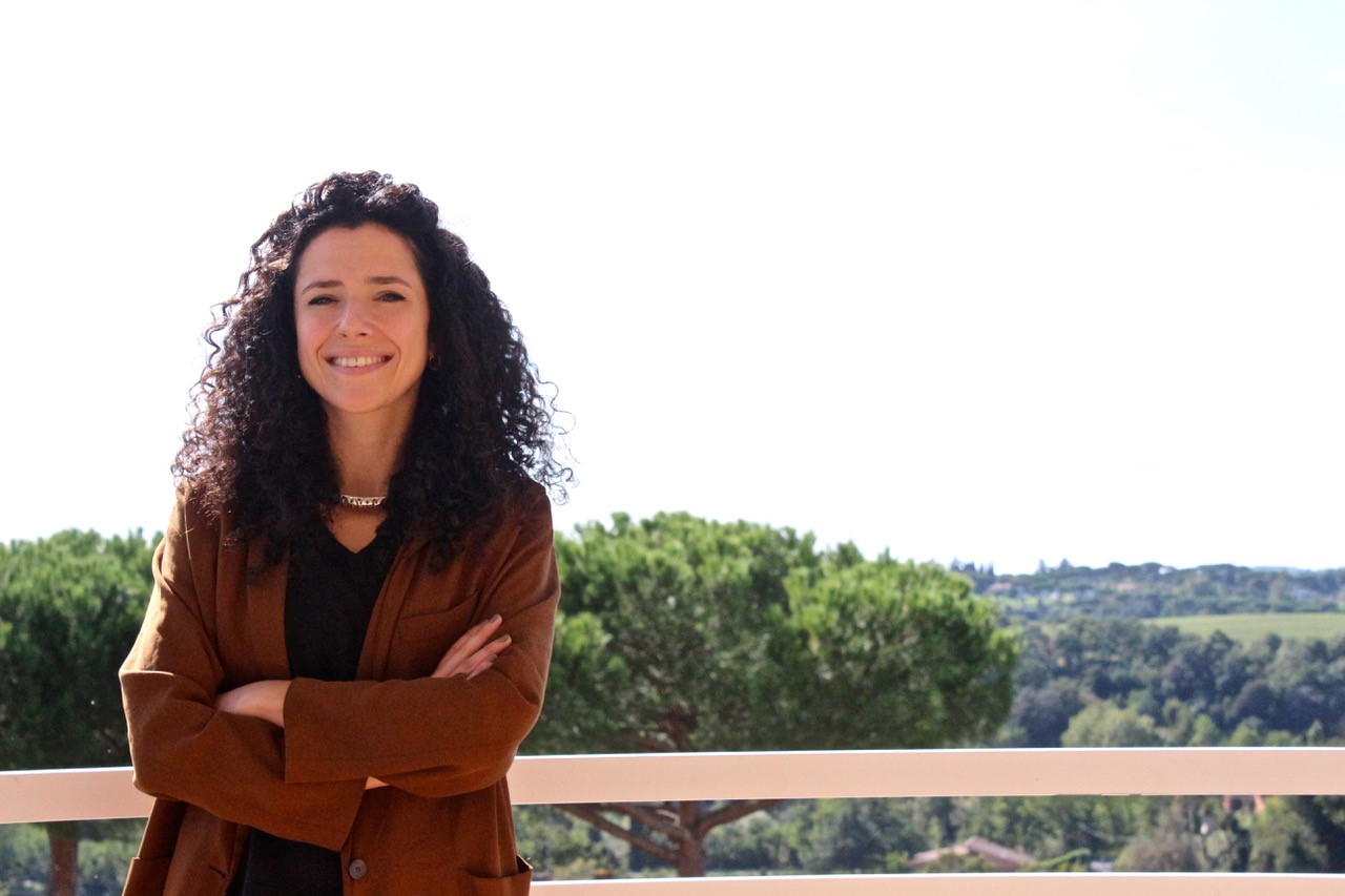
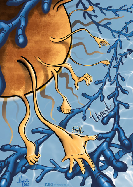
Our eighth profile in the series features Marzia Munafò, a postdoctoral researcher currently based in Rome.
Where are you originally from and what do you work on now?
I am originally from Rome. I recently completed my PhD in Medical Sciences at the University of Cambridge UK and I am now a postdoctoral fellow at EMBL Rome. I am a molecular biologist by training and I have worked mostly on RNA biology. During my PhD I investigated how small RNAs protect the genome from transposon activity, using Drosophila as a model organism. For my postdoc I am now focusing on understanding epigenetic inheritance and transposon regulation in mammals.
Where you always going to be a scientist?
I would say that curiosity in general has always played an important role in my life. I was an inquisitive, curious child who really enjoyed learning about the world. What initially sparked my interest for biology was learning about Mendel’s laws of inheritance in secondary school but I didn’t really consider becoming a scientist until much later. It was towards the end of high school when I realised that the one job that would fulfil my curiosity and passion for learning would be a career in research. Having realised that, biology was a natural choice for me.
And what about art – have you always enjoyed it?
Absolutely yes. Drawing is the thing that I remember loving ever since I was a child. I was always drawing my favourite characters from comics, books or movies and even more from my imagination. At some point I really wanted to become a comic book illustrator!
What or who are your artistic influences?
I think my drawing “style”, so to speak, owes a lot to the Disney comics I used to read in my childhood/teenage years and more generally to the visual aesthetics of cartoons and anime from the 90s. I also deeply love fantasy literature and often draw characters and scenery from books, with The Lord of the Rings being my main inspiration above all.
Speaking of scientific illustrations, one of the artists I admire the most is David Goodsell. I find his way of drawing the crowded interior of a cell so elegant and thought-provoking!
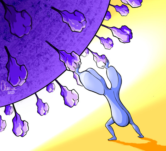
Neutralising antibodies fighting against SARS-CoV2.
How do you make your art?
Nowadays I mostly use digital media for my illustrations. Digital art is truly amazing, it offers so many opportunities and I have just scratched the surface of its potential. When I approach a new artwork I usually spend most of the time thinking about the message I want to convey. If I am working for someone else I try to grasp the main scientific concepts and do a bit of research to see how people typically represent that something. Once I have a rough idea of what I am aiming for, I move to the actual painting. I find it somewhat liberating to stare at the white canvas and just go with the flow, without knowing what the final result will look like. It’s a nice contrast with my daily routine as a researcher, where everything is much more schematic and there is no room for improvisation. Nonetheless, in my view science is also a creative process. Thinking outside the box to formulate new hypotheses or devising innovative technologies requires some degree of creativity, so the two worlds are not so far apart.
“I find it somewhat liberating to stare at the white canvas and just go with the flow“
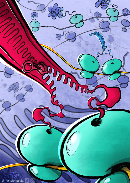
Co-co assembly: a novel mechanism of co-translational assembly of protein complexes (reference: Bertolini et al., 2021, Science)
Does your art influence your science at all, or are they separate worlds?
Finding a way to combine my creativity and my scientific education has definitely helped me to grow as a scientist and also consolidated my enthusiasm for research. First, having this side-project has helped me to balance out a lot of the stress during the final years of PhD. It was very important to have my own happy place to switch the brain off and recharge batteries. Secondly, I like thinking about scientific concepts without constraints and just letting my imagination go. It’s a different approach to science, as I don’t have to be rigorous but I can just take the main facts and shift them into a new perspective. I enjoy having my imagination take the lead and drawing without strictly adhering to canonical imagery. At the end of the day, doing this makes me even more fascinated by the wonders of biology!
I don’t have a clear plan of where I’m going with my scientific illustrations, but I definitely want to perfect my skills in digital painting and challenge myself with something new, like animation or 3D. Communicating science to peers and public is something I deeply care about, so I’m hoping that my illustrations can help people come closer to biology.
We’re looking for new people to feature in this series throughout the year – whatever kind of art you do, from sculpture to embroidery to music to drawing, if you want to share it with the community just email thenode@biologists.com (nominations are also welcome!).
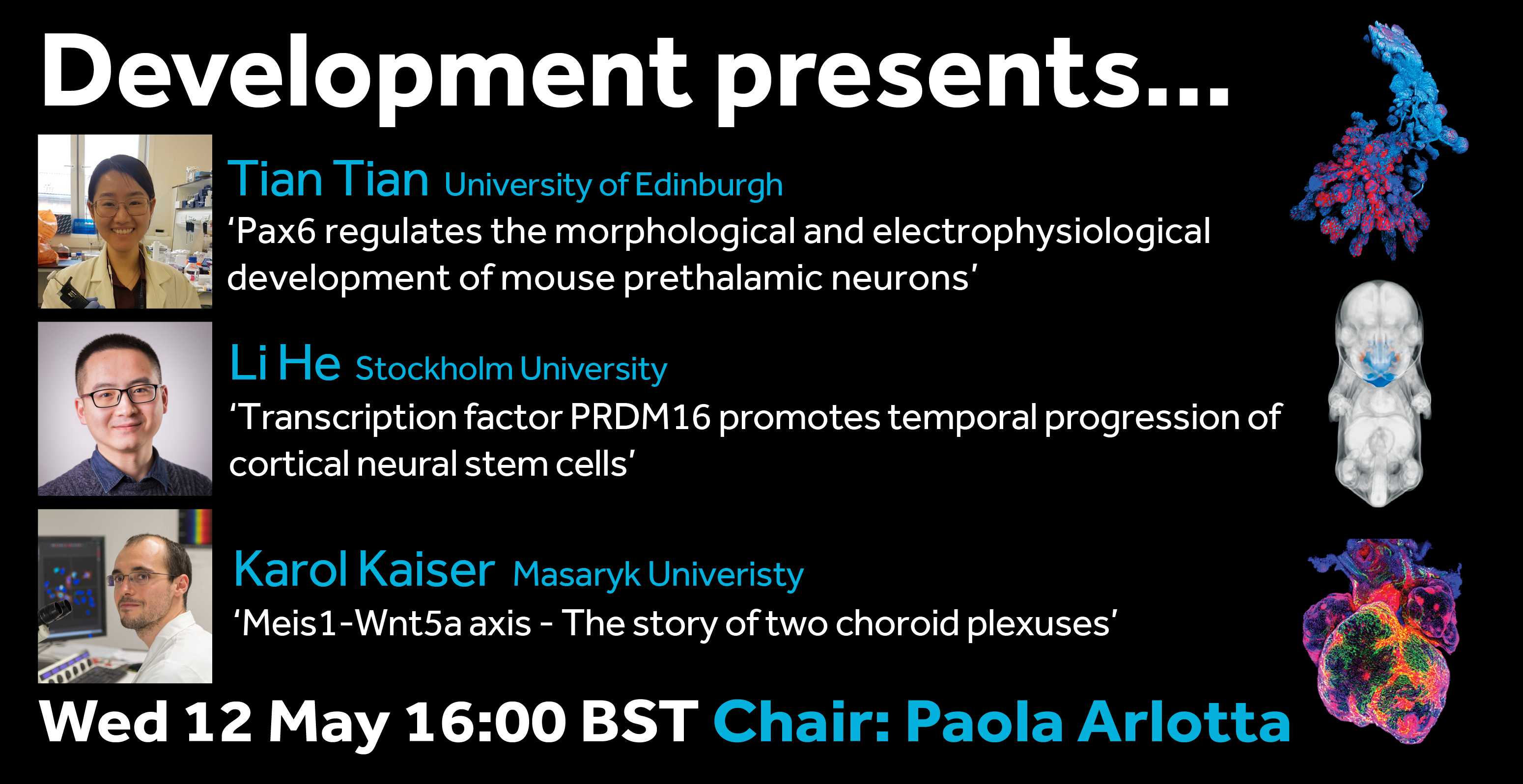
The eighth webinar in our Development presents… series will be chaired by Development Editor Paola Arlotta (Harvard Stem Cell Institute), who has invited three exciting talks on the topic of mammalian brain development.
Tian Tian (formerly a PhD student in David Price’s lab and now a postdoc in Matthew Nolan’s group at the University of Edinburgh) ‘Pax6 regulates the morphological and electrophysiological development of mouse prethalamic neurons’
Li He (PhD student in Qi Dai’s lab at Stockholm University) ‘Transcription factor PRDM16 promotes temporal progression of cortical neural stem cells’
Karol Kaiser (formerly a postdoc in Vitezslav Bryja’s lab at Masaryk Univeristy now a postdoc in Fiona Doetsch’s labat the University of Basel) ‘Meis1-Wnt5a axis – The story of two choroid plexuses’
The webinar will be held in Remo, our browser-based conferencing platform. After the talks you’ll have the chance to meet the speakers and other participants at virtual conference tables. If you can’t make it on the day, talks will be available to watch after the event on the Node. You can also sign up to our mailing list for email alerts.
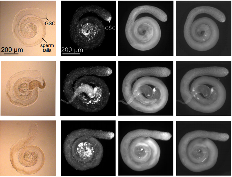
Scientists led by Dr Jason Heaney from Baylor College of Medicine in Texas, USA, have found that a failure in the development of sperm cells can lead to the formation of testicular tumours in mice. These findings, published in the journal Development, provide new clues for understanding how testicular cancer arises in mammals.
Testicular cancer is the most common type of cancer to affect men between the ages of 15 and 49, and around 95% of these cases are caused by testicular germ cell tumours. Although testicular germ cell tumours typically manifest after puberty, problems in the embryonic development of germ cells can transform them into cancer cells that form tumours later in life.
In the embryo, germ cells initially have the potential to form many different cell types, a characteristic called pluripotency. Normally, germ cells lose this ability as embryonic development progresses and they become restricted to only form sex cells – sperm in males and eggs in females – in a process known as sex determination. Male sex determination also coincides with the embryonic period when the cancer cells that form testicular germ cell tumours can develop.
As Dr Jason Heaney, Associate Professor at the Department of Molecular and Human Genetics at Baylor College of Medicine revealed, the timing between cancer cell formation and sex determination is more than just coincidental: “previous work from our lab and others indicated that defects in this sex-specific switch may play a central role in facilitating the initiation of testicular germ cell tumours. In this study, we set out to test whether testicular germ cell tumours arise from germ cells that do not begin the sex-specific differentiation process and retain features of pluripotent cells.”
To test this hypothesis, Dr Heaney together with Dr Nicholas Webster and team explored whether a gene called Nanos2, which is needed for sex-determination, also prevented embryonic cancer cells developing in mice. “NANOS2 plays a key role in the sex-specific development of embryonic germ cells by suppressing the female (egg) fate and promoting the male (sperm) fate,” explained Dr Heaney.
Using a strain of mice that spontaneously develop germ cell tumours, the researchers showed that some germ cells lacked NANOS2. Not only did the NANOS2-deficient germ cells fail to mature into sperm, but they remained pluripotent and, crucially, were more likely to transform into embryonic cancer cells. The scientists also characterised the NANOS2-deficient germ cells and uncovered important changes in cell behaviour, which could be used to identify and treat cancer cells in the future. “Our work reveals changes in gene expression when germ cells transform into cancer cells that suggest alterations in metabolism and cell division, which could be used for targeted therapies,” said Dr Heaney.
Although it is currently unclear what role NANOS2 plays in the development of human testicular germ cell tumours, this study highlights an important link to the process of male sex determination, providing a new direction for future research. “Our studies provide functional evidence for a mechanism – disrupted male germ cell fate determination – through which these genes cause testicular germ cell tumours in humans,” said Dr Heaney, “future studies will use genetically modified mice to explore how genes associated with germ cell tumours in humans influence germ cell transformation.”
This press release was released by Development (paper link).
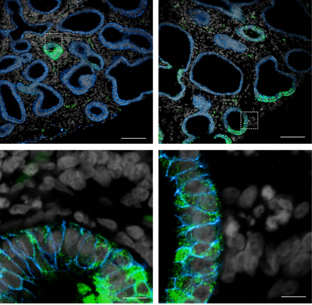
The placenta forms the interface between the maternal and foetal circulatory systems. As well as ensuring essential nutrients, endocrine and immunological signals get through to the foetus to support its development and growth, the placenta must also protect it from the accumulation of potentially toxic compounds. A study from Cécile Demarez, Mariana Astiz and colleagues at the University of Lübeck in Germany now reveals that the activity of a crucial placental gatekeeper in mice is regulated by the circadian clock, changing during the day-night cycle. The study, which has implications for the timing of maternal drug regimens, is published in the journal Development.
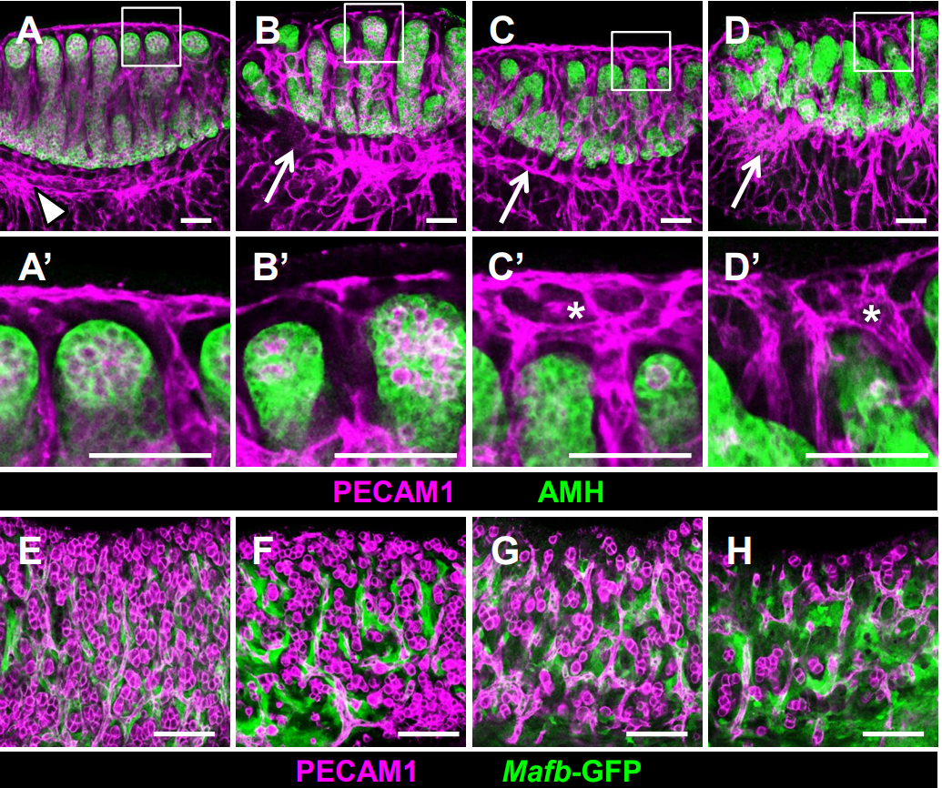
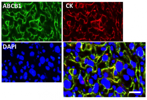
Immunofluorescence reveals that the ABCB1 protein (in green) localises to the labyrinth zone of the mouse placenta.
The circadian clock translates time-of-day information into physiological signals through rhythmic regulation of downstream genes. In this study, the researchers discover that in the labyrinth zone of the mouse placenta, a tissue functionally equivalent to the human chorionic villi, clock genes are expressed in a 24h rhythm. Importantly, they show that this placental clock is responsible for regulating the expression and activity of ABCB1, a drug efflux transporter with hundreds of known substrates.
An important prediction of this work is that the time-of-day of maternal treatment could be an important factor to consider to avoid non-desirable effects for the foetus during pregnancy.
“Pharmacological treatments are mostly avoided by pregnant women but in certain circumstances there is no other option,” says Dr. Mariana Astiz. “An example would be maternal treatment with antiretrovirals (many of which are in fact substrates of ABCB1). So, choosing the correct time of day to take drugs like these might reduce the ammount of drug reaching the baby and hence the possible negative effects in the short and long-term.”
Dr. Astiz hopes this study will provide a stimulus to design studies that specifically test hypotheses about the placental circadian clock in humans. “This is definitely a very exciting and rapidly advancing field of research.”
When I wrote the #DevBiolWriteClub rules, I made Rule #2 simply restate Rule #1. This, of course, was a cheap ploy by the author to make the reader compare him favorably to Brad Pitt. But there is a serious intent behind it.
Becoming a better writer takes dedication and it takes hard work, but mainly it takes patience. Let’s imagine you read my first post on the Node last Spring, or that you’ve been allowing me to hector you on Twitter. By now, you’ve put a year of effort into developing your craft as a writer. Seen any impact yet? Perhaps you’ve got more words down on the page, or maybe you’re a bit more pleased with your writing. Maybe, but it’s more likely that any progress you’ve made is pretty much intangible at this point. That’s the nature of writing, and it’s a major part of why it is such a frustrating endeavor.
But trust me, simply practicing is by far the best thing you can do for your writing. If you have an active project, try to write at least a little every day. If you are between writing projects, read more and take the time to notice the writing while you read it. That said, there’s no question that we all hit a wall sometimes. Learning to get some outside help is critical.
Thus, we finally come to Rule #5: You can’t do it alone.
Now, getting help can take two forms. The simpler way is to read books about writing. Notice that I said “books,” plural. I wrote about this in my last post, so I won’t repeat it here, except to say there are LOTS of great books about writing. Go read them. But also know that no book will ever do for you what an engaged reader can do, so let’s talk about the real meat of Rule #5. Show your work to others and get their feedback.
Many years ago, Frank Conlon sent me a great essay in The New Yorker by Atul Gawande. The essay pointed out that essentially all professional athletes will be coached throughout their entire careers, as will most opera singers. But surgeons aren’t. Scientists aren’t, either. So, you need to nurture your own stable of informal coaches.
It turns out that I am both lazy and ambitious. It’s an odd combination but it has made me very good at asking for help. In terms of writing, getting help started early. One of my first “coaches” was Mr. Mike Cullinan, an English teacher loved and feared by generations of students at my high school in Houston. He once gave me a 10 on a paper. Out of 100, yes. You see, he graded content and writing separately and averaged the scores. Various transgressions of grammar or diction had fixed point values (25 points for each run-on sentence). So, despite a high score for content, I had scored a negative 75 for writing. I had to re-write it. I had to re-write most things I wrote for him. And you know what? The writing always got better. Every single time.
By the time I became a PI, I had become religious about seeking advice on anything I write. I actually got the first R01 grant I applied for as a PI. (It was fun to briefly boast of my 100% NIH success rate, but the euphoria was short-lived; I didn’t get the second one.) What’s important to understand, though, is that I spent an entire year writing that grant. Of course, I also had to order equipment, hire people, and go to new faculty orientation. But I worked on the grant, bit by bit, most days. For a year. I revised and revised and revised. Along the way, that one grant application took in the serious feedback of five faculty members, three developmental biologists (Richard Harland, Paul, Krieg, David Parichy) and two cell biologists (Terry O’Halloran, Arturo De Lozzane). It was an awesome learning experience and it generated one of the tightest pieces of grant writing I’ve ever produced.
As I have aged, I find myself more lazy than ambitious, and I sometimes submit writing that no one else has read. It almost always goes badly. Luckily, however, old habits die hard, and I still usually seek input from an outside reader. I wrote an essay in Developmental Cell in 2019. that is probably my favorite piece of writing. But let me tell you, it started poorly. I sent an early draft to Lila Solnica-Krezel, and her response was something like, “Oh, you can’t possibly think of publishing this! It’s awful!” She was absolutely correct. You see, I knew what I wanted to say. It was clear in my head. But the points were wholly lost on my reader. Clearly, I was not yet able to say what I wanted to say. I went back to work, started almost entirely from scratch. It was over a year before I completed the essay and sent it off. Then, I was lucky enough to have Marie Bao handle the essay as Editor at Dev. Cell. She liked the idea and found it important, but the essay needed work. Entire sections had to be cut, other shad to be focused, still others expanded. We went back and forth through several rounds of revision. When the piece was finally published, I was proud of it and I was even more delighted that it was well-received. But honestly, it was a team effort.
I tell this story because it illustrates perhaps the most important and most challenging part of getting feedback on your writing. You have to do it early. The key mistake I see writers make is to wait until the very final stages to get feedback. They want to give their reader a “polished draft,” usually because they are concerned about what the reader will think of their rough work. This presents a host of problems:
First, by the time you’re in the final stages, there is often very little time to make serious revisions. Edits of spelling and grammar, sure; but real change? If you read a friend’s grant that is due in 48 hours and your thought is that all of Aim 1 sucks, what do you do? You correct the typos and perhaps utter a small prayer. You certainly don’t say “replace all of Aim 1.” But what if you had seen that grant three weeks before it was due? Now, it’s true that papers do not come with deadlines, but let’s be honest: Every piece of writing has an expiration date, as the author’s patience with the project inevitably wanes.
Second, by the time you have gotten to what you consider is a polished draft, you obviously like it! And, simply because of the cumulative effect of effort, the more work you put into a piece of writing, the more invested you become. This creates a fatal problem: the longer you wait for feedback, the less you will be willing to change, the less likely you will be to really listen to feedback.
Finally, a more subtle point. If you wait until the end of the process, you will get editorial feedback, but you’ll learn nothing about your craft as a writer. This is especially important for trainees showing their work to mentors. Given the greater experience, your mentor is very likely the better writer. Thus, by sharing your very rough work early in the process, you can get feedback not just on what you’ve written, but also on your writing process. And remember, like it or not, you are a writer, and you need to be serious about getting better at your craft.
So that’s it. Toughen up and show your writing, however rough or embarrassing, to other people and get their feedback. It’s uncomfortable, but it really, really works.
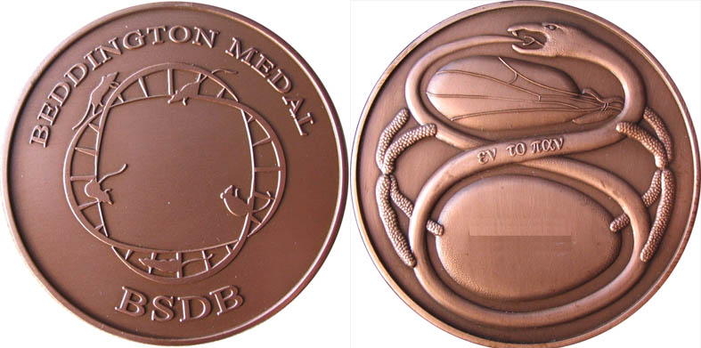
The Beddington Medal is the British Society for Developmental Biology’s major commendation to promising young biologists, awarded for the best PhD thesis in Developmental Biology defended in the year previous to the award.
The design of the medal, mice on a stylised DNA helix, is from artwork by Rosa herself.
In 2020 the Beddington Medal was awarded to Wajid Jawaid, who did his PhD with Berthold Göttgens and Jenny Nichols at the Wellcome-MRC Stem Cell Institute, University of Cambridge. After the cancellation of the spring meeting in 2020, Wajid is going to present his work today at the BSDB/Genetics Society 2021 meeting, right before the 2021 winner (who we’ll also interview soon). In advance, we caught up with Wajid to find out more about his life in science. Be sure to also check out the profile of Wajid – including words from Jenny Nichols and a list of Wajid’s selected publications – over on the BSDB site.
Where were you born and where did you grow up?
Birmingham is my home town and where I was born and schooled. My family are still there and I love visiting regularly.
When did you first get interested in science?
I’ve always been interested in how things work since as far back as I can remember and have had a keen interest in science since school. My eyes were really opened during my first year at University in Aberdeen when I found the treasure trove ‘Pubmed’ during a special study module on road traffic accidents. I couldn’t believe the amount of information that was freely available to anyone with an internet connection.
You came into your PhD from paediatric surgery – why did you decide to move into research, and why with Berthold Göttgens and Jenny Nichols in particular?
I became interested in paediatric surgery after my elective at British Columbia Children’s Hospital in Vancouver which exposed me to rare congenital anomalies and their surgical correction or at least amelioration. The process of reading and learning about the embryology that forms the basis of normal development and how it can go wrong consolidated my interest and I decided I wanted to become a Paediatric surgeon.
My interest in science and embryology drove me to apply for an Academic Clinical Fellowship (ACF) post in Paediatric Surgery based at Alder Hey hospital. There I met two wonderful mentors Professor Paul Losty and Mr Edwin Jesudason. As part of my ACF I visited Dr Emma Rawlin’s lab at the Gurdon Institute. This was my first exposure to lab based research and I soon became aware of the importance of integrating multiple and large sources of data. At the time I also became aware of Professor Jenny Nichol’s work in early embryogenesis, embryonic stem cells and pluripotency.
The Wellcome Trust kindly agreed to allow me to extend my PhD by taking 1 year to do a MPhil in Computational Biology at the Department of Applied Mathematics and Theoretical Physics co-ordinated by Dr Stephen Eglen and Dr Boris Adryan. This was a critical year that gave me the skills that I would use through-out my PhD. During the MPhil I attended a talk given by Professor Bertie Gottgens where he presented his most recent dataset and the concept of computational reconstruction of a pseudo-time developmental trajectory from single cell resolved qPCR data. I approached him and together we developed a project with Professor Jenny Nichols.
Tell us about your PhD project: what were the main questions you were trying to answer?
The development of new technologies allowing application of ‘-omics’ methods to single- cells was paving the way to understanding cell biology at much higher resolutions than previously possible. One application was in elucidating the journey of a progenitor cell as it became sequentially fate restricted until it finally reached its destination cell type. Despite the advances in technology it had not been possible to follow this journey across all genes/transcripts over time in-vivo. Single cell RNA sequencing was in its infancy but had the potential to achieve computational reconstructions of these journeys if single cells could be harvested from mouse embryos and retain their transcriptome. At the time it was not clear whether single cell transcriptomic data could be gathered at sufficient precision from dissociated embryos to allow cell type identification and lineage reconstruction.
The overarching theme of this body of work was to develop methods to reconstruct ordered ontogenic trajectories through sequentially sampled cross-sectional data at gastrulation in mice. The main focus was using single-cell resolved, transcriptomic data collected during early mouse embryonic development. Where available this was supplemented with limited, hand selected cell surface proteomic measurements.
The main aims were: 1. Identify cell populations; 2. Trace biologically plausible trajectories; 3. Identify novel molecular pathways; and 4. Develop models that can faithfully simulate cell progression along trajectories
This method of lineage reconstruction best fitted with retrospective lineage tracing. Lineages were traced based on the assumption that within a window of developmental time, cells with the most similar transcriptional signatures were related by lineage.
And what do you think your key discoveries were?
Our first experiment was to take early mouse embryos at gastrulation at four time points between E6.5 and E7.75. In the 3 later time-points cells were sorted to select for Flk1+ or CD41+ mesodermal cells using and An early key finding was that single cell resolved embryonic cell type could be accurately determined. Having identified cell types we were able to the identify sub-populations within the endothelial cluster and focus our analysis on a set of genes associated with erythromyeloid progenitors. In this way we were able to identify the activation of the leukotriene branch of the arachidonic acid pathway within this subset of endothelium. This reminded me of biochemistry from my pre-clinical years and its association with asthma so I was easily able to recall an inhibitor Zileuton. We went on to validate its role in haematopoiesis in an in vitro model of haematopoiesis. Another interesting observation Zileuton itself a derivative of hydroxyurea induces foetal haemaglobin and may have a role in the treatment of sickle cella anaemia. This finding then potentially suggests the mechanism of action of Zileuton.
Given advances in stem cells and organoids, what do mice still have to tell us about early mammalian development?
There have been great achievements in stem cell biology and organoids but they are still a long way off the gold standard the developing embryo which can at relatively high efficiency develop from a single cell to a recognisable organism with cells arranged in functional organ units. This complex process can not yet be faithfully recapitulated in any stem cell or organoid system.
By studying this process in nature we may be able to adjust our culture systems to improve the fidelity of both stem cell and organoid models of normal physiology and disease.
If you took one abiding memory with you from your PhD, what would it be?
Progress through a PhD is full of ups and downs. The moment that I am most fond of is when I was developing a neural network to model a bifurcating developmental trajectory to endothelial and blood fates. This had worked very well compared to a linear regression model. To test it, I wanted to perform a gene knock-out in Tal 1 which should have resulted in failure in erythroid generation. Unfortunately this was not working and I was ready to change tact when I realised I was dealing with qPCR data where activation was associated with a low cycle count – in my case it meant for knockout I should have been using a value of 14 rather than zero. When I corrected this error the network finally reproduced the experimental findings.
What are you doing post-PhD?
At the moment I have focused on completing clinical training. In the meantime I am also preparing to apply for a post-doc clinical fellowship to combine clinical and research work. My aim in the long term is to combine both research and clinical work. Clinical work often raises questions and challenges providing important research questions.
Where do you think developmental and stem cell biology will be in ten years?
I hope we will capture more the complex interactions and higher order abstractions of these interactions beyond pathways, linking the genome and its structure to function and anatomy. In 10 years, I hope that we are at the stage where our understanding of the information in the genome and the functional modules is sufficient to not only describe how organs develop but also how we can make changes to the programs of development to prevent disease phenotypes. So that in time, rather than disrupting development we can generate our own programs de novo.
Integration of multi-omics data with spatial context may help us move from the concept of differentiating a stem cell to more committed fates to generating complex structures of multiple cell types that can be used as substitutes for organs without artificial or de-cellularised scaffolds. Some organoid systems are already taking an early step in this direction.
When you’re not in the lab, what do you do for fun?
Over the last few years through my children going to a football club, I have become a volunteer coach at this local football club. More recently I’ve got my self a motorbike which I love to ride and fix up.
In the latest episode of Genetics Unzipped we’re finding out how researchers are unlocking the information hidden within the human genome using new technologies like CRISPR gene editing and artificial intelligence with the aim of developing better medicines and getting them faster to the patients who need them.
If you enjoy the show, please do rate and review on Apple podcasts and help to spread the word on social media. And you can always send feedback and suggestions for future episodes and guests to podcast@geneticsunzipped.com Follow us on Twitter – @geneticsunzip

 (No Ratings Yet)

.jpg)