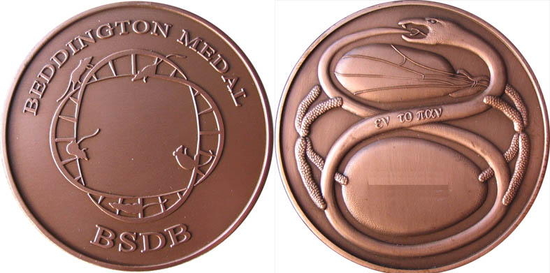
The Beddington Medal is the British Society for Developmental Biology’s major commendation to promising young biologists, awarded for the best PhD thesis in Developmental Biology defended in the year previous to the award.
The design of the medal, mice on a stylised DNA helix, is from artwork by Rosa herself.
In 2020 the Beddington Medal was awarded to Wajid Jawaid, who did his PhD with Berthold Göttgens and Jenny Nichols at the Wellcome-MRC Stem Cell Institute, University of Cambridge. After the cancellation of the spring meeting in 2020, Wajid is going to present his work today at the BSDB/Genetics Society 2021 meeting, right before the 2021 winner (who we’ll also interview soon). In advance, we caught up with Wajid to find out more about his life in science. Be sure to also check out the profile of Wajid – including words from Jenny Nichols and a list of Wajid’s selected publications – over on the BSDB site.
Where were you born and where did you grow up?
Birmingham is my home town and where I was born and schooled. My family are still there and I love visiting regularly.
When did you first get interested in science?
I’ve always been interested in how things work since as far back as I can remember and have had a keen interest in science since school. My eyes were really opened during my first year at University in Aberdeen when I found the treasure trove ‘Pubmed’ during a special study module on road traffic accidents. I couldn’t believe the amount of information that was freely available to anyone with an internet connection.
You came into your PhD from paediatric surgery – why did you decide to move into research, and why with Berthold Göttgens and Jenny Nichols in particular?
I became interested in paediatric surgery after my elective at British Columbia Children’s Hospital in Vancouver which exposed me to rare congenital anomalies and their surgical correction or at least amelioration. The process of reading and learning about the embryology that forms the basis of normal development and how it can go wrong consolidated my interest and I decided I wanted to become a Paediatric surgeon.
My interest in science and embryology drove me to apply for an Academic Clinical Fellowship (ACF) post in Paediatric Surgery based at Alder Hey hospital. There I met two wonderful mentors Professor Paul Losty and Mr Edwin Jesudason. As part of my ACF I visited Dr Emma Rawlin’s lab at the Gurdon Institute. This was my first exposure to lab based research and I soon became aware of the importance of integrating multiple and large sources of data. At the time I also became aware of Professor Jenny Nichol’s work in early embryogenesis, embryonic stem cells and pluripotency.
The Wellcome Trust kindly agreed to allow me to extend my PhD by taking 1 year to do a MPhil in Computational Biology at the Department of Applied Mathematics and Theoretical Physics co-ordinated by Dr Stephen Eglen and Dr Boris Adryan. This was a critical year that gave me the skills that I would use through-out my PhD. During the MPhil I attended a talk given by Professor Bertie Gottgens where he presented his most recent dataset and the concept of computational reconstruction of a pseudo-time developmental trajectory from single cell resolved qPCR data. I approached him and together we developed a project with Professor Jenny Nichols.
Tell us about your PhD project: what were the main questions you were trying to answer?
The development of new technologies allowing application of ‘-omics’ methods to single- cells was paving the way to understanding cell biology at much higher resolutions than previously possible. One application was in elucidating the journey of a progenitor cell as it became sequentially fate restricted until it finally reached its destination cell type. Despite the advances in technology it had not been possible to follow this journey across all genes/transcripts over time in-vivo. Single cell RNA sequencing was in its infancy but had the potential to achieve computational reconstructions of these journeys if single cells could be harvested from mouse embryos and retain their transcriptome. At the time it was not clear whether single cell transcriptomic data could be gathered at sufficient precision from dissociated embryos to allow cell type identification and lineage reconstruction.
The overarching theme of this body of work was to develop methods to reconstruct ordered ontogenic trajectories through sequentially sampled cross-sectional data at gastrulation in mice. The main focus was using single-cell resolved, transcriptomic data collected during early mouse embryonic development. Where available this was supplemented with limited, hand selected cell surface proteomic measurements.
The main aims were: 1. Identify cell populations; 2. Trace biologically plausible trajectories; 3. Identify novel molecular pathways; and 4. Develop models that can faithfully simulate cell progression along trajectories
This method of lineage reconstruction best fitted with retrospective lineage tracing. Lineages were traced based on the assumption that within a window of developmental time, cells with the most similar transcriptional signatures were related by lineage.
And what do you think your key discoveries were?
Our first experiment was to take early mouse embryos at gastrulation at four time points between E6.5 and E7.75. In the 3 later time-points cells were sorted to select for Flk1+ or CD41+ mesodermal cells using and An early key finding was that single cell resolved embryonic cell type could be accurately determined. Having identified cell types we were able to the identify sub-populations within the endothelial cluster and focus our analysis on a set of genes associated with erythromyeloid progenitors. In this way we were able to identify the activation of the leukotriene branch of the arachidonic acid pathway within this subset of endothelium. This reminded me of biochemistry from my pre-clinical years and its association with asthma so I was easily able to recall an inhibitor Zileuton. We went on to validate its role in haematopoiesis in an in vitro model of haematopoiesis. Another interesting observation Zileuton itself a derivative of hydroxyurea induces foetal haemaglobin and may have a role in the treatment of sickle cella anaemia. This finding then potentially suggests the mechanism of action of Zileuton.
Given advances in stem cells and organoids, what do mice still have to tell us about early mammalian development?
There have been great achievements in stem cell biology and organoids but they are still a long way off the gold standard the developing embryo which can at relatively high efficiency develop from a single cell to a recognisable organism with cells arranged in functional organ units. This complex process can not yet be faithfully recapitulated in any stem cell or organoid system.
By studying this process in nature we may be able to adjust our culture systems to improve the fidelity of both stem cell and organoid models of normal physiology and disease.
If you took one abiding memory with you from your PhD, what would it be?
Progress through a PhD is full of ups and downs. The moment that I am most fond of is when I was developing a neural network to model a bifurcating developmental trajectory to endothelial and blood fates. This had worked very well compared to a linear regression model. To test it, I wanted to perform a gene knock-out in Tal 1 which should have resulted in failure in erythroid generation. Unfortunately this was not working and I was ready to change tact when I realised I was dealing with qPCR data where activation was associated with a low cycle count – in my case it meant for knockout I should have been using a value of 14 rather than zero. When I corrected this error the network finally reproduced the experimental findings.
What are you doing post-PhD?
At the moment I have focused on completing clinical training. In the meantime I am also preparing to apply for a post-doc clinical fellowship to combine clinical and research work. My aim in the long term is to combine both research and clinical work. Clinical work often raises questions and challenges providing important research questions.
Where do you think developmental and stem cell biology will be in ten years?
I hope we will capture more the complex interactions and higher order abstractions of these interactions beyond pathways, linking the genome and its structure to function and anatomy. In 10 years, I hope that we are at the stage where our understanding of the information in the genome and the functional modules is sufficient to not only describe how organs develop but also how we can make changes to the programs of development to prevent disease phenotypes. So that in time, rather than disrupting development we can generate our own programs de novo.
Integration of multi-omics data with spatial context may help us move from the concept of differentiating a stem cell to more committed fates to generating complex structures of multiple cell types that can be used as substitutes for organs without artificial or de-cellularised scaffolds. Some organoid systems are already taking an early step in this direction.
When you’re not in the lab, what do you do for fun?
Over the last few years through my children going to a football club, I have become a volunteer coach at this local football club. More recently I’ve got my self a motorbike which I love to ride and fix up.
In the latest episode of Genetics Unzipped we’re finding out how researchers are unlocking the information hidden within the human genome using new technologies like CRISPR gene editing and artificial intelligence with the aim of developing better medicines and getting them faster to the patients who need them.
If you enjoy the show, please do rate and review on Apple podcasts and help to spread the word on social media. And you can always send feedback and suggestions for future episodes and guests to podcast@geneticsunzipped.com Follow us on Twitter – @geneticsunzip
Recently we reported the unexpected ability of fish mutants to develop limb-like bones in their pectoral fins (Hawkins et al., 2021). However, the most critical element of the study—finding these mutants in the first place—receives relatively little attention in the paper. Here I describe our efforts to find these monsters lurking inside the unassuming zebrafish.
The nuts and bolts of fins and limbs
The transition from fins to limbs is a defining transformation in vertebrate history and has served as a pivotal study system for comparative anatomy, paleontology, biomechanics, and developmental biology (Clack, 2009). Insight from each of these fields has illuminated different facets of how a relatively simple ancestral fin evolved into the complex arms and legs of tetrapods. Modern fins and limbs look quite different from one another but revealed in the fossil record are intermediate forms that connect their disparate morphologies (Jessen, 1972; Shubin et al., 2006; Zhu and Yu, 2009). By comparing the gene programs active in fins and limbs, we can ask which patterning mechanisms are common to both appendages, which mechanisms are derived in each, and which could be responsible for the changes in form found in evolution.
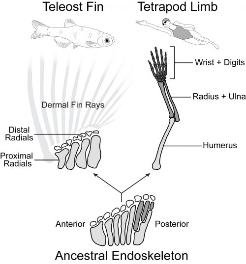
Tetrapod limbs have many long bones that articulate end on end away from the shoulder with distinct regions such as the upper arm (humerus), forearm (radius and ulna), and hand (Figure 1). In contrast, the pectoral fins of teleost fishes have just four long bones set side by side (proximal radials) followed distally by some small nodular bones (distal radials) and the dermal fin rays (Arratia, 1999). Non-teleost ray-finned fishes such as the bowfin have a slightly more impressive endoskeleton with additional articulations in the posterior part of the fin. Paleontological evidence suggests that the common ancestor of ray-finned fishes and tetrapods had a pectoral configuration much like that of the bowfin (Jessen, 1972, Zhu and Yu, 2009). In this scenario, teleosts such as the zebrafish represent a reduction of the ancestral appendage skeleton, while limbs exhibit its impressive elaboration (Coates, 1995).
Figure 1. Pectoral appendage anatomy is variable across the bony fishes. From the ancestral configuration that exhibited moderate elaboration in the posterior fin (bottom), teleosts (left) simplified the endoskeleton such that it consists of a row of proximal radials followed by small nodular distal radials. In contrast, in the lineage leading to tetrapods (right), the fin was elaborated through the addition of distal long bones to form a limb with a three-part structure containing the upper arm (humerus), forearm (radius and ulna), and hand (wrist and digits). Anterior to left, distal to top in skeletal schematics.
Forming an impressive body of work spanning the last four decades, developmental geneticists across the globe have discovered and characterized the manifold genes and pathways that control the growth and patterning in the nascent limb, leading to a deep understanding of the signaling ligands, receptors, and transcription factors necessary to make a normal appendage (Capdevila and Izpisúa Belmonte, 2001). Surprisingly, despite the morphological differences, many of these key limb patterning pathways are also expressed in developing teleost fins and play analogous (or conserved) functional roles (Mercader, 2007). There are differences in the expression and signaling function of some of these important players, but on the whole fin buds and limb buds behave quite like one another, and there is not one clear genetic factor present in limbs and absent in fins that is sufficient to imbue ‘limb-ness.’
Fishing for fin mutants
While assessing the role of candidate limb genes in growing fins has yielded critical insights into fin development, my colleagues Katrin Henke, Matthew Harris, and I decided to investigate the genes that can modify the zebrafish fin pattern using forward genetics. In a forward genetics approach, mutations are made at random and the investigator screens through mutated animals to pick out individuals with an interesting phenotype (Patton and Zon, 2001). Once an interesting mutant is found, we then work backwards using genetic mapping to determine which gene was mutated to cause the phenotype. The beauty of forward screens is that they let the organism tell you which genes are important to the process you want to know about. Sometimes you find an allele of a known essential regulator that has been extensively characterized, other times you find a gene that hasn’t been studied at all.
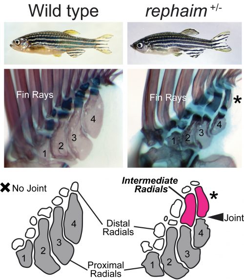
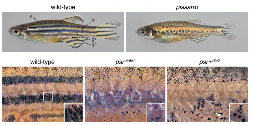
Katrin conducted several prodigious screens with a focus on mutants that affect the formation of the adult skeleton, and isolated hundreds of novel mutants (Henke et al., 2017). One mutant she picked out because of its modified pigmentation and dysmorphic fin rays was of particular interest to me and Matthew. Upon observing the internal skeleton under the dissecting scope, Matthew saw that the fin endoskeleton was affected and suggested I take a look. Shockingly, instead of having just the four long bones set side by side, this mutant had additional long bones forming in the distal endoskeleton that articulated with the proximal elements with a joint analogous to an elbow (Figure 2). This mutant didn’t just have fins, but what it had were not quite limbs either: it grew “flimbs.” At the time my dissertation project was focused on craniofacial mutants, but after this discovery (and Katrin’s blessing) my focus shifted to this fascinating mutant with limb-like fins. All good mutants need a name, and my friend and Old Testament scholar Maria suggested rephaim, a race of biblical giants fabled to have extra digits on their hands and feet.
Figure 2. Novel long bones form in the distal endoskeleton of rephaim mutants. Top panels show the external appearance of wild-type and rephaim mutant fish. Middle panels contain micrographs of pectoral fins stained red for bone and blue for cartilage. The proximal radials are numbered 1 through 4, and the new bones in the mutant are indicated with an asterisk. Bottom panel schematizes the endoskeletons of wild-type and rephaim fins and indicates the position of the intermediate radials and novel joint in the rephaim mutant.
After receiving a name, mutants need to be mapped to determine which gene is affected. Genetic mapping of mutations used to be a long and involved process using chromosomal markers to track linkage and recombination. In early zebrafish screens, a mutant line would be crossed to a wild-type fish from a different genetic background, and PCR-based methods would be used to find variable genomic positions and track down regions of DNA that segregated with the mutant phenotype (Knapik et al., 1996). In the last 15 years, however, the advent of next-generation sequencing made it possible to sequence mutants and their wild-type siblings to quickly identify genomic regions that associate with the mutant phenotype. To map rephaim, we utilized a whole-exome mapping approach to determine genomic regions that likely contain the causative mutation (Bowen et al., 2012). The mapping data gave us two putative regions that could contain the rephaim mutation, one on chromosome 4 and one on chromosome 9. Chromosome 4 has a reputation as being a nightmare for mapping, replete with inversions and transposons, and the implicated interval didn’t contain any interesting limb patterning genes. I did not like chromosome 4. On the other hand, the interval on chromosome 9 contained the HoxD cluster, a battery of genes with critical roles in appendage patterning and particularly implicated in the differential patterning of fins and limbs (Sordino et al., 1995; Freitas et al., 2012; Woltering et al., 2014). Not only would a mutation in a HoxD gene fit my expectations of what could cause a phenotype like rephaim, it would make subsequent analysis of the mutant phenotype much easier and fit well within the existing fin-to-limb literature. I even thought I might finish my dissertation early.
This, however, was not to be. Linkage analysis definitively ruled out chromosome 9 and the HoxD cluster. The initial mapping signal that I had pinned my hopes on was due to a block of genetic homogeneity that was shared between mutants and wild-type siblings, meaning that both mutants and wild-type animals had the same alleles in this region and thus could not contain the causative mutation. Meanwhile, additional recombination analysis strengthened the association of rephaim with chromosome 4 and narrowed the linkage interval to a small window containing just one coding mutation in a gene called wiskott-aldrich syndrome protein like-b (waslb). Unlike my precious HoxD cluster, the waslb gene was not a known regulator of limb development, and everything I saw in the literature gave the impression that it was a “housekeeping” gene: ubiquitous expression, essential functions in actin metabolism, and involvement in myriad cellular pathways (Snapper and Rosen, 1999). Around this time, we were also mapping a second mutant with a similar phenotype, a fish called wanda (van Eeden et al., 1996; Haffter et al., 1996). The causative mutation for wanda mapped to the gene vav2, a similarly unexciting locus from a skeletal patterning perspective (Hornstein et al., 2004). I felt the path to my PhD lengthening in real time.
X marks the spot on a genetic treasure map
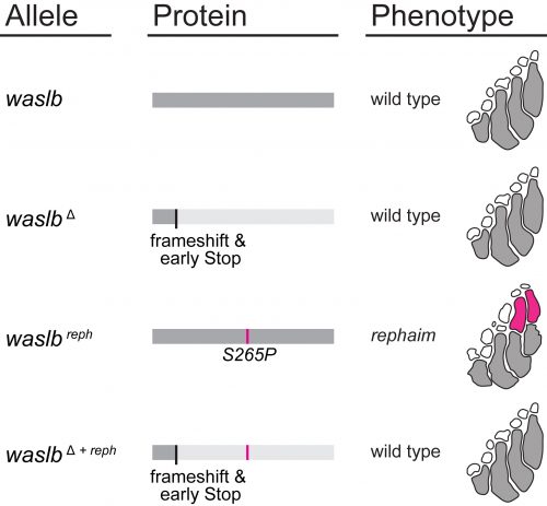
Nevertheless, all the mapping data pointed to waslb and vav2, so these genes demanded our attention. Mapping implicated these genes, but we still needed to experimentally confirm their role in the flimb phenotype. We used CRISPR-Cas9 to make loss-of-function alleles, but even homozygous null mutants had a wild-type phenotype (Figure 3). Next we tried injecting mutant mRNA into the embryo but saw no effect, likely due to the late appearance of the phenotype. In a final push to demonstrate the causative nature of the waslb and vav2 mutations, we used CRISPR to create frameshift lesions in cis to the candidate mutations and knockout the mutant alleles specifically. When the mutant copies of waslb and vav2 were removed, we rescued the phenotype and reverted the mutants to wild-type fin patterning. There was no doubt, mutations in waslb and vav2 cause the flimb phenotype. But this left us with a bigger question, how in the world are these genes changing skeletal patterning? I came up with an axiom to sooth myself: “if good science raises more questions than it answers, then the best science must raise only questions and answer nothing.”
Figure 3. Rescue experiments demonstrate that a mutation in waslb causes the rephaim phenotype. CRISPR-Cas9 was used to generate null alleles in wild-type and rephaim mutant waslb. Removing wild-type alleles (waslbΔ) had no effect on fin patterning. However, creating a frameshift and early stop upstream of the S265P mutation (waslbΔ+ reph) prevented the formation of intermediate radials and rescued wild-type patterning.
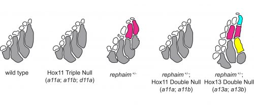
However, the wealth of limb patterning knowledge established by developmental geneticists was able to guide our inquiry. As mentioned earlier, Hox genes have critical functions in the patterning and growth of limb bones along the proximal-distal axis, and recent studies had revealed that Hox13 was required for the formation of the most distal structures in fins just like in limbs (Nakamura et al., 2016). We thought that the new bones in rephaim might also share this distal Hox13 regionality, and crossed rephaim into a Hox13-null genetic background. To our surprise, we found that loss of Hox13 actually enhanced the flimb phenotype and resulted in the formation of even more bones along the distal aspect of the endoskeleton (Figure 4). Hoxa13 is known to negatively regulate Hoxa11 expression in the limb (Kherdjemil et al., 2016), and we thought the enhanced phenotype might be the result of derepression of Hox11 genes. Around this time we also were analyzing limb-specific Wasl knockout mice, and observed limb defects similar to those seen in Hoxa11 mouse mutants. Intriguingly, Hox11 genes are also required for the normal development for the bones in the middle region of the limb, the radius and ulna (Davis et al., 1995).
Figure 4. Genetic interaction between rephaim and Hox genes suggests limb-like patterning mechanisms function in the fin. While loss of hoxa11a, hoxa11b, and hoxd11a has no effect on fin patterning in the wild-type background, removal of these genes prevents the formation of intermediate radials in rephaim mutants. In contrast, removing hoxa13a and hoxa13b from rephaim mutants enhances the phenotype and results in the formation of additional intermediate radials. The requirement of Hox11 genes is shared between intermediate radials and the limb forearm.
Following these clues, we generated null alleles of the hoxa11 and hoxd11 paralogs in the zebrafish. While loss of these genes had no effect on fin patterning in a wild-type background, we found that loss of hoxa11a and hoxa11b prevented the formation of the extra bones in rephaim mutants (Figure 4). Moreover, we generated knock-in hoxa11b reporter zebrafish and found that rephaim and wanda mutants cause the upregulation of hoxa11b expression. These results were quite interesting: even though the Hox11 paralogs are not required for normal fin patterning in zebrafish, they still possess the ability to specify the formation of an intermediate long bone position along the proximal-distal axis of the appendage skeleton. This suggests that the capacity to specify ‘middle’ and ‘distal’ regions is not unique to limbs, but was present in the common ancestor of ray- and lobe-finned fishes. Although not expressed in teleosts, this developmental potential has been retained in a latent state and can be redeployed by simple perturbations.
Wasl, Hox, and Beyond
This forces me to wonder what other latent limb patterning mechanisms that might reside in the developing fin bud, and how waslb is able to activate at least some of them. The mechanistic connection between waslb, vav2, and Hox regulation is an open question. In part, we know that waslb mediates the formation of F-actin foci that colocalize with Hox-positive cells in the distal fin (Hawkins et al., 2021). Given its roles in cell motility it could be that waslb effects the migration of these cells, but there are many other possibilities. Wasl also directly regulates transcription (Wu et al., 2006), modulates Wnt (Lyubimova et al., 2010) and TGFB (Lefever et al., 2010) signaling, and is directly involved in the colinear activation of the HoxB cluster (Ferrai et al., 2009). Then again, there could be another pathway that we do not yet understand. I will go out on a flimb here and say the zebrafish still has more to tell us about these mechanisms…as long as we are willing to trust the mapping.
Arratia, G. (1999). The monophyly of Teleostei and stem-group teleosts. Mesozoic Fishes, 2, 265–334.
Bowen, M. E., Henke, K., Siegfried, K. R., Warman, M. L., & Harris, M. P. (2012). Efficient mapping and cloning of mutations in zebrafish by low-coverage whole-genome sequencing. Genetics, 190(3), 1017–1024.
Capdevila, J., & Izpisúa Belmonte, J. C. (2001). Patterning mechanisms controlling vertebrate limb development. Annual Review of Cell and Developmental Biology, 17, 87–132.
Clack, J. A. (2009). The Fin to Limb Transition: New Data, Interpretations, and Hypotheses from Paleontology and Developmental Biology. Annual Review of Earth and Planetary Sciences, 37(1), 163–179.
Coates, M. I. (1995). Limb evolution. Fish fins or tetrapod limbs–a simple twist of fate? Current Biology: CB, 5(8), 844–848.
Davis, A. P., Witte, D. P., Hsieh-Li, H. M., Potter, S. S., & Capecchi, M. R. (1995). Absence of radius and ulna in mice lacking hoxa-11 and hoxd-11. Nature, 375(6534), 791–795.
Ferrai, C., Naum-Onganía, G., Longobardi, E., Palazzolo, M., Disanza, A., Diaz, V. M., Crippa, M. P., Scita, G., & Blasi, F. (2009). Induction of HoxB transcription by retinoic acid requires actin polymerization. Molecular Biology of the Cell, 20(15), 3543–3551.
Freitas, R., Gómez-Marín, C., Wilson, J. M., Casares, F., & Gómez-Skarmeta, J. L. (2012). Hoxd13 contribution to the evolution of vertebrate appendages. Developmental Cell, 23(6), 1219–1229.
Haffter, P., Odenthal, J., Mullins, M. C., Lin, S., Farrell, M. J., Vogelsang, E., Haas, F., Brand, M., van Eeden, F. J., Furutani-Seiki, M., Granato, M., Hammerschmidt, M., Heisenberg, C. P., Jiang, Y. J., Kane, D. A., Kelsh, R. N., Hopkins, N., & Nüsslein-Volhard, C. (1996). Mutations affecting pigmentation and shape of the adult zebrafish. Development Genes and Evolution, 206(4), 260–276.
Hawkins, M. B., Henke, K., & Harris, M. P. (2021). Latent developmental potential to form limb-like skeletal structures in zebrafish. Cell. https://doi.org/10.1016/j.cell.2021.01.003
Henke, K., Daane, J. M., Hawkins, M. B., Dooley, C. M., Busch-Nentwich, E. M., Stemple, D. L., & Harris, M. P. (2017). Genetic Screen for Postembryonic Development in the Zebrafish (Danio rerio): Dominant Mutations Affecting Adult Form. Genetics, 207(2), 609–623.
Hornstein, I., Alcover, A., & Katzav, S. (2004). Vav proteins, masters of the world of cytoskeleton organization. Cellular Signalling, 16(1), 1–11.
Jessen, H. L. (1972). Fossils and Strata, Schultergurtel und Pectoralflosse bei Actinopterygiern. Lethaia, 5(3), 344.
Kherdjemil, Y., Lalonde, R. L., Sheth, R., Dumouchel, A., de Martino, G., Pineault, K. M., Wellik, D. M., Stadler, H. S., Akimenko, M.-A., & Kmita, M. (2016). Evolution of Hoxa11 regulation in vertebrates is linked to the pentadactyl state. Nature, 539(7627), 89–92.
Knapik, E. W., Goodman, A., Atkinson, O. S., Roberts, C. T., Shiozawa, M., Sim, C. U., Weksler-Zangen, S., Trolliet, M. R., Futrell, C., Innes, B. A., Koike, G., McLaughlin, M. G., Pierre, L., Simon, J. S., Vilallonga, E., Roy, M., Chiang, P. W., Fishman, M. C., Driever, W., & Jacob, H. J. (1996). A reference cross DNA panel for zebrafish (Danio rerio) anchored with simple sequence length polymorphisms. Development , 123, 451–460.
Lefever, T., Pedersen, E., Basse, A., Paus, R., Quondamatteo, F., Stanley, A. C., Langbein, L., Wu, X., Wehland, J., Lommel, S., & Brakebusch, C. (2010). N-WASP is a novel regulator of hair-follicle cycling that controls antiproliferative TGF{beta} pathways. Journal of Cell Science, 123(Pt 1), 128–140.
Lyubimova, A., Garber, J. J., Upadhyay, G., Sharov, A., Anastasoaie, F., Yajnik, V., Cotsarelis, G., Dotto, G. P., Botchkarev, V., & Snapper, S. B. (2010). Neural Wiskott-Aldrich syndrome protein modulates Wnt signaling and is required for hair follicle cycling in mice. The Journal of Clinical Investigation, 120(2), 446–456.
Mercader, N. (2007). Early steps of paired fin development in zebrafish compared with tetrapod limb development. Development, Growth & Differentiation, 49(6), 421–437.
Nakamura, T., Gehrke, A. R., Lemberg, J., Szymaszek, J., & Shubin, N. H. (2016). Digits and fin rays share common developmental histories. Nature, 537(7619), 225–228.
Patton, E. E., & Zon, L. I. (2001). The art and design of genetic screens: zebrafish. Nature Reviews. Genetics, 2(12), 956–966.
Shubin, N. H., Daeschler, E. B., & Jenkins, F. A., Jr. (2006). The pectoral fin of Tiktaalik roseae and the origin of the tetrapod limb. Nature, 440(7085), 764–771.
Snapper, S. B., & Rosen, F. S. (1999). The Wiskott-Aldrich syndrome protein (WASP): roles in signaling and cytoskeletal organization. Annual Review of Immunology, 17, 905–929.
Sordino, P., van der Hoeven, F., & Duboule, D. (1995). Hox gene expression in teleost fins and the origin of vertebrate digits. Nature, 375(6533), 678–681.
van Eeden, F. J., Granato, M., Schach, U., Brand, M., Furutani-Seiki, M., Haffter, P., Hammerschmidt, M., Heisenberg, C. P., Jiang, Y. J., Kane, D. A., Kelsh, R. N., Mullins, M. C., Odenthal, J., Warga, R. M., & Nüsslein-Volhard, C. (1996). Genetic analysis of fin formation in the zebrafish, Danio rerio. Development , 123, 255–262.
Woltering, J. M., Noordermeer, D., Leleu, M., & Duboule, D. (2014). Conservation and divergence of regulatory strategies at Hox Loci and the origin of tetrapod digits. PLoS Biology, 12(1), e1001773.
Wu, X., Yoo, Y., Okuhama, N. N., Tucker, P. W., Liu, G., & Guan, J.-L. (2006). Regulation of RNA-polymerase-II-dependent transcription by N-WASP and its nuclear-binding partners. Nature Cell Biology, 8(7), 756–763.
Zhu, M., & Yu, X. (2009). Stem sarcopterygians have primitive polybasal fin articulation. Biology Letters, 5(3), 372–375.
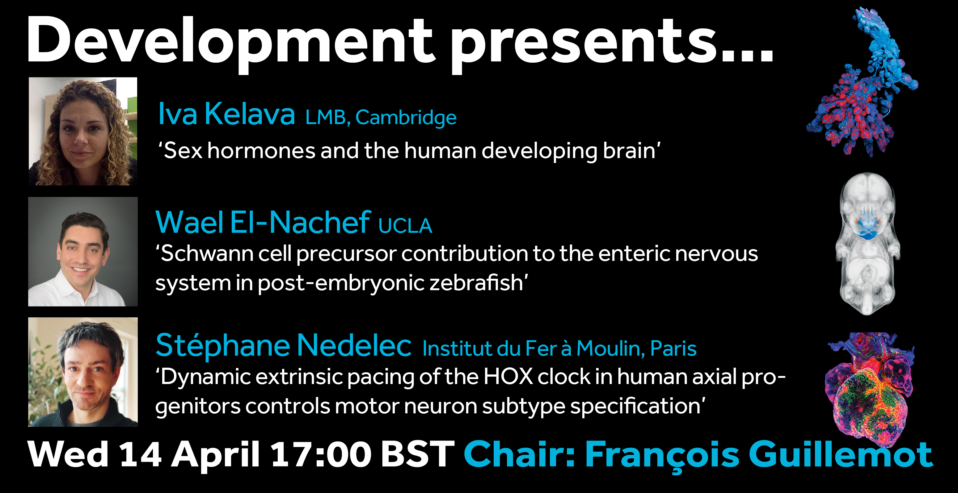
On Wednesday 14 April Development welcomed three researchers with interests in developmental neurobiology to our seventh Development presents… webinar.
Below you’ll find each of the talks, plus a Q&A chaired by Development Editor François Guillemot. The next #DevPres webinar will be held on 12 May 2021, and chaired by Paola Arlotta – subscribe to our mailing list for updates.
Iva Kelava (LMB Cambridge) – ‘Sex hormones and the human developing brain’
Wael El-Nachef (UCLA) – ‘Schwann cell precursor contribution to the enteric nervous system in post-embryonic zebrafish’
Wael’s paper was published last year in Development (you can also find a link to an interview we did with Wael and Marianne Bronner at the bottom of the abstract)
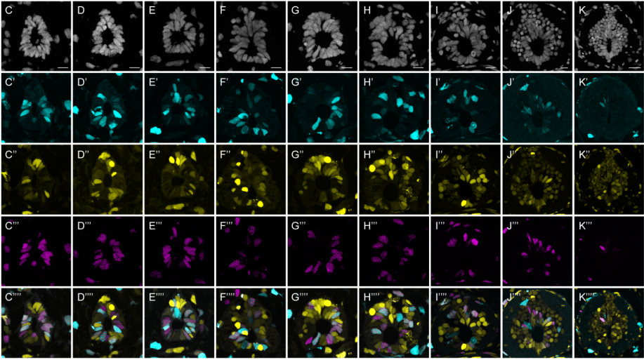
Stéphane Nedelec (Institut du Fer à Moulin) – ‘Dynamic extrinsic pacing of the HOX clock in human axial progenitors control motor neuron subtype specification’
Stéphane’s paper – featuring co-first authors Vincent Mouilleau and Célia Vaslin – was recently published in Development (you can also find a link to an interview we did with Vincent, Célia and Stéphane at the bottom of the abstract).
Our seventh profile in the series features Dorotea Fracchiolla, who works as a Project Manager in Frankfurt and is also a scientific illustrator.
Where are you originally from and what do you work on now?
I’m originally from Ruvo di Puglia, a town in the province of Bari, Puglia, South Italy. I worked as a Postdoctoral fellow in the field of in vitro reconstitution of Autophagy at the University of Vienna in Austria. In January 2021 I started working at the Max Planck Institute for Biophysics in Frankfurt (Germany) as a Project Manager of an International research collaboration that aims at studying Parkinson’s Disease pathogenesis. In April last year I started my project as a scientific illustrator and founded Art&Science, which gave me the chance to work with many scientists worldwide on art projects in different fields.
Has science always been an important part of your life?
I’ve always been a curious person and with some difficulty in finding THE subject to study. At school I liked everything and was always excited to start reading about new things. In this sense I think I’ve always been a scientist in everyday life. Later on, the actual scientific training provided me with material to feed my curiosity and exercise critical thinking.
And what about art?
Drawing, painting, crafting and trying out new materials were my preferred activities as a child/teenager. Soon enough I started photography, too. All these activities had an aspect in common: sensing. In other words, exploring the world around me, experiencing the new, interacting.
What or who are your artistic influences?
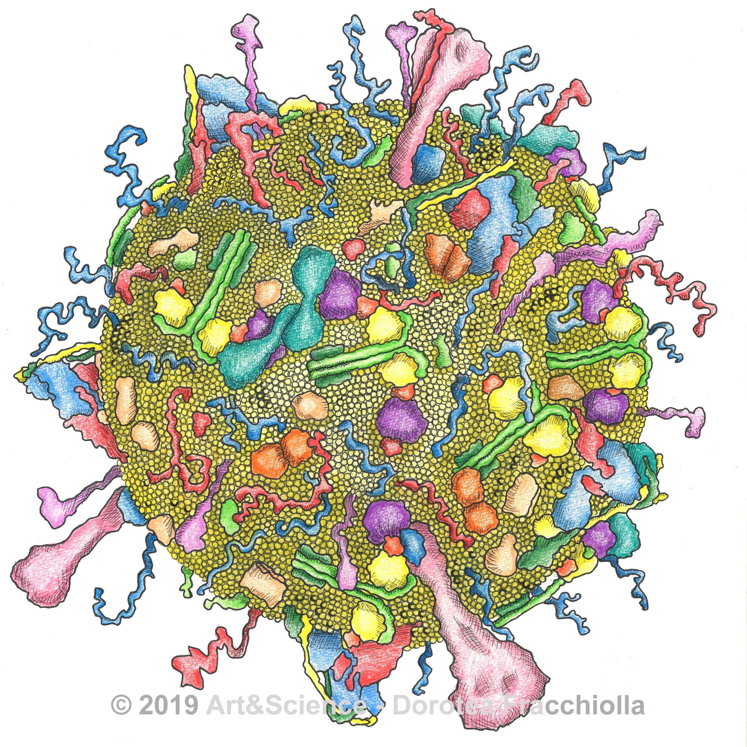
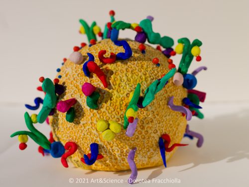
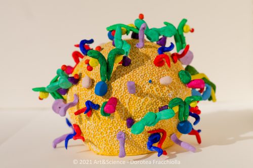
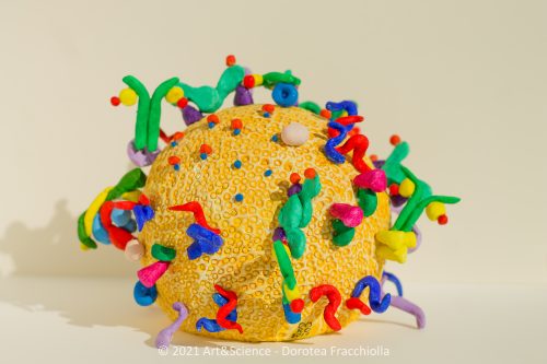
During my studies I have always been attracted by the personality and life of Leonardo da Vinci, an artist and a scientist in one person. As for him, my place of inspiration is Nature. In the beginning, I used to draw things at a macroscopic scale that I had observed while exploring, then I ended up depicting the molecular mechanisms of nano machines like those scientists study in labs. When it comes to style, I think I cannot underplay the influence on me of my place of origin, in its light and bright colors. One example: here in Puglia there is a tradition to make colored clay whistles as local art pieces. Among others, I have recently built a clay model of a set of proteins that participate in the formation of autophagosomes in cells. I think it grossly resembles in style these artcrafts.
How do you make your art?
For me, making art comes after creating an idea in my mind. The creation cannot precede the study and understanding of the subject. I start from reading and while doing so I put together images in my head that finally make up the puzzle. If something is not clear I keep reading until the full picture is completed. My motto is: if you can draw it, you understood it in the first place.
Does your art influence your science at all, or are they separate worlds?
In my opinion, art and science are tightly connected and rely on each other. This is why I named my activity as an illustrator ‘Art&Science’. Very often, while studying a new topic, I tend to first visualize things and make schematics of concepts. Simplification is my approach to understanding. That’s when the art comes into play: it depicts the core message and makes it simple for the eye to grasp.
“Art depicts the core message and makes it simple for the eye to grasp”
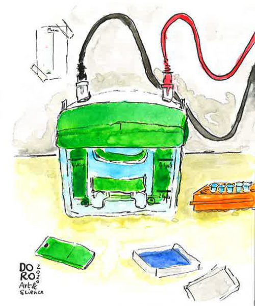
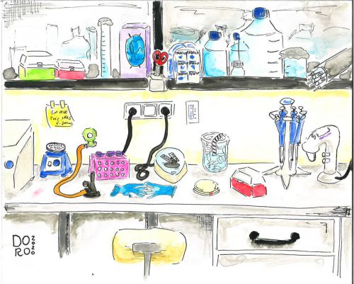
Tell us about the work you’ve shared with us.
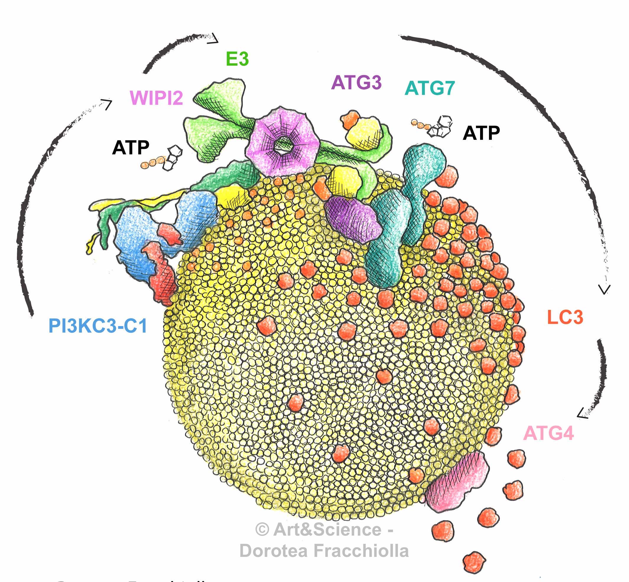
One project turns around the topic of autophagosome formation. I started drawing these pieces during my PhD when I learnt hardcore biochemistry in the Laboratory of Prof. Sascha Martens at the University of Vienna while studying autophagy in his lab. The entire team was focused on getting recombinant purified components of the autophagy machinery and studying their properties in vitro. The goal was to understand how the different proteins work together to build autophagosomes. Scientific research needs to be coupled to a certain degree of imagination in order for questions to arise. I always liked to visualize things and my preferred way to convey a message is drawing.
Supported by structural biology data, I started off sketching how I thought the molecular machinery looks like, and that’s how the “Autophagosome Biogenesis” project arose.
Clay model, top view
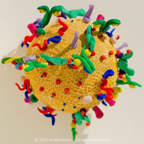
Clay model, top view, close up
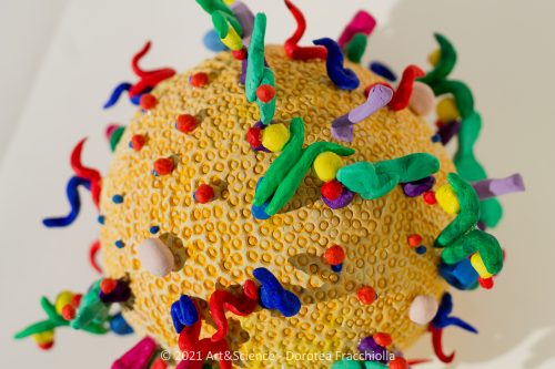
Clay model, side view
Clay model, side view 2
Clay model, front view
I first made the drawing and then I created a 3D clay model as shown in the gallery above.
This model was then used as a basis for a stop motion animation (for more information about this movie see my website).
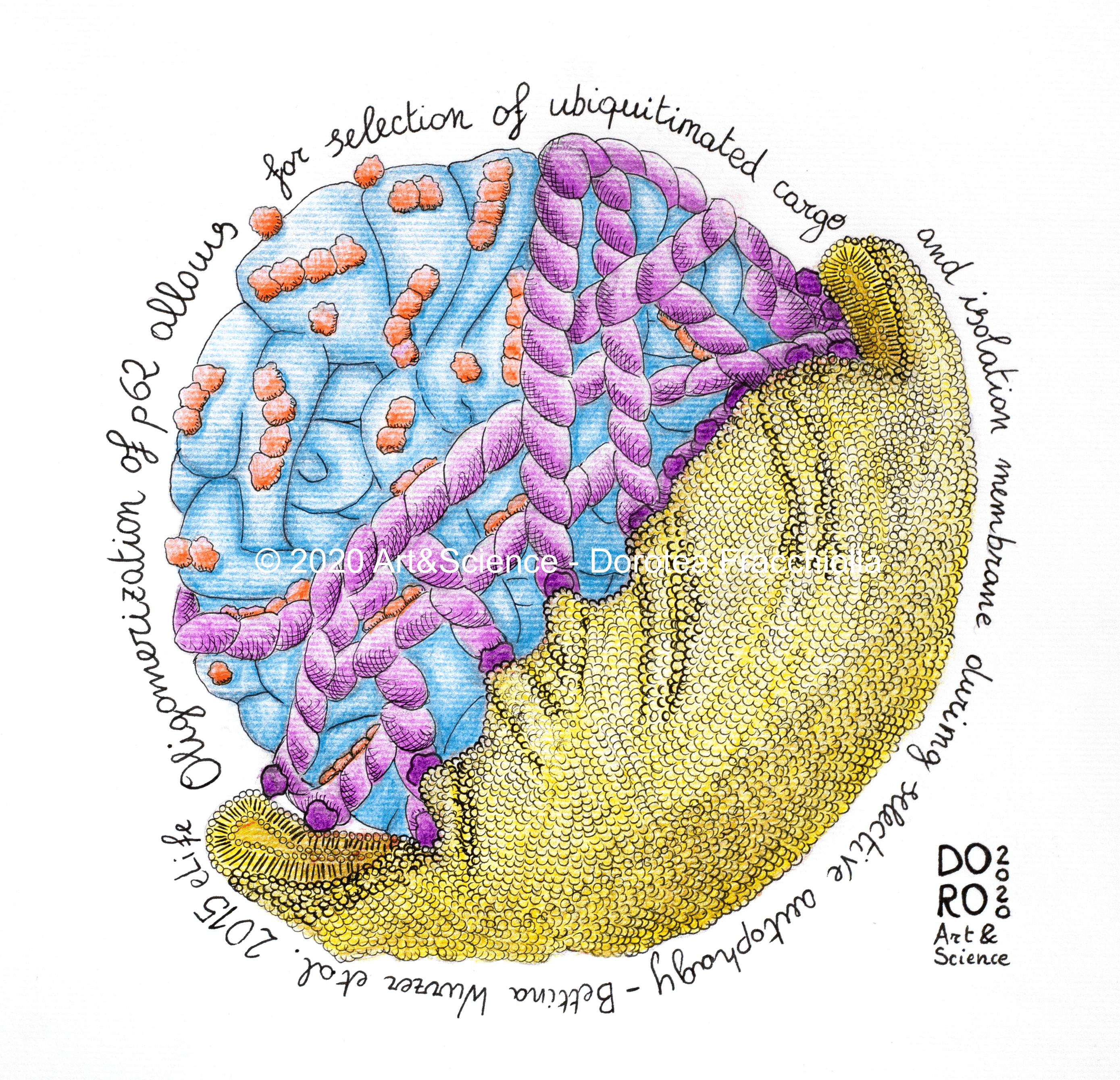
Next is my Ub-p62-cargo illustration: like a sticky sugar muffin that gets stuck in its paper wrap, ubiquitin-tagged misfolded protein aggregates are tightly attached to LC3-positive isolation membranes via p62 during selective autophagy in human cells. I created this for the following publication: https://elifesciences.org/articles/08941
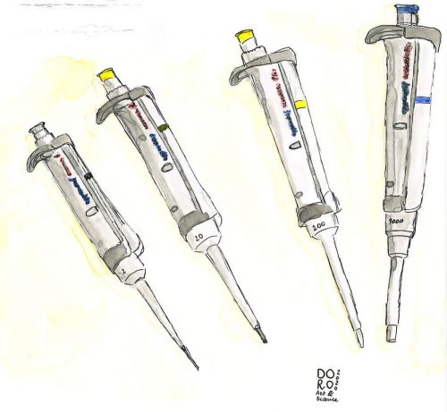
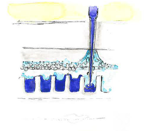
Finally, this gallery shows a lab life series done with acquerells. I created this one during lockdown as well because I was a bit nostalgic for the lab.
What are you thinking of working on next?
Like everyone, I have a dream in the drawer: I would love to illustrate a book, a short collection of recent scientific discoveries made into figures for students. The idea comes not only from the fun I’d have in engaging with such a project but also from my belief that if you make something interesting for students, they will enjoy studying it. At school, drawing things was my way to make things simple for myself – when things are simple it is easier to understand them, and in turn appreciate them.
We’re looking for new people to feature in this series throughout the year – whatever kind of art you do, from sculpture to embroidery to music to drawing, if you want to share it with the community just email thenode@biologists.com (nominations are also welcome!).
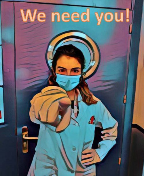
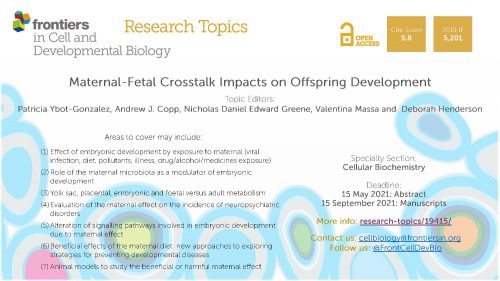
We are looking for researchers to participate in the special issue “Maternal-Fetal Crosstalk Impacts on Offspring Development” in Frontiers in Cell and Developmental Biology journal.
In this episode we’re taking a look at the history of gene editing, from the early days of restriction enzymes in the 1960s through to the CRISPR revolution and the very latest base editing techniques.
But while these tools are undeniably powerful and hold great promise for treating disease, with great power comes great responsibility: what are the acceptable limits of genome engineering in humans, and will we see more CRISPRd babies in the future?
If you enjoy the show, please do rate and review on Apple podcasts and help to spread the word on social media. And you can always send feedback and suggestions for future episodes and guests to podcast@geneticsunzipped.com Follow us on Twitter – @geneticsunzip
The proteome of remyelination is different from that of developmental myelination Joana Paes de Faria, Maria M. Azevedo, Damaris Bausch-Fluck, Ana Seixas, Helena S. Domingues, Maria A. Monteiro, Patrick G.A. Pedrioli, Eduarda Lopes, Rui Fernandes, Chao Zhao, Robin J. M. Franklin, Bernd Wollscheid, Joao B. Relvas, Laura Montani
Epiblast morphogenesis is controlled by selective mRNA decay triggered by LIN28A relocation Miha Modic, Igor Ruiz de Los Mozos, Sebastian Steinhauser, Emiel van Genderen, Silvia Schirge, Valter Bergant, Joel Ryan, Christopher B Mulholland, Rupert Faraway, Flora C Y Lee, Tajda Klobučar, Juliane Merl-Pham, Stephanie M Hauck, Micha Drukker, Sebastian Bultmann, Heinrich Leonhardt, Heiko Lickert, Nicholas M Luscombe, Derk ten Berge, Jernej Ule
A pro-endocrine pancreatic transcriptional program established during development is retained in human gallbladder epithelial cells Mugdha V. Joglekar, Subhshri Sahu, Wilson KM Wong, Sarang N. Satoor, Charlotte X. Dong, Ryan J Farr, Michael D. Williams, Prapti Pandya, Gaurang Jhala, Sundy N.Y. Yang, Yi Vee Chew, Nicola Hetherington, Dhan Thiruchevlam, Sasikala Mitnala, Guduru V Rao, Duvvuru Nageshwar Reddy, Thomas Loudovaris, Wayne J. Hawthorne, Andrew G. Elefanty, Vinay M. Joglekar, Edouard G. Stanley, David Martin, Helen E. Thomas, David Tosh, Louise T. Dalgaard, Anandwardhan A. Hardikar
DNA methylation clocks show slower progression of aging in naked mole-rat queens Steve Horvath, Amin Haghani, Nicholas Macoretta, Julia Ablaeva, Joseph A. Zoller, Caesar Z. Li, Joshua Zhang, Masaki Takasugi, Yang Zhao, Elena Rydkina, Zhihui Zhang, Stephan Emmrich, Ken Raj, Andrei Seluanov, Chris G. Faulkes, Vera Gorbunova
Stiffness Regulates Intestinal Stem Cell Fate Shijie He, Peng Lei, Wenying Kang, Priscilla Cheung, Tao Xu, Miyeko Mana, Chan Young Park, Hongyan Wang, Shinya Imada, Jacquelyn O. Russell, Jianxun Wang, Ruizhi Wang, Ziheng Zhou, Kashish Chetal, Eric Stas, Vidisha Mohad, Marianna Halasi, Peter Bruun-Rasmussen, Ruslan I. Sadreyev, Irit Adini, Richard A. Hodin, Yanhang Zhang, David T. Breault, Fernando D. Camargo, Ömer H. Yilmaz, Jeffrey J. Fredberg, Nima Saeidi
Mapping origins of variation in neural trajectories of human pluripotent stem cells Suel-Kee Kim, Seungmae Seo, Genevieve Stein-O’Brien, Amritha Jaishankar, Kazuya Ogawa, Nicola Micali, Yanhong Wang, Thomas M. Hyde, Joel E. Kleinman, Ty Voss, Elana J. Fertig, Joo-Heon Shin, Roland Bürli, Alan J. Cross, Nicholas J. Brandon, Daniel R. Weinberger, Joshua G. Chenoweth, Daniel J. Hoeppner, Nenad Sestan, Carlo Colantuoni, Ronald D. McKay
Cortex cis-regulatory switches establish scale colour identity and pattern diversity in Heliconius Luca Livraghi, Joseph J. Hanly, Steven M. Van Belleghem, Gabriela Montejo-Kovacevich, Eva S. M. van der Heijden, Ling Sheng Loh, Anna Ren, Ian A. Warren, James J. Lewis, Carolina Concha, Laura H. López, Charlotte Wright, Jonah M. Walker, Jessica Foley, Zachary H. Goldberg, Henry Arenas-Castro, Michael W. Perry, Riccardo Papa, Arnaud Martin, W. Owen McMillan, Chris D. Jiggins
OpenCell: proteome-scale endogenous tagging enables the cartography of human cellular organization Nathan H. Cho, Keith C. Cheveralls, Andreas-David Brunner, Kibeom Kim, André C. Michaelis, Preethi Raghavan, Hirofumi Kobayashi, Laura Savy, Jason Y. Li, Hera Canaj, James Y.S. Kim, Edna M. Stewart, Christian Gnann, Frank McCarthy, Joana P. Cabrera, Rachel M. Brunetti, Bryant B. Chhun, Greg Dingle, Marco Y. Hein, Bo Huang, Shalin B. Mehta, Jonathan S. Weissman, Rafael Gómez-Sjöberg, Daniel N. Itzhak, Loic A. Royer, Matthias Mann, Manuel D. Leonetti
The seventh webinar in our Development presents… series will be chaired by Development Editor, François Guillemot (The Francis Crick Institute), who has brought together three exciting talks on the development of the nervous system.
Wednesday 14 April 2021 – 17:00 BST (GMT+1)
Iva Kelava (Postdoc in Madeline Lancaster’s lab at the Laboratory of Molecular Biology) ‘Sex hormones and the human developing brain’
Stéphane Nedelec (from the Institut du Fer à moulin) ‘Dynamic extrinsic pacing of the HOX clock in human axial progenitors control motor neuron subtype specification’
The webinar will be held in Remo, our browser-based conferencing platform – after the talks you’ll have the chance to meet the speakers and other participants at virtual conference tables. If you can’t make it on the day, talks will be available to watch for a couple of weeks after the event; details will be posted on the Node or you can sign up to our mailing list for email alerts.
Feel free to share this poster with your colleagues:

 (5 votes)