Drs. Hillary Maddin and Tetsuto Miyashita are seeking motivated students and fellows to join our Carleton-based team in Ottawa, Canada. Successful candidates will participate in a newly funded project aimed at understanding various aspects of the development and evolution of the vertebrate skeleton. In particular, projects will focus on investigating skull and limb development in model and non-model species of amphibians. Collaborators for these projects include Chris Joslin (Carleton), James Hanken (Harvard), and Ryan Kerney (Gettysburg). MSc, PhD, and postdoctoral positions available.
Carleton University has research partnerships with Canadian Museum of Nature, which houses natural history collections of over 14 million specimens, and the University of Ottawa, which offers an extensive network of biomedical and life science labs and the state-of-the-art next-generation sequencing facility. Ottawa is a vibrant, multicultural, and affordable city embraced in the green belt. Browse some overview and Google Image search summary of life in Canada’s capital.
The veins are the vascular networks of plant leaves, functioning as channels for transport of signals and nutrients. A new paper in Development investigates how the spatial regulation of auxin transport contributes to vein patterning in Arabidopsis. We caught up with first author Priyanka Govindaraju and her supervisor Enrico Scarpella, Associate Professor at the Department of Biological Sciences, University of Alberta in Edmonton, Canada, to find out more about the work.
Priyanka (L) and Enrico (R)
Enrico, can you give us your scientific biography and the questions your lab is trying to answer?
ES: I graduated in Biological Sciences at the University of Milan, but I spent my fifth undergraduate year at Leiden University as an exchange student. That’s when I met Annemarie Meijer and Harry Hoge, who would later become my PhD supervisors. Every day I am grateful for the life-changing opportunity they gave me and for how patient they had been – before I could start my PhD with them, I had to go back to Italy, finish my last year of undergraduate studies (university degrees took longer then) and complete a year of civil service (in lieu of the mandatory military service to which I objected).
Early in my PhD – I think it was toward the end of the first year – I fell in love with vascular tissues, especially the mesmerizing networks of veins in the leaves. So after my PhD in Mathematical and Natural Sciences from Leiden University, I moved to Toronto, where Thomas Berleth had generously given me the opportunity to do a postdoc with him.
A little more than a year into my postdoc, Thomas selflessly passed on to me an ad for a position in the Department of Biological Sciences at the University of Alberta. I interviewed there toward the end of my second year of postdoc. Though early in my postdoc, the search committee and the department took a chance on me and offered me an Assistant Professor position, which I took on a year later. I have been an Associate Professor there since 2010, and I have just now applied for Full Professor. The questions my lab tries to address are the same that have been haunting me since my graduate days: how do veins form and how are they assembled into networks?
And Priyanka – how did you come to work in Enrico’s lab and what drives your research today?
PG: During my undergrad, I came to be interested in plant developmental biology. I was looking for labs in which to continue my training as graduate student, and therefore I was doing a lot of background research about the prospective research groups. One of my undergrad supervisors had suggested that I have a look at the leaf vein development research done in the Scarpella lab, and I was very much interested and encouraged by the lab’s research. I also thought it would provide me with the platform to learn techniques such as imaging and molecular biology, and I was very curious and motived to know how veins form: I wanted to solve a small portion of the huge puzzle. I was happy when I saw that there were vacancies in the lab and contacted Enrico showing my interest in the research. The experience that I have gained from Enrico and the lab will certainly help me with my career.
How has your research been affected by the COVID-19 pandemic?
ES: Well, my lab – like all the labs at our university that do not work on COVID-19 – was shut down on March 15, and, until the end of May, we only had permission to maintain essential plant collections – basically, to water plants. Since the beginning of June, we have received permission to resume research part-time – only a maximum of two people can be in the lab at any given time. We currently do not know when we will be able to resume full-time research, but given time my research will recover. Right now, I am more concerned about that of my trainees and how I can minimize the impact this delay in research will have on their career.
Before your paper, how were auxin and its transporter PIN1 thought to regulate vein development?
PG & ES: We thought that, in the epidermis of the leaf, PIN1 would transport auxin toward discrete points. From these ‘convergence points’ of auxin transport in the leaf epidermis, PIN1 would transport auxin into the inner tissues of the leaf, where PIN1-mediated auxin transport would select the cell paths that would later become veins.
Can you give us the key results of the paper in a paragraph?
PG & ES: Contrary to our expectations, we found that reintroducing PIN1 in the epidermis of leaves that lack PIN1 failed to rescue the mutant vein pattern. Furthermore, removing PIN1 from the epidermis of normal leaves, or of leaves that lack PIN1’s most closely related proteins, had no effect on vein patterning. By contrast, reintroducing PIN1 in the inner tissues of leaves that lack PIN1, or PIN1 and its most closely related proteins, rescued the mutant vein patterns. Finally, eliminating PIN1 from the inner tissues of normal leaves led to abnormal vein patterns.
How do you think vascular PIN1 works to promote vein patterning?
PG & ES: We think that the concept at the core of the Canalization Hypothesis formulated by Tsvi Sachs more than 50 years ago still offers the best starting point to understand how PIN1 in the inner tissues of the leaf may promote vein patterning. The Canalization Hypothesis proposes a positive feedback between the auxin that flows through a segment of a cell’s plasma membrane and the capacity of that cell to transport auxin in that same direction. The pre-existing vasculature – at first, the midveins of the cotyledons (i.e. the embryonic leaves) for the first two post-embryonic leaves we analysed in our paper – would act as an auxin sink that orients toward itself auxin transport, mediated by PIN1 and related proteins, in the inner tissues of the developing leaves. Because of the postulated positive feedback of auxin transport on itself, broad domains of auxin transport in the inner tissues of the developing leaf would be gradually refined into narrow paths of auxin transport – the canals the hypothesis refers to. And these narrow paths of auxin transport would define the positions where veins would later form.
How fortunate that we never run out of research questions and opportunities!
But, as usual, the devil is in the details because we still don’t know, for example, how auxin finds, as efficiently as it seems to be doing, a sink located cells away. Nor do we know how a cell senses the auxin that flows through it or how some auxin transport paths end up connecting two sinks to each other. How fortunate that we never run out of research questions and opportunities!
Do you think the epidermis plays any role at all in the process?
PG & ES: Yes, we do, and indeed our results do not rule out a role for the leaf epidermis, or for epidermal auxin, in vein patterning.
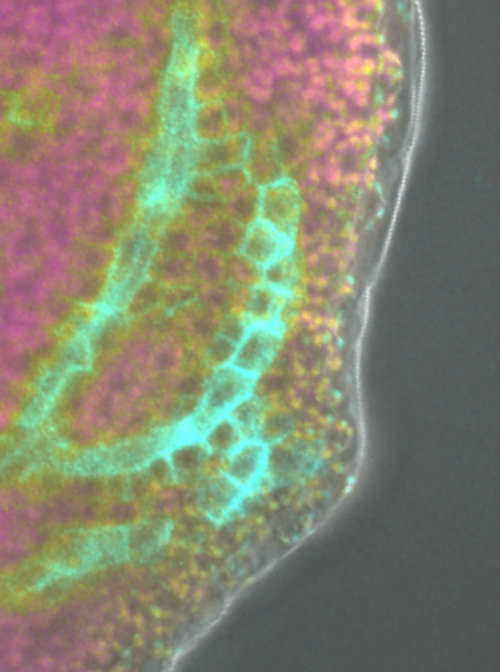
PIN1::cPIN1:GFP expression in a pin1 mutant.
When doing the research, did you have any particular result or eureka moment that has stuck with you?
PG I found that PIN1 expression in the epidermis was not sufficient to rescue the vein pattern defects of pin1 mutants but was sufficient to rescue their inflorescence defects; conversely, PIN1 expression in the inner tissues was sufficient to rescue the vein pattern defects, but not the inflorescence defects. This is the moment that stuck with me: when I found that there is a mechanistic difference in leaf vein patterning and inflorescence morphogenesis.
And what about the flipside: any moments of frustration or despair?
PG: I would not exactly term it as despair, but when I had started the research work, based on previous reports, I was using PIN1 expression in the epidermis as a control. And indeed, PIN1 expression in the epidermis did rescue the inflorescence defects of pin1 mutants. But when I found that the same PIN1 expression in the epidermis did not rescue the vein pattern of pin1 mutants, I suddenly felt that something was wrong as it was not going in the way I predicted it would go.
I understand you’ve since left the Scarpella lab – what are you up to now?
PG: There is a lot of uncertainty due to COVID-19. There were certain exams that I was planning to take and now they have been delayed indefinitely. Right now, I am planning to work as a research fellow and look for labs where I could potentially do a PhD in the near future.
Where will this work take your lab?
ES: We are currently testing hypotheses of the possible role of the leaf epidermis in vein patterning.
Finally, let’s move outside the lab – what do you like to do in your spare time in Alberta?
PG: When I was new to Alberta, in my spare time I preferred to explore the city and walk around the river valley, which was very close to the university. I constantly made a lot of plans, before I finally made it to beautiful lakes and the National Parks in Alberta. Apart from traveling, I enjoy reading books and do gardening during summer.
ES: I love spending time with my wife and my daughter – I particularly enjoy talking, walking and playing with them. I love the ocean, but there isn’t one in Alberta, so I make do with the swimming pool – though, for obvious reasons, I have not been able to see the ocean or its surrogate in a while. And I love listening to and playing music – especially with my daughter – reading, thinking and writing.
Heat map of cell growth in a developing Arabidopsis sepal (outermost floral organ). Fast growth is shown in warm colors and slow growth in cool colors. Note that the growth of cells is highly variable, while growth of the whole sepal is robust. Image courtesy of Dr. Lilan Hong.
An NIH-funded postdoctoral position is available in Dr. Adrienne Roeder’s group at Cornell University to research robustness of organ size in Arabidopsis. Development is remarkably reproducible, generally producing the same organ with invariant size, shape, structure, and function in each individual. Robustness is the ability of cellular systems to adjust and develop correctly sized and shaped organs in the face of stochastic fluctuations, and environmental and genetic variations. In the absence of robustness, these perturbations would cause large variations in organ size and shape. To determine how robustness emerges, we identified mutants with enhanced variability in organ size or shape (vos), thus disrupting robustness. Our goal is to elucidate mechanisms generating robustness by using these vos mutants in combination with live imaging of growing sepals, image processing to quantify growth, computational modeling, biomechanical tests, molecular genetics, and genomics. Our recent publications on the vos1 and vos2 mutants show that precise timing of the initiation and termination of organ development are critical to robustness of organ size (Hong et al., 2016 Developmental Cell 38, 15-32; Zhu et al., 2020 Nature Plants 6, 686-698). Please visit our laboratory website (http://roeder.wicmb.cornell.edu) for more information and a full list of publications. This postdoctoral project will focus on determining how coordination of growth across the three dimensions of the organ contributes to robustness in size and shape.
The ideal candidate will have a strong track record and extensive lab experience in plant molecular biology, genetics, and/or confocal imaging. The starting date is flexible from March to August 2021. Applications will be reviewed starting on January 11th until the position is filled. Competitive salary commensurate with experience and skills, as well as a generous benefits package will be offered. Interested applicants should send a PDF including a CV, a brief description of future research interests, and contact information for three references to Dr. Adrienne Roeder (ahr75@cornell.edu). Please include “Robustness Postdoc 2020” in the subject line. Informal inquiries are welcome.
Diversity and Inclusion are a part of Cornell University’s heritage. We are an employer and educator recognized for valuing AA/EEO, Protected Veterans, and Individuals with Disabilities.
POSTDOCTORAL POSITION is immediately available to follow up on our recent published work identifying that lymphatic-secreted signals are required for organ growth and repair using a variety of available mouse models (https://www.nature.com/articles/s41586-020-2998-x). Highly motivated individuals who recently obtained a PhD. or MD degree and have a strong background in mouse cardiovascular, molecular and developmental biology are encouraged to apply. Interested individuals should send their curriculum vitae, a brief description of their research interests, and the names of three references to:
Guillermo Oliver, Ph.D
Thomas D Spies Professor of Lymphatic Metabolism
Director Center for Vascular and Developmental Biology
Northwestern University Feinberg School of Medicine
Northwestern University is an Equal Opportunity, Affirmative Action Employer of all protected classes, including veterans and individuals with disabilities. Women, underrepresented racial and ethnic minorities, individuals with disabilities, and veterans are encouraged to apply. Hiring is contingent upon eligibility to work in the United States.
A position is available for a Post Doctoral Associate 1 in the Ameen Lab, within the Section of Digestive Diseases at Yale School of Medicine. Dr. Ameen is a pediatric gastroenterologist and physician scientist. She is Professor Pediatrics, Cellular and Molecular Physiology, and has been investigating CFTR in the intestine for over 25 years. Collaborative studies are ongoing with Dr. Kaelyn Sumigray in the Department of Genetics. This is a wonderful opportunity to learn complementary skills from the laboratory of a physician-scientist and basic stem cell geneticist.
A major goal of the Ameen laboratory is understanding the regulation of CFTR chloride channels in the native intestine and its relevance to intestinal diseases. Studies are predominantly conducted in animal models, including normal rat, transgenic mouse models of diseases where CFTR is implicated. We also conduct studies in human intestinal organoids and cultured human intestinal cells. Studies in the Sumigray lab focus on intestinal stem cell biology. Collaborative project with the Sumigray lab is using single cell RNA-sequencing and cutting edge techniques to examine and characterize a unique enterocyte subtype in the intestine and its relevance to Cystic Fibrosis. Yale is a very collaborative environment with many opportunities to interact with scientists and physician-scientists on campus.
Techniques used in the laboratory include: PCR, qPCR, tail clipping, animal husbandry, cell and organoid culture, Ussing Chamber electrophysiology in tissues and cells, immunofluorescence staining of cells and tissues, fluorescence and confocal microscopy imaging, immunoblot analysis of rodent tissue lysates, co-immunoprecipitation using tissue lysates, Ussing chamber electrophysiology.
Interested candidates please contact the PI: Nadia Ameen, Professor Pediatrics, Cellular and Molecular Physiology at Nadia.Ameen@yale.edu
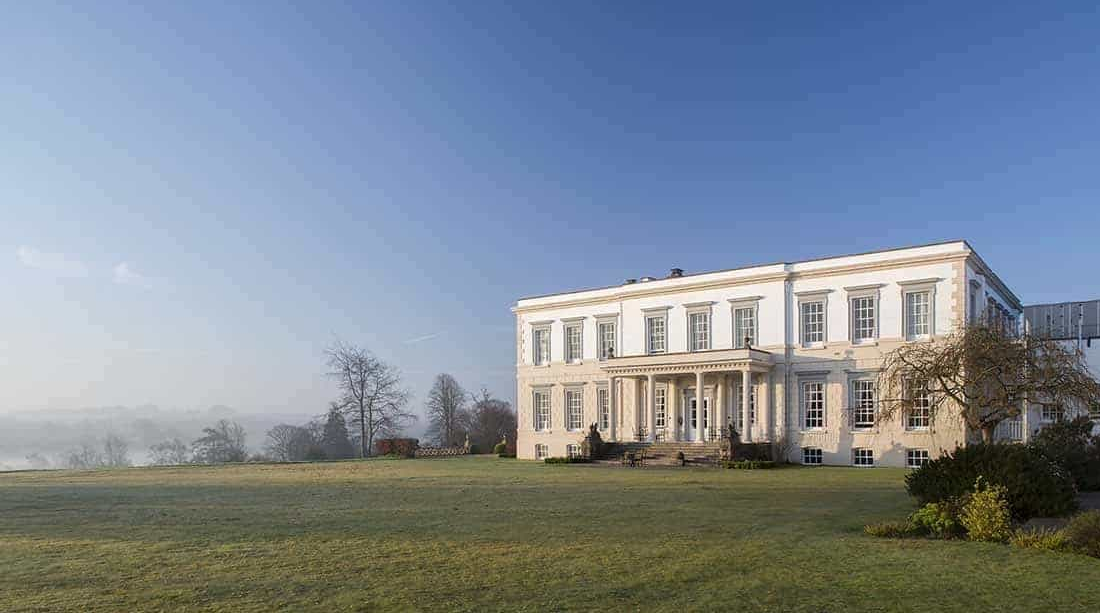
In July 2021, a Workshop from The Company of Biologists will bring together experts in human cell fate engineering and experts in the manipulation and characterization of single cells, with the goal of understanding the molecular steps underlying the transformation of one cellular identity into another. The Workshop is organised by Marisa Karow, Samantha Morris and Barbara Treutlein and will be held in Buxted Park in East Sussex.
There are 10 funded places for early-career researchers to attend this Workshop along with the 20 speakers – a fantastic opportunity to interact with the leaders in the field (see the speaker list below.
The application deadline for this Workshop is 16 December 2020.
preLights is a preprint highlighting service that is centred around a community of early-career researchers. Launched in 2018, this initiative has gained significant attention from researchers as well as the publishing industry, being nominated for an ALPSP Award for Innovation in Publishing in 2019. As preLights nears its 1,000th post, we are looking for the right person to join us for the next phase of community building and the site’s growth and development.
Joining an experienced and successful publishing team, this is an exciting opportunity for an enthusiastic and motivated team player to take a step into publishing or for someone already working in publishing to extend their interest in online communities. Please see the full job description for further details.
To apply, or for more information, contact recruitment@biologists.com. Applications should be received by 31 December 2020, although late applications may be considered. Interviews will be virtual and are expected take place in January. Applicants should be eligible to work in the UK.
preLights, a preprint highlighting service that is centred around a team of early-career researchers, is looking for a new Community Manager.
Launched in 2018, preLights has gained significant attention from researchers as well as the publishing industry, being nominated for an ALPSP Award for Innovation in Publishing in 2019. As we near our 1,000th post, we are looking for the right person to join us for the next phase of community building and the site’s growth and development.
Joining an experienced and successful publishing team, this is an exciting opportunity for an enthusiastic and motivated team player to take a step into publishing or for someone already working in publishing to extend their interest in online communities. Please see the full job description for further details.
To apply, or for more information, contact recruitment@biologists.com. Applications should be received by 31 December 2020, although late applications may be considered. Interviews will be virtual and are expected to take place in January.
The Company of Biologists is also looking for a Features and Reviews Editor for the journal Disease Models & Mechanisms, see the job description here.
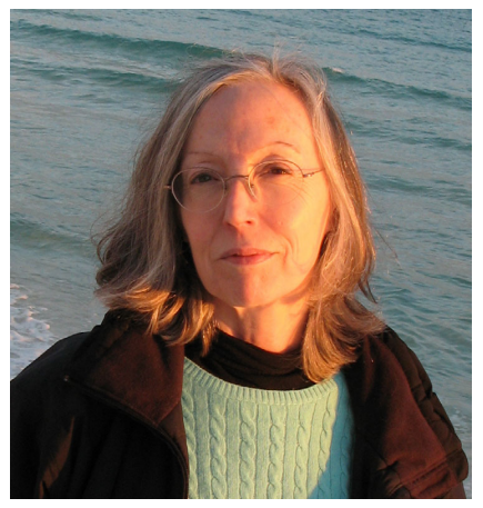
We at the Node and Development were greatly saddened to hear about the recent death of Kathryn Anderson, Professor & Chair of the Developmental Biology Program at the Memorial Sloan Kettering Cancer Center in New York,. Kathryn’s lab worked on the genetic pathways directing embryonic patterning and morphogenesis in the mouse embryo.
We also point readers towards this tribute from Tamara Caspary on Twitter – the dozens of replies show how highly the community thought of Kathryn.
I am heartbroken that Kathryn Anderson passed away this morning- at home and in her sleep. My thoughts are with her husband, her lab members and all my colleagues who loved her as I did. 1/n
— Tamara Caspary (she, her, hers) (@TamaraGenes) December 1, 2020
I was lucky enough to interview Kathryn for Development in 2016 – the year she won the SDB Edwin Conklin Medal. She was a generous interviewee and I can still remember sitting in Boston with her, bonding over our shared love of Radiohead!
Earlier in the year, we were similarly saddened to learn about the death of José Luis Gómez-Skarmeta, PI at the Centro Andaluz de Biología del Desarrollo in Seville who worked on vertebrate gene regulation, evolution and morphogenesis.
At the recent SEBD virtual meeting, a session on gene regulation was held to honour José Luis, and the society also announced the launch of the ‘José Luis Gómez-Skarmeta Awards for scientific excellence in Developmental Biology’. This year, the award were won by Manuel Irimia and Alvaro Rada-Iglesias.
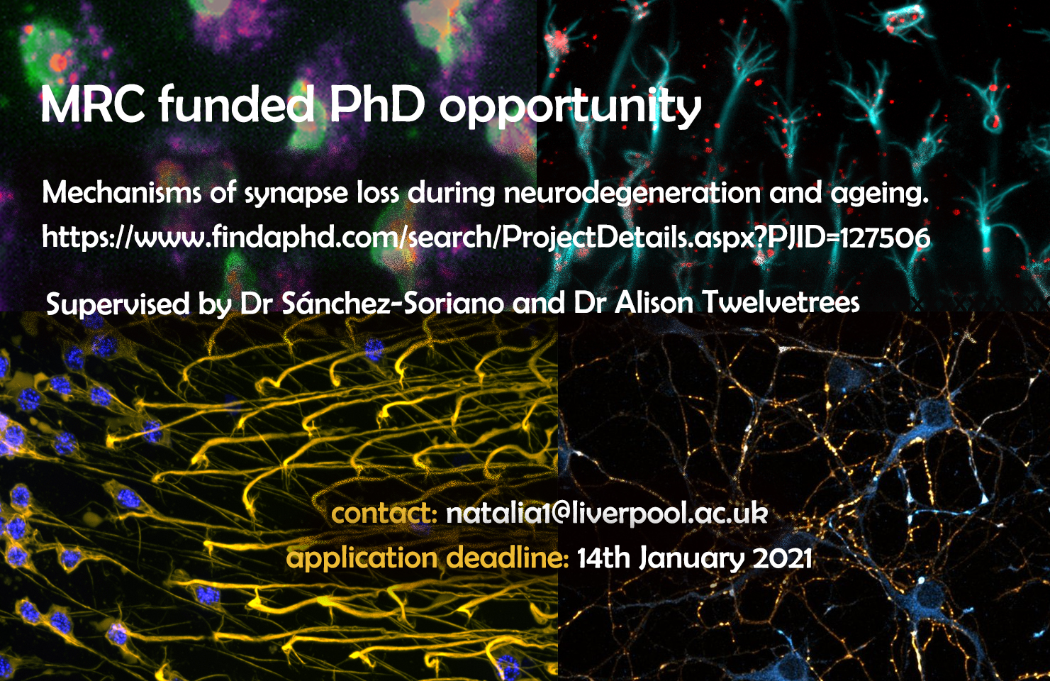
Exciting PhD project supervised by Dr Sanchez-Soriano and Dr Alison Twelvetrees through the MRC DiMeN Doctoral Training Partnership on mechanisms of synapse loss during neurodegeneration and ageing.
The aim of this studentship is to understand the processes of ageing and neurodegeneration, through the study of mechanisms of synaptic loss. You will be part of a multidisciplinary collaboration between two experienced groups at the Institute of Systems, Molecular & Integrative Biology (ISMIB, University of Liverpool) and the Sheffield Institute for Translational Neuroscience (SITraN, University of Sheffield).
Nerve cells are organised into complex neuronal networks, wiring the body or brain regions over distances up to a meter away in humans. For this, neurons extend long and thin processes called axons. At the tip of these axons, neurons establish synapses, specialised neuronal cell junctions which contain complex machinery for rapid transmission of signals to partner cells. The maintenance of this synaptic machinery fails during ageing and in disease, and the resulting synaptic malfunction is an important cause for cognitive, sensory and motor decline. Maintaining synapses requires transport of synaptic proteins from the cell body to the distant synapses up to a meter away. The Jun-Kinase (JNK) signalling pathway is a key regulator of this process. Importantly, physiological changes such as oxidative stress typically occurring during ageing and neurodegeneration, alter JNK activation patterns. The goal of this project is to understand how the JNK pathway regulates the transport and precise delivery of synaptic components and how it links to synapse loss occurring during ageing and disease.
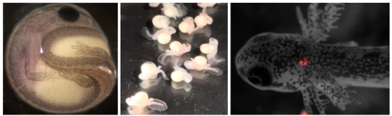
This studentship represents a unique opportunity to integrate in vivo models of ageing and neurodegeneration capitalising on the brain of the fruit fly Drosophila as a highly efficient model, together with mouse and rat neuronal models and in vitro reconstitutions assays. Using these systems, you will study the role of JNK during the regulation of intracellular transport and synaptic decay. You will receive training by the two supervisory groups in neuronal cell biology (fly neurons in culture and in vivo in the adult Drosophila brain, primary neuronal culture from mouse and rat), in genetic strategies, in quantitative live imaging of cultured neurons and whole tissue, in analytical methods, techniques required for in vitro reconstitution of transport assays with complementary quantitative analysis. Understanding the causes of synapse decay during ageing or disease is crucial to providing new avenues for therapeutic intervention.
Creative individuals with an eye for detail are encouraged to apply. The successful applicant will be based in the Institute of Systems, Molecular & Integrative Biology supervised by Dr Sánchez-Soriano (sanchezlab.wordpress.com/research), whilst working closely with the SITraN lab, Department of Neuroscience in Sheffield under the supervision of Dr Alison Twelvetrees (www.twelvetreeslab.co.uk). Applications from candidates, ideally with some background in cell biology, genetics, neuroscience and/or biomedical sciences are welcome. Interested applicants should contact Dr Sanchez-Soriano to discuss the project: n.sanchez-soriano@liverpool.ac.uk.
 (3 votes)
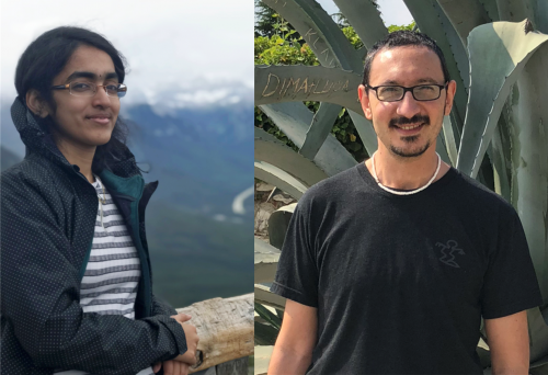
 (No Ratings Yet)
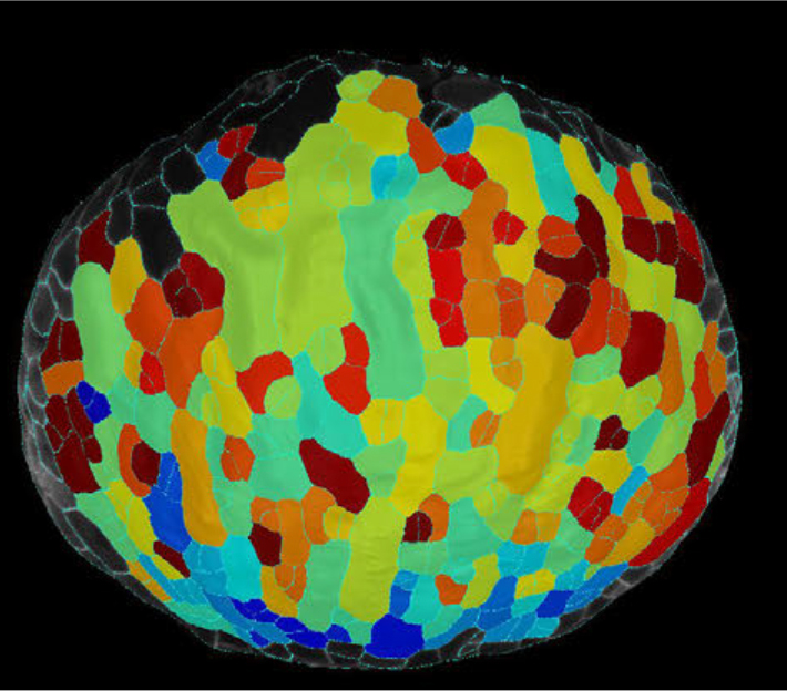
 (3 votes)