A research scientist position is available in the research group of Dr Anthony Gavalas. The group investigates the role of signaling pathways and metabolism in the late stages of endocrine pancreas development, the application of novel signals for the conversion of human pluripotent stem cells into functional beta cells and the function of adult pancreas stem cells. A combination of directed pluripotent stem cell differentiation, genomics, in vivo genetic analyses and ex vivo experiments including organoids is being used (https://www.digs-bb.de/research/research-groups/anthony-gavalas/).
The successful candidate will have either a M.Sc. or Ph.D. degree in Biology and related disciplines, extensive experience in cell culture, preferably with embryonic and pluripotent stem cells, very good organizational skills and strong molecular biology background. Experience with mouse handling and genetics will be considered as an asset.
The lab is located in the Center for Regenerative Therapies Dresden (CRTD) with full access to state of the art core facilities for Deep Sequencing, including single cell RNA Seq, Genome Engineering, Imaging and FACS analysis.
The salary will be according to the TV-L scale commensurate with experience and qualifications. The contract will be initially for two years with the possibility for renewal. Applicants are requested to send their CVs along with names and emails of at least two referees to Dr Anthony Gavalas (Anthony.Gavalas@tu-dresden.de), before August 15th, 2019.
In this article I share with you a more personal, chronological account of how our story unfolds (recently published in Nature), and highlight some key events and insights that help guide the direction of the study, which are not described in the publication. Readers are welcome to refer to the publication for more technical details.
Quantifying blastocoel pressure in vivo
The defining feature of mammalian development is the formation of a blastocyst with a fluid-filled cavity, termed blastocoel. However, the physiological function of blastocoel in mammalian blastocysts, and the influence of fluid pressure on tissue mechanics, embryos size and fate specification remain largely unknown. Trained as an experimentalist in cell mechanics, my first thought when I conceived the project was to devise a method to characterize the spatiotemporal change in the blastocoel pressure during mouse blastocyst development. Surprisingly very few studies have quantified lumen pressure in embryonic development. Precise quantification of the blastocoel pressure will allow us to address if the pressure grows or decreases with blastocyst expansion (the latter case analogous to a balloon, where its internal pressure drops as it inflates, which is why it is easier to blow up a large balloon). Initially we tried to characterize the pressure indirectly by using the Laplace law, where pressure is equal to the tissue surface tension divided by the cavity size. However we found that the measured pressure scales with the pipette size, which cannot be the case. This leads us to explore alternative methods to directly quantify the lumen pressure. Inspired by the works of Ryan Petrie, who used a micropressure probe to measure intracellular pressure1, we wondered whether we could apply a similar technique to mouse blastocysts. Bringing with me some mouse blastocysts and starfish oocytes (as a control, thanks to Johanna Bischof!), I then made a daring trip to Ryan’s lab at Drexel University in Pennsylvania to test the feasibility of the device in measuring blastocoel pressure (Fig. 1a). I still remember how thrilled we were when we made our first successful measurement (Fig. 1b), which was found to be surprisingly high (~1500 Pa), certainly higher than the known cytoplasmic pressure2. Overall it proved to be an extremely fruitful trip, all thanks to Ryan’s generosity and patience. Here I would also like to acknowledge the assistance of A.K. Hadjantonakis for providing us with ‘back-up’ mice during the trip.
Figure 1.Direct quantification of mouse blastocyst pressure.a. Ryan Petrie and me in front of a micropressure setup in his lab (Drexel University, Pennsylvania). This visit adds much synergy to the project, especially during its early phase. b. Using a micropressure probe (0.5 μm needle was injected into the blastocoel, as shown on the right), we directly measured the blastocoel pressure in a mouse blastocyst for the first time, and were surprised to detect a high value! We also had some fun with measuring cytoplasmic and even nucleoplasmic pressure in Starfish oocytes. Scale bar, 20 μm.
Blastocyst collapse is triggered by mitosis rather than random rupture of cell-cell junctions
While we found that the pressure and the trophectoderm (TE) cortical tension grow with blastocyst maturation, we also observed that the blastocysts exhibit regular patterns of collapse followed by re-expansion. Surprisingly few studies have tackled what precisely drives this dynamics, a topic I proposed to study in my first year lab retreat in the Hiiragi lab (I called it ‘lumen breathing’, a term that raised many eyebrows in the lab!). My natural hypothesis, similar to what many in the field have speculated, is that these events are likely stochastic and are driven by random rupture of TE cell-cell junctions, when the cell tension exceeds the threshold cell-cell adhesion strength. By this time we had already observed that the TE cells in the reduced embryos (e.g. quarter embryo is derived from a single blastomere from a four-cell stage embryos) have a higher cortical tension than that of the full embryos at early blastocyst stage, so the reduced embryos should show more frequent collapse events, which proves to be the case! While this shows clearly that higher cortical tension leads to more frequent collapse dynamics, it still does not answer what ‘triggers’ the collapse. Interestingly, we found that blastocyst collapse is not triggered by some random events of junctional rupture. Instead they are triggered by mitosis of mural TE cells, which we carefully confirmed with a series of pharmacological and embryological perturbations. This experience taught me to always check my assumptions carefully – Nature always has new surprises for us!
Mechanosensing is crucial for blastocyst development
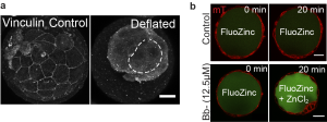
While it provides great satisfaction for me to understand how the blastocyst regulates its size, we wondered if lumen size control has important functions in blastocyst development (which is one thing I learned in a biological lab: to ask not just how, but also why). One question we asked was whether the growth in lumenal pressure impacts the development of TE cell-cell junctions, which are crucial for tissue integrity (adherens junctions) and blastocyst sealing (tight junctions). Intuitively we thought that relaxing cell contractility and TE tension should lead to a larger cavity (as observed in epithelial dome3), but to our surprise we found a smaller cavity when we abolished actomyosin contractility. This suggests that there could be an active response from the TE epithelium that ensures proper lumen expansion. Indeed we found that increased cortical tension leads to an active recruitment of junctional proteins (such as vinculin, Fig. 2a) that seals the tight junctions, which possibly functions to counterbalance the higher pressure building up during blastocyst maturation. This is a beautiful example of positive feedback control that exists so abundantly in developmental processes. However, to confirm this, we still needed to observe these events directly. Here we struggled for a few months. We tried loading fluorescently labelled dextran into the blastocoel and tracing the leakage sites, which proved futile since the very weak signal at the leakage site was ‘drowned’ out by the strong background signal in the blastocoel. Finally, during a joint retreat with Carl-Philipp Heisenberg’s lab, I learnt of a cool technique to monitor junctional leakage based on Fluozinc leakage (thanks to C. Schwayer, T. Higashi and A. Miller, whose work was recently published4). This method finally gave us a good signal-to-noise ratio to unambiguously confirm leakage due to transient reduction of cortical tension (Fig. 2b).
One question that arose early on in our study (also commented by a keen reviewer) is if cell stretching caused by lumen expansion promotes cell division in TE cells, as observed in other epithelia5. To our disappointment, we found no difference in the cell cycle length between the more-stretched TE cells in late blastocysts and those of the less-stretched ones in early blastocysts, which suggest that stretch-induced cell division may not play a role in mouse preimplantation development.
Figure 2. Lumenal expansion promotes junctional maturation. a. Immunostaining of vinculin in mechanically-deflated embryos at E4.25 (dotted lines denote cavities). Vinculin being mechanosensitive to cell stretching, disassembles from the tight junctions upon reduction in lumen size and cell cortical tension. Scale bars: 20 μm. b. Tight junction permeability assay assesses tight junction sealing. Blastocysts treated with Bb- (bottom) showed enhanced signals in the cavity due to impaired tight junction sealing and ZnCl2 penetration.
Towards a theoretical model
At this point we approached Teresa Ruiz-Herrero and L. Mahadevan from Havard to see if we can build a theoretical model that recapitulates our experimental findings. Building on their previous model6, it did not take long before they came up with the simulations that could explain the key features of our experiments. However the true merit of a model is to make predictions that could be tested experimentally. A key prediction of the model is that the steady-state blastocyst size is governed by the ratio of tissue yield stress and tissue elasticity, as well as the initial tissue size. To verify this prediction, we embryologically manipulated the initial tissue size by making reduced and amplified blastocysts. Knowing the tissue yield stress from the measured blastocoel pressure and tissue thickness, we were able to extract the tissue elasticity. With the help of Martin Bergert and Alba Diz-Muñoz at EMBL, we were able to use the atomic force microscopy to measure the tissue elasticity of TE, which nicely falls within the range predicted by the model.
Lumen size impacts tissue architecture and cell fate
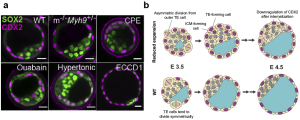
Early on in the project we postulated that another function of lumen may be its regulation of tissue architecture and cell fate specification. After all, the lumen takes a significant portion of the entire embryo, and its presence must impact how the cells are allocated to the inside versus the outside of the blastocyst. In fact it is not difficult to think that given the same tissue volume (cell number x cell volume), a blastocyst with a smaller cavity would have more cells allocated to the inner layer, compared to one with a larger cavity, purely by geometric argument. This means that the ICM-to-TE cell ratio should be higher in blastocysts with a reduced cavity size. This is indeed what we see with both pharmacological and genetic perturbation studies (Fig. 3a). Still, it remained unclear what is the cellular mechanism driving this change in ICM/TE ratio. We then considered three scenarios: a). less ‘asymmetric’ division of inner cells that give rise to daughter cells that end up on the outer layer, b). more asymmetric division of outer TE cells to generate an inner daughter cell, and c). active cell migration from the outside (polar TE) to the inside (ICM). While discussing with Dimitri Fabrigès, an expert in lightsheet microscopy within the lab, I learnt that scenario a) and c) are almost never observed during blastocyst expansion phase. I therefore focused on investigating scenario b). At this point I recalled some works in the past where people were able to track the TE lineage in preimplantation stage7, using a DII dye that stays with the outer cells through subsequent generations. Furthermore, combining this with live marker CDX2-GFP x H2B-mCherry, I was able to reliably track the movement and final position of the inside and the outside cells. Using this approach, we indeed observed more DII-labelled cells in the ICM of blastocyst with reduced cavity size, than those in the WT. Importantly, we also found that the internalized cells, following asymmetric TE division, eventually lose their CDX2 signal and adopt an inner cell fate, consistent with our hypothesis based on scenario b) (Fig. 3b). Our results are consistent with some classic papers by Rossant et. al. showing that TE and ICM cell fates remain plastic during the blastocyst expansion phase (16-32 cell stage)8,9. In my view, this is a beautiful finding because it reveals that lumen pressure can influence the division pattern of cells around it, and thereby lead to changes in cell position and eventual fate specification (through the HIPPO pathway). While it is known that cell shape and force can guide spindle orientation10, this is the first time we observe that the lumen pressure could directly influence the division pattern of cells in vivo and impact their cell fate specification.
Figure 3. Lumenal pressure couples cell positioning and fate specification.a. Immunostaining of late blastocyst stage (E4.25) wild-type (WT), m-/-Myh9+/-, CPE-, ouabain-, hypertonic-, and ECCD1-treated embryos showing TE (CDX2, magenta) and epiblast (SOX2, green) fate. Scale bars: 20 μm. b. Schematic representing how lumenal expansion impacts cell positioning and fate acquisition. Reduced lumenal expansion increases the frequency of asymmetric division of outer cells, which generates a TE- and an ICM-forming cell, with the latter eventually acquiring ICM fate.
Outlook and open questions
This work opens up several new directions, even beyond mouse mechanobiology. First, in terms of technique, the micropressure probe allows us to directly measure hydrostatic pressure in vivo, independent of the geometry of the sample, which is often required for indirect determination of pressure (e.g. round cells as in Laplace’s Law). Furthermore this device allows us to quantify spatial difference in pressure within a tissue (for example, we measured a higher pressure in the nucleus compared to the cytoplasmic pressure in starfish oocytes). In the future it will be exciting to investigate the role of fluid pressure in organ development, such as in lungs and kidneys, and in other model systems such as sea animals (body extension) and organoids (symmetry breaking). Second, I think this study really highlights a critical role of lumen pressure in tissue mechanics (apart from the conventional key players such as actomyosin contractility or cell-cell adhesion). In the future it will be interesting to investigate if lumenal pressure plays a similar role in guiding cell division and cell positioning in other developmental processes.
Are there other evidence that lumen pressure may trigger mechanosensing or mechanotransduction in mouse blastocysts? We could hypothesize a few. For example, does cell stretching due to lumen expansion lead to YAP signaling in the TE cells, which then act to stabilize its CDX2 expression? Also, it is known that mural TE cells eventually ‘invert’ their polarity as it enters the peri-implantation stage, where the lumen-facing side becomes apical and the outer surface becomes basal. It is believed that this facilitates the mural TE cells to adhere to the surrounding tissue as the blastocyst embeds itself into the uterine wall. Does luminal pressure play a role here?
One exciting aspect is if lumenal expansion also plays a role in the second lineage segregation during the mid-to late blastocyst stage, where the ICM further segregates into the epiblast and the primitive endoderm11. Indeed, using a combination of pharmacological and biophysical approach, work in our lab has shown that lumen expansion is required for proper EPI-PrE cell fate specification, as well as their spatial segregation12, suggesting that the lumen may provide mechanical and biochemical cues (pressure, contact-free surface, signalling niche) to impact cell fate specification. This provides further evidence that lumen morphogenesis is an integral part of tissue patterning in mammalian preimplantation development.
I feel extremely privileged to have the opportunity to work with many great scientists along this incredible journey. These experiences truly enriched me on both the professional and personal level.It also taught me the benefits of engaging in an interdisciplinary approach early on in the project, which also means taking risks, get out of your comfort zone, and approaching people with the right expertise to learn new techniques and concepts. In the long run this also allows one to build a strong network with fellow scientists, and many will go on to become close friends, as in my case.
References
Petrie, R. J., Koo, H. & Yamada, K. M. Generation of compartmentalized pressure by a nuclear piston governs cell motility in a 3D matrix. Science 345, 1062–1065 (2014).
Stewart, M. P. et al.Hydrostatic pressure and the actomyosin cortex drive mitotic cell rounding. Nature 469, 226–231 (2011).
Latorre, E., Kale, S., Casares, L., Gomez-gonzalez, M. & Uroz, M. Active superelasticity in three-dimensional epithelia of controlled shape. Nature 563, 203–208 (2018).
Stephenson, R. E. et al.Rho flares repair local tight junction leaks. Dev. Cell 48, 445–459 (2019).
Gudipaty, S. A. et al.Mechanical stretch triggers rapid epithelial cell division through Piezo1. Nature1–12 (2017).
Ruiz-Herrero, T., Alessandri, K., Gurchenkov, B. V., Nassoy, P. & Mahadevan, L. Organ size control via hydraulically gated oscillations.Development 144, 4422–4427 (2017).
Krupa, M. et al.Allocation of inner cells to epiblast vs primitive endoderm in the mouse embryo is biased but not determined by the round of asymmetric divisions (8-16- and 16-32-cells). Dev. Biol. 385, 136–148 (2014).
Rossant, J. & Lis, W. T. Potential of isolated mouse inner cell masses to form trophectoderm derivatives in vivo. Dev. Biol. 70, 255–261 (1979).
Rossant, J. & Vijh, K. M. Ability of outside cells from preimplantation mouse embryos to form inner cell mass derivatives. Dev. Biol. 482, 475–482 (1980).
Xiong, F. et al.Interplay of cell shape and division orientation promotes robust morphogenesis of developing epithelia. Cell 159, 415–427 (2014).
Chazaud, C. & Yamanaka, Y. Lineage specification in the mouse preimplantation embryo. Development 143, 1063–1074 (2016).
Ryan, A. Q., Chan, C. J., Graner, F. & Hiiragi, T. Lumen expansion facilitates epiblast-primitive endoderm fate specification in the mouse blastocyst formation. BioRxiv https://doi.org/10.1101/575282
In a previous blog, I have highlighted several ways to visualize the cell-to-cell heterogeneity from time-lapse imaging data. However, I have ignored that data is often rescaled in a way that reduces variability. For time-lapse imaging data, it is common to set the initial fluorescence intensity to 1 (or 100%). As a consequence, any changes in the fluorescence are displayed as deviations from unity. This rescaling method is often indicated as “normalization”. A definition of normalization would be “the rescaling of data to facilitate comparison”. Below, several methods of data normalization are highlighted. The examples use experimental data from fluorescence spectroscopy or imaging but rescaling methods are widely applied to all sorts of data.
Part 1: Normalization by initial value
Normalization based on the initial signal is useful when perturbations of a steady state are studied. In this type of experiment, the unperturbed system is monitored for some time, after which the perturbation (e.g. addition of an agonist or optogenetic stimulation) is applied. The initial signal, or more strictly, the signal at t=0, is defined as I0 (also often abbreviated as F0). Instead of this strict definition, we often use the signal from a ‘baseline’. The baseline is the average signal that is acquired in the period before the stimulation. By using an average, instead of a single value, we obtain a better estimate of I0. There are several ways to use the I0 to perform normalization, as will be discussed below.
Fold change: Division by initial value (I/I0)
One way to normalize fluorescence intensity data from time-lapse imaging is by dividing the intensity at every time-point (I) by the fluorescence intensity of the first time point (I0). One application of this normalization method is for analyzing and comparing photostability. The intensity at t=0 is set to 1 (or 100%) and the effects of illumination on the intensity (usually a reduction according to an exponential decay) are followed over time. The initial fluorescence is set to 1 to get rid of the absolute intensity that reflects the protein concentration. This is acceptable, since photobleaching rates do not depend on initial concentration of intensity.
Another application is in detecting changes in location of fluorescent biosensors (Postma et al 2003). The fluorescence signal in the cytoplasm is monitored over time and a reduction in the signal reflects depletion from the cytoplasm and accumulation elsewhere. To account for different expression levels between cells, these data are often normalized to the baseline value, setting the initial fluorescence to 1. The normalization enables the comparison (or averaging) of the data from different cells (this would be difficult or impossible if the data are not treated in this way). A similar normalization method is used to average and compare the response of FRET based biosensors (Reinhard, 2017)
Figure 1: Data from a time-lapse imaging experiment that measures the fluorescence intensity of the calcium sensitive biosensor GCaMP6s in HeLa cells. At t=40s, the cells were stimulated with histamine which results in an increase of intensity, reflecting an elevated calcium concentration. The raw data is shown on the left, with I0 and Imax indicated in the graph. The effect of two normalization methods, showing ’fold change’ and ‘relative intensity’ is shown in the middle panel and right panel.
Difference: subtraction of initial value (I-I0)
In the previous method, the fold change is obtained by dividing the data by I0. Instead of this relative change, the absolute change in intensity can be determined. To this end, the intensities are subtracted by the intensity at t=0 (I0). The same considerations for I0 apply. This normalization is used when the absolute difference from the baseline is of interest.
Relative change: the difference divided by the initial fluorescence (ΔI/I0)
This rescaling method can be applied to data from timelapse experiments, to show the relative change compared to the baseline. Using the relative change has the advantage (just like the fold change) that the initial intensities are equal and therefore can be used to average multiple measurements.
The relative change is commonly used for the display of calcium changes measured with a fluorescent biosensor (figure 1). In some cases it is multiplied by 100 to depict the change as a percentage (relative to I0). The rescaling method for the ‘relative change’ is related to the method for the fold change that was treated before. To understand the relation we can rewrite ΔI/I0:
ΔI/I0 = (I-I0) / I0 = (I/I0) – 1
Since I/I0 is defined as the ‘fold change’, the ‘relative change’ normalization is essentially the same as I/I0, but offset by one. Knowing this relationship, it is straightforward to convert data between the ‘fold change’ and ‘relative change’.
Z-score: the difference divided by the standard deviation of the initial value ((I-I0)/SD(I0))
The standard deviation of the baseline reflects the variability of the initial signal. The change in intensity relative to this variability is the Z-score. The Z-score indicates the number of standard deviations that the signal differs from the initial signal (Segal 2018) and reflects how well signals can be detected. For instance, if there is a large standard deviation, a small change in the signal will be hard to detect. The Z-score is of interest for assay development, where the detection of an effect needs to be optimized.
Part 2: Normalization by minimal and/or maximal values
The previous normalization methods all use I0 for the normalization. This is useful for data from timelapse experiments. Other normalization methods use other values, for instance Imax and/or Imin. Two methods will be treated below.
Set maximum to 1: I/Imax
Normalization of values based on the maximal value is common for the rescaling of absorbance and emission spectra from spectroscopy (for examples see Mastop et al, 2017). The shape of a spectrum is usually the feature of interest instead of their amplitude. The normalization is done by dividing each value by the maximal value, to enable the comparison of spectral shapes.
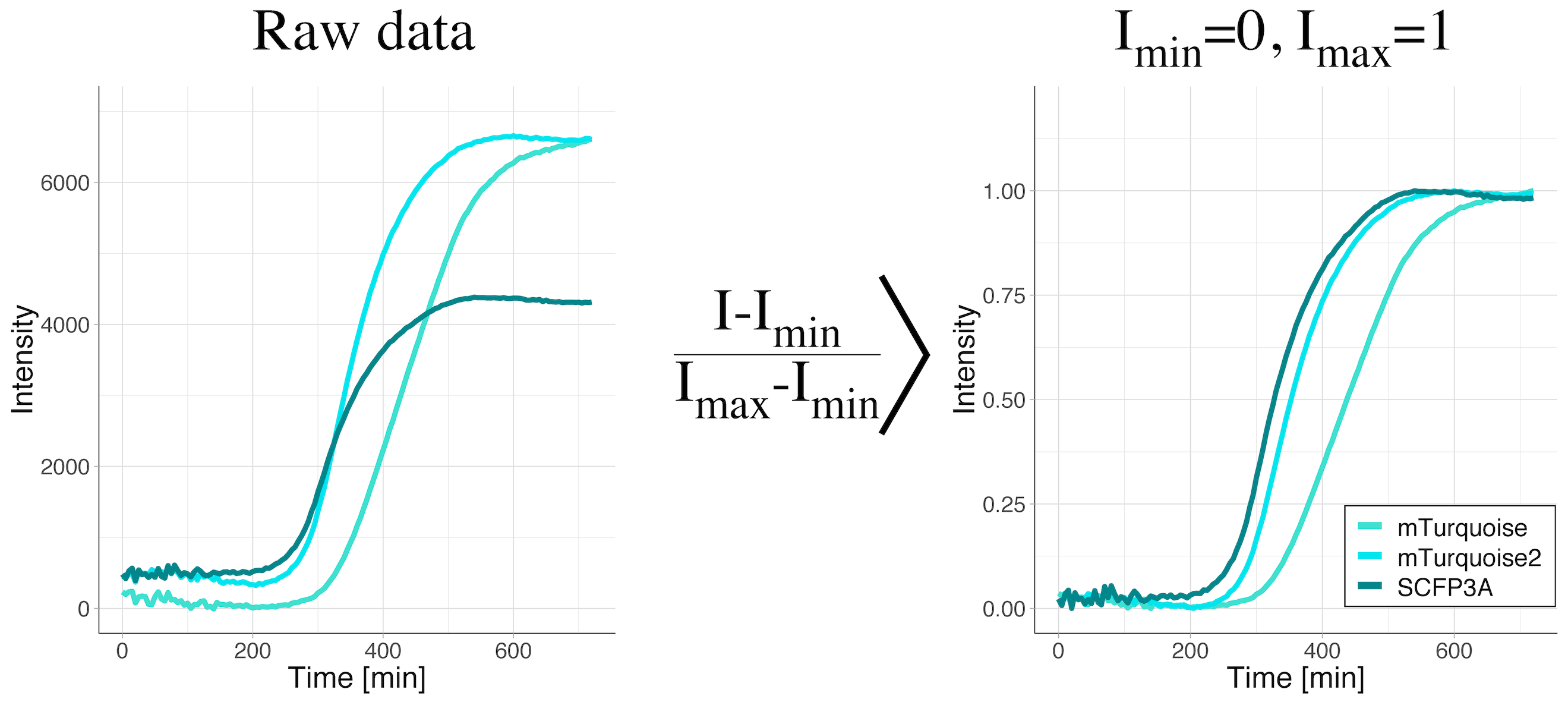
Rescale between 0 and 1: (I-Imin)/(Imax-Imin)
When data is rescaled based on the maximum and minimum value, the minimal value is set to zero and the maximal value is set to unity. As a consequence, the information about the absolute values is lost. Still, the shape of the curve is kept. Therefore, this rescaling method facilitates the comparison of the shapes of the curves. This method is applied when changes in the signal are relevant, but their absolute values are not. It is a valuable transformation when dynamics are compared. It is also used for dose-response curves, where the midpoints (value on the y-axis that is exactly between the minimal and maximal value) of different conditions are compared. After rescaling, the midpoint corresponds to a value of 0.5 on the y-axis.
We have used this type of rescaling to compare maturation kinetics across different fluorescent proteins (Goedhart, 2012). The rescaling gets rid of the differences in fluorescence intensity and enables direct visual comparison of maturation kinetics (figure 2).
Figure 2: Data from a time-lapse measurement of the fluorescence intensity of bacterial cultures to evaluate fluorescent protein maturation rates. The bacteria were producing the cyan fluorescent proteins SCFP3A, mTurquoise or mTurquoise2. The raw data shows a clear difference in the fluorescence levels (left panel). Only after rescaling the intensity between the minimal and maximal values (right panel), the difference in maturation rates become visible. The maturation rate increases in the order mTurquoise < mTurquoise2 < SCFP3A.
Final Words
Data normalization is a meaningful data manipulation method as it facilitates comparisons. It should be realized however, that information is lost by the data rescaling. Whether this reduction of information is acceptable should be carefully evaluated. For instance, we have seen cases where a cellular response to a biosensor depended on the absolute intensity (protein concentration). Hence, it is advisable to examine both normalized and raw data for trends.
Implementation
Data normalization is readily performed by different applications and we have used R and microsoft Excel. The rescaling methods discussed here are implemented in the webtool PlotTwist: https://huygens.science.uva.nl/PlotTwist/. The R code for the webtool is available on Github.
Shout out
I like to thank all colleagues for the discussions (IRL or in the twitterverse) that helped to improve the PlotTwist webtool and especially the people that contributed suggestions for normalization methods.
Organizers pattern surrounding tissues via secreted morphogens that specify different cell states as a function of concentration. Wolpert’s French Flag model is commonly used to describe how morphogen gradients specify different fates. Our recent study integrates tail organizer signaling with control of morphogenesis during vertebrate body elongation (Das, Jülich, Schwendinger-Schreck et al., 2019). This figure shows a French Flag in the background, and a reconstruction of tracks of cell motion in the zebrafish tailbud (posterior is down and anterior is up). The tail organizer is green, the posterior neural tube is magenta, the presomitic mesoderm is cyan, and the domain containing the neural-mesodermal progenitors is red. We found that Bmp signaling is confined to the posterior of the organizer, but that cell motion in the posterior neural tube is affected by organizer signaling. Mechanical perturbation of the organizer has similar long-range effects. We hypothesized that local perturbation of signaling and cell motion in the organizer is propagated by a cell to cell relay (yellow) to affect cell motion in the posterior neural tube. We called this effect mechanical information, and propagation of mechanical information beyond the range of morphogen signaling enables embryonic organizers to expand their sphere of influence.
Organization of embryonic morphogenesis via mechanical information
This study started as a side project during Jamie Schwendinger-Schreck’s doctoral research. Andrew Lawton and Nicolas Dray, a former graduate student and postdoc in the lab, respectively, had developed methods for systematically analyzing cell motion in the zebrafish tailbud. Andrew’s thesis research described the effects of inhibition of Wnt and Fgf signaling on cell motion and found that regulation of tissue fluidity is essential for normal body elongation. Bmp signaling is also localized in the posterior tailbud, and prior studies in Xenopus and zebrafish had linked Bmp signaling and the transcription factor eve1 with ‘tail organizer’ activity, i.e. manipulation of organizer signaling could cause tail duplications. Jamie performed these initial experiments with help from Andrew and Nico. She found that even though Bmp signaling was localized to the posterior tailbud, after perturbing the organizer, she observed effects on cell motion far from the Bmp signaling domain. This seemed pretty interesting, but we were stuck with no physical explanation of our observation and no direct experimental validation that the effects were truly non-autonomous to the Bmp signaling domain. Meanwhile, Jamie graduated, as students are wont to do, and there was no one in the lab working on the project.
Dipjyoti Das, a theoretical physicist who goes by ‘Dip’, joined the lab as a postdoc, and with our collaborator Thierry Emonet, who has expertise in fluid mechanics, developed a 3D computational model of the elongating tailbud. In silico experiments are faster and less resource intensive than wet lab experiments, and we wanted to see if we could explain the long-range effects that Jamie observed using the computational model. Corey O’Hern and Mark Shattuck are theoretical soft matter physicists with whom we also collaborate. Their expertise in the physics of granular matter guided our in silico experiments, and Dip observed signatures in the tailbud simulations analogous to pressure waves traveling through granular matter, i.e. similar to someone tapping on one end of a table and a second person sensing the vibrations at the opposite end of the table. The model led us to look for this signature in our in vivo data, and remarkably, we observed the same phenomenon in the in vivo cell motion data.
Once we had a physical explanation for our observation, we needed additional in vivo experiments test the idea that a localized change in cell signaling could instigate a relay effect that alters cell movement indirectly and far from the signaling domain. Optogenetics was the obvious way to proceed. Dörthe, a Research Scientist in the lab, spent the better part of a year trying unsuccessfully to establish an optogenetic perturbation. She’s an experimental maestro, so when she couldn’t get the optogenetics to work, we had hit another wall.
Dörthe thought that, given more time, she could get optogenetics to work. That may very well be true, but the project was already taking years longer than anticipated. Here, we needed to avoid having the perfect be the enemy of the good. We had some alternative ideas for experiments, but they wouldn’t be as elegant as an optogenetic perturbation. These experiments were still technically challenging, and Dörthe got them to work after considerable trouble-shooting. Emilie Guillon, a postdoc in the lab, and Dip helped with the data analysis. These data supported the hypothesis that perturbation of the tail organizer has indirect effects far from the organizer signaling domain. The effect travels at a rate too rapid to be mediated by transcription and translation, and we hypothesize that it is propagated as a relay from cell to cell as the cells migrate during body elongation.
Two of the joys of being a scientist are seeing a complicated multi-year project to completion and discovering something new. This project started with the intent of Jamie getting a ‘quick’ additional paper to round out her dissertation. As it often goes in science (and in life), reality intervened and things did not go according to plan. After Jamie graduated, we received an NIH grant to pursue this research, and that grant ended in 2017 as we began writing the first draft of the manuscript. Jamie is now a Scientific Project Manager at 10x Genomics. Dip is starting his own lab as an Assistant Professor at the IISER, Kolkata. Dörthe is on a well-deserved vacation hiking in the arctic circle. After I submit this blog post, I will go on a literal fishing expedition with friends from college. We still do not know how the mechanical information is propagated nor how general this mechanism is. When I return from vacation, I look forward to being surprised by the next experiment. Who knows to where it might lead?
Das, D.*, Jülich, D.*, Schwendinger-Schreck, J.K.*, Guillon, E., Lawton, A.K., Dray, N., Emonet, T., O’Hern, C.S., Shattuck, M.D. and Holley, S.A. 2019. Organization of embryonic morphogenesis via mechanical information. Dev Cell, 49: 829-839. * equal contribution
With its growing adoption in the laboratory, an electronic lab notebook, or ELN, can be a useful tool to aid research, whether in academia or industry. But I found there is limited information on the practicalities of an ELN in a wet lab. Wet labs are a messy business, so strict guidelines are put in place to prevent spillages and contamination. It may seem absurd to use digital software while conducting experiments with innumerous hazards, but do the benefits of using an ELN in a wet lab outweigh the risks?
The majority of wet labs use paper lab books, few of which transfer the information from paper to an ELN. This is largely due to hazards involved in a wet lab, where the use of laptops or desktops is ill-advised. It is much easier to simply use a paper lab book, to keep all live results jotted down on paper. Furthermore, if a wet lab is using an ELN, scientists still have to use a paper lab book and copy some research notes into their ELN software at a later date.
A large issue with using an ELN in the wet lab is the inaccessibility of protocols. It is effortless to bring a paper lab book into the lab to see past results or double check some protocol results. But there is much more difficulty when it comes to ELNs since you can’t bring your laptop into the lab.
Nevertheless, the inability to bring a laptop into a wet lab isn’t the be all and end all. Some wet labs use tablets that are shared by several scientists, allowing protocols and previous research notes to be easily accessed while working.
The need for stringently organized lab books in wet labs can make ELNs seem more appealing. Many labs have strict controls on the data’s structure, to ensure its integrity and compliance. Some ELNs offer a dedicated templates section in which scientists can store and reuse their laboratory protocols and/or SOPs. Moreover, since those protocols are usually shared amongst several scientists, an ELN can help to guarantee that each colleague has access and is using the correct protocol. This maintains a consistent project structure, for easier research organization.
Moreover, by having a system in which team members can collaborate in real time, I saw supervisors providing immediate feedback through the use of an ELN. No more meetings had to be scheduled for supervisors to check content from paper lab books. That is the beauty of storing laboratory data online in an electronic lab book. Projects can be accessed from anywhere in the world. This also means that when a group member leaves a project, the data will not be lost with the person carrying the lab book.
As the lab is becoming more digitized, ELNs harness the simplicity of data transference. I could simply drag and drop digital data from microscopy images straight into an ELN. Hence removing the need to print and stick these results into a paper lab book. Moreover, with an ELN, data is more organized and easily accessible through advanced search and filter options.
ELN technology has recently advanced to the point where voice recognition can be converted into text, and there are countless other advancements in ELNs that are making the wet lab much more productive and efficient. For example, as the integration of laboratory data directly into ELNs becomes more likely to occur, the benefits of ELNs will be hard to miss up on in the wet lab. If you work in a wet lab and are searching for a solution, this comprehensive guide of the best electronic lab notebooks may prove useful.
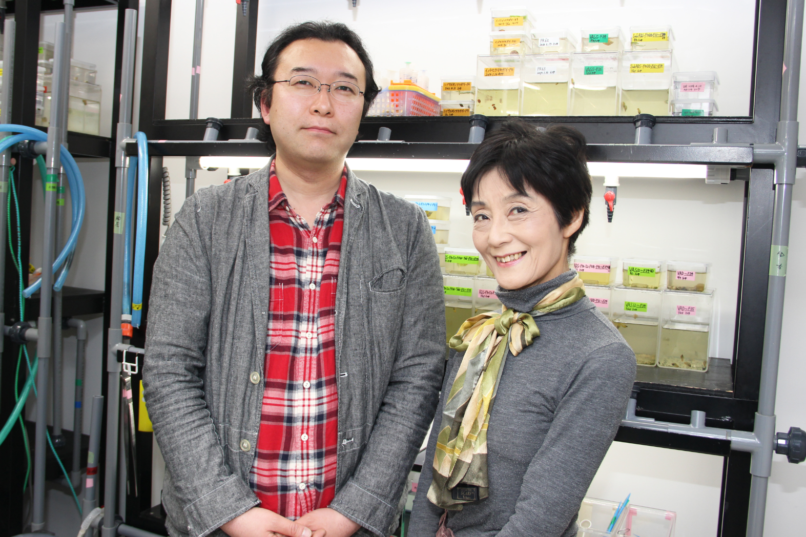
During development, mechanical forces sculpt tissues into myriad forms. Actomyosin contractility generated within the cell has an increasingly appreciated role in this process, but how tissue forces relate to the physical properties of the extracellular matrix is still poorly understood, particularly at longer time scales and the whole tissue level. A new paper in Development addresses these issues using Drosophila leg development as a model, taking advantage of an ex vivo culturing method. We caught up with first author Amsha Proag and last author Magali Suzanne, group leader at the Centre for Integrative Biology in Toulouse, France, to hear more about the story.
Amsha and Magali
Magali, can you give us your scientific biography and the questions your lab is trying to answer?
MS My main motivation is to push the limits of our knowledge, probably like many scientists. I discovered Drosophila during my PhD and was amazed by the power of this model organism. I then focused on how tissues acquire their shape or morphogenesis, an aspect that still fascinates me. I grew up surrounded by artists, and particularly sculptors, and this probably had an influence on the fascination I have for tissue shapes. When I settled my lab in 2011, we were initially working on a particular aspect of morphogenesis, the impact of cell death on the final shape of a tissue. We discovered that dying cells mechanically influence their surroundings and this led us to the biomechanics field. We are now interested more broadly in how mechanical signals are integrated at the tissue level to generate new shapes.
And Amsha, how did you come to work in the Suzanne lab, and what drives your research?
AP What first attracted me was hearing Dr Suzanne explaining some of her findings at a conference, which demonstrated how tissue shape arose from exquisite mechanical communication between cells. I remember asking her after the talk about the mechanisms behind this spatial patterning, which strongly appealed to my background in soft matter physics. The interplay between physical forces and adaptive biological behaviour remains a fascinating topic for me and a major question in developmental biology.
The interplay between physical forces and adaptive biological behaviour remains a fascinating topic
Why are cultured Drosophila leg discs a useful system to understand developmental mechanics? Was much known about the forces driving eversion before your work?
AP & MS First, the leg disc constitutes an isolated tissue that is able to pursue its development ex vivo, and is thus directly accessible to live microscopy and micro-manipulation. Second, it is small enough for multiscale investigation of morphogenesis (integrating cytoskeleton dynamics, single cell behaviour and tissue-scale mechanics).
Although little was known before our work on leg disc eversion, a few papers described the process of wing disc eversion, showing on one hand that Myosin II was important for peripodial epithelium (PE) opening and eversion, and on the other hand that extracellular matrix (ECM) was being degraded in order to allow disc eversion. The novelty in our paper, following the dynamics of both the ECM and the cell monolayer of the PE, is the discovery of their physical and mechanical uncoupling during leg disc eversion.
Can you give us the key results of the paper in a paragraph?
AP & MS The leg disc is enclosed in the PE, a thin tissue that opens, contracts and is removed to allow the leg to evert. Our work shows that the tension produced by the growing leg disc on the PE is at first mainly borne by the ECM. But as the leg elongates, the ECM and cell layer are progressively uncoupled and tension builds up in the cell monolayer. Then, each layer of the peripodial epithelium withdraws by a different mechanism. The ECM layer is opened by local proteolysis and its tension completes its removal, whereas the cell monolayer opens and is removed by Myosin-II-dependent contraction, independently of ECM degradation.
How do you think the PE cells stay alive and seemingly happy without attachment to the ECM?
AP & MS This came as a surprise at first: seeing holes form in the ECM layer while the PE cells remained cohesive told us here was something worth investigating. Indeed, single adherent cells will tend to undergo apoptosis when cultured on non-adhesive or soft substrates. However, PE cells remain attached to each other after the detachment from the ECM. In addition, apoptotic markers only appear at the free edge of the PE, where cells have lost half their intercellular adhesion as well as their contractility. Hence, we hypothesise that intercellular adhesion compensates for basal adhesion by providing sufficient survival signals, through a combination of adherens junction signalling and mechanotransduction. This in vivo behaviour recalls recent observations on cultured cell suspended monolayers and calls for characterisation as a general property of epithelia.
Cell-cell junctions in the peripodial epithelium were visualised with fluorescent α-catenin and colour-coded according to their orientation.
When doing the research, did you have any particular result or eureka moment that has stuck with you?
AP The first time I saw individual collagen structures, fibre bundles, changed my perception of the ECM basement membrane. I grasped how thin it was compared to my previous view of ECM as an amorphous gel. It made me consider other properties of the layer, such as the propensity to form holes, but also the ability to resist tension. It was also a nice demonstration of the benefits of looking at the same system using different setups.
And what about the flipside: any moments of frustration or despair?
AP Investigating a living tissue requires some patience as several technical requirements have to be met simultaneously and even successful experiments tend to bring out the complexity of the tissue. Thankfully, these challenges also act as spurs.
So what next for you after this paper?
AP I plan on developing quantitative approaches for the life sciences, mainly through automated image and data analysis. In addition, I wish to contribute to bridging biology and physics and am currently working on undergraduate teaching material to this aim.
Where will this work take the Suzanne lab?
MS Until now, we were focusing on single cell dynamics and their influence on their direct surroundings, at a local scale. The study of peripodial epithelium dynamics opens new doors in considering biomechanics at the tissue scale.
Finally, let’s move outside the lab – what do you like to do in your spare time in Toulouse?
AP A little badminton, a lot of reading. I am also involved in the Human Library organisation, which aims at removing barriers between people through conversation.
MS Toulouse is a very nice place for hiking with the Pyrenees close by. There is also a number of festivals I enjoy a lot such as the Short Film festival, the image festival ‘MAP’, or the jazz festival ‘Jazz sur son 31’.
Of all the charts being ridiculed at WTFviz, many get shamed for their lack of a zero-baseline. When teaching DataViz, zero-baselines are invariably a topic of debate. The rules about zero baselines are necessary are often unclear. Therefore, let’s quickly recap.
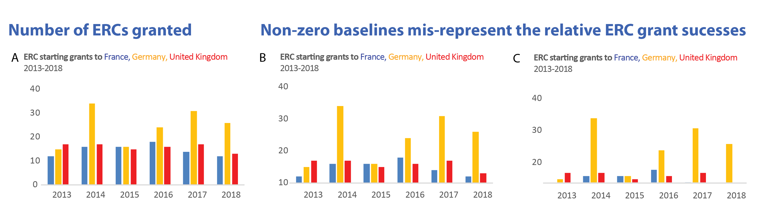
Bar charts: always show zero
When we ecnode amounts by length, as done in bar charts, the zero-baseline is critical to reading the data. A bar twice as long represents that the category has twice the amount of counts. The number of the prestigious ERC starting grants to German host institutes roughly doubled from 2013 to 2014, correctly encoded by a bar twice the size in 2014 (A).
When the y-axis does not start at zero (B), the increase from 2013 to 2014 is over-emphasized and looks 4 to 5 times bigger. In (C) the baselines even starts above the first data point. This leaves entire bars out of the graph. The result is, that it appears as if only Germany received ERC funding in 2018.
Non-zero baselines (and also axis-breaks in bar charts!) skew the relative difference between categories and mislead. But non-zero baselines are often used to save space, not to intentionally mislead. Then, the chart could simply be shown with a less overall height. This option maintains the relative bar sizes faithfully. When reading bar charts we are always interested in relative, not absolute size differences among our categories. (And I learned that Israel is part of the ERC funding consortium!)
Number of submitted ERC grants varies a lot across countries. Varying the physical height of the plot faithfully maintains the relative differences.
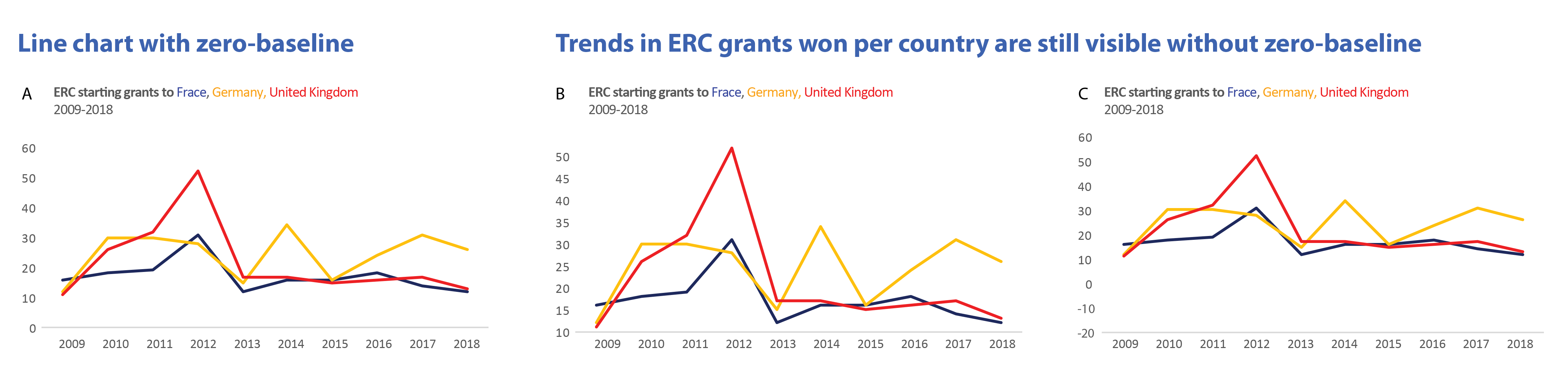
Line charts are happy without zero
The situation is entirely different for line charts. We use them to show trends, e.g. increase or decrease in categories over time. The rate of change is encoded by the line slope relative to the horizon. For this, its distance to zero is not critical. Even without the zero-baseline we see that ERC success in Germany fluctuates, while UK and France have stable funding rates. And, no matter where the zero-baseline is, why does the UK have such a curious funding peak in 2012, what happened there!?
For understanding trends in line charts, we do not critically need to see the zero baseline.
Sometimes showing zero is misleading
Showing a zero-baseline is sometimes misleading in line charts. Think of a fever curve with a human body temperature scale from 0-100 ˚C. Such a scale would prevent us from seeing a life-threatening increase of 1˚, from 39 to 40˚C in a patient. Similarly, showing global temperatures at a scale from 0 to 120˚C results in an entirely flat line. It was used by opponents of climate research to hide man-made global temperature changes. (And an outcry at twitter swiftly followed).
Line chart misleading BECAUSE of a zero-baseline. Tweet: @EcoSenseNow, 23rd April 2019
Distributions: it depends on the data
When showing statistical summaries, again the zero often is not necessary to be visible. We are interested in the shape of the data (normal or bimodal), it’s median, and outliers. How far the majority of data points are from zero is not usually of interest as long as all data is shown. Instead, the relative distance of individual data points from each other are key.
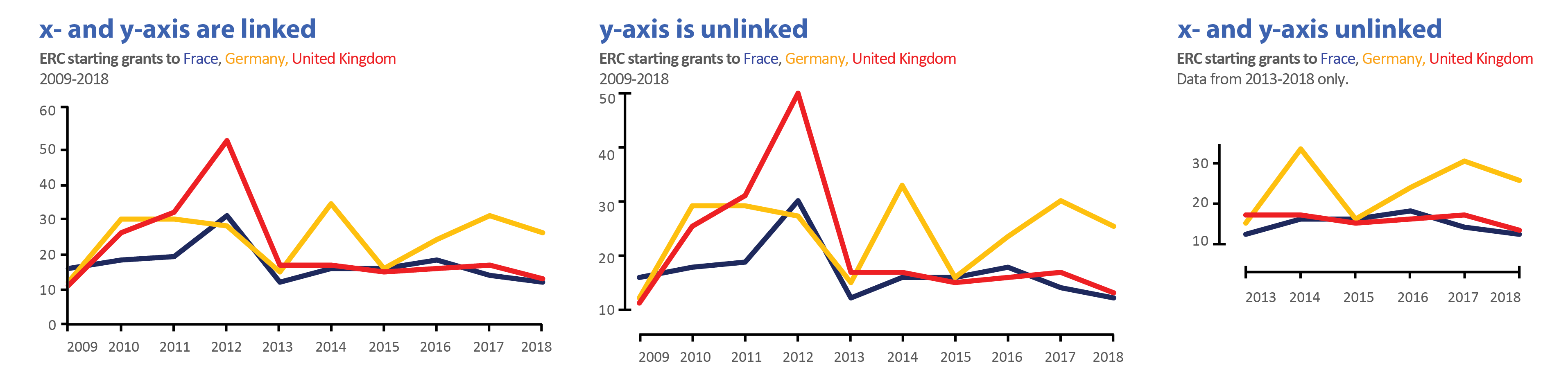
Good practice for non-zero baselines
When using non-zero baselines, the common practice is to unlink the x- and y-axes. For educational purposes I cut data from the right example. This is a dangerous territory and in some cases may be misleading the audience. In this example, I effectively hide the early lead of the UK in winning ERCs!
One possibility to alert readers to a non-zero baseline in your charts.
What did Watson and Crick discover? Rosalind Franklin’s notes…
In this episode from our centenary series exploring 100 ideas in genetics, we’re unravelling the story of the double helix, cracking the triplet code, and sketching out a Punnett square.
If you enjoy the show, please do rate and review and spread the word. And you can always send feedback and suggestions for future episodes and guests to podcast@geneticsunzipped.com
We are looking for a lab technician to assist in research on muscle stem cells, development, regeneration, disease, and evolution. More details about our research can be found at http://www.kardonlab.org/. Technician will assist in management of a mouse colony and conduct supervised research (leading to publications). Technician must be reliable, well organized, detail-oriented, excited about research and committed to working in our lab for at least two years. Prior lab experience is preferred (although not necessarily required), and class work in biology and enthusiasm for science is essential. Lab is located at the University of Utah in Salt Lake City, affording amazing opportunities for science and outdoor recreation. Looking for someone to start July-August 2019.
Contact Gabrielle Kardon (gkardon@genetics.utah.edu) with CV, list of references, and a brief statement about why you are interested in the position. BS or BA required.
One of the most obvious examples of left-right asymmetry in animal bodies comes from snails: in most species or strains, the shells coil dextrally, but some coil sinistrally. The control of coiling is genetic and begins in the early embryo. Previous work has implicated the formin diaphanous in the regulation of snail shell chirality, and a new paper in Development now decisively proves its involvement, thanks to the first application of CRISPR/Cas9 gene knockouts in molluscs. We caught up with the author team behind the paper: Masanori Abe and his supervisor Reiko Kuroda, Professor at Chubu University in Japan (recently moved from Tokyo University of Science), to find out more.
Masanori and Reiko
Reiko, can you give us your scientific biography and the questions your lab is trying to answer?
RK During my PhD at the University of Tokyo and in my first postdoc period under Professor Stephen Mason at King’s College London, I studied chemistry and carried out research on X-ray crystallography and spectroscopy of chiral transition-metal complexes. I then moved to the Biophysics Department and worked on DNA-carcinogen/anti-cancer drug interactions using X-ray crystallography, computer graphics and molecular biology. I was appointed Associate Professor and then Professor in the Department of Chemistry, Graduate School of Arts and Sciences of the University of Tokyo, and then recently, Professor at Tokyo University of Science, and very recently Professor at Chubu University. As I am interested in chirality, my attention was inevitably drawn to the molecular mechanisms of snail coiling. I was a late starter in developmental biology, but I have now been working in the field for the last 20 years.
In 1999, I coined the term ‘chiromorphology’, which expresses the concept of my research to link macroscopic and microscopic morphological phenomena in both biology and chemistry domains through chirality. Eventually, I hope to understand the impact and the origin of the homochiral biological world [in which almost all proteins contain only L-amino acids and RNA/DNA contain D-(deoxy)ribose]. In the area of crystallography, I try to understand how billions of molecules gather to form either chiral or non-chiral crystals, and how chirality is generated, recognized, transferred and/or amplified in the solid state. In the area of spectroscopy, I have developed chiroptical spectrophotometers, which can analyze samples in their condensed phases, such as the crystalline, gel or membrane state, without the contamination of artefact signals that can arise from intrinsic macroscopic anisotropy. We have used them to explore the dynamics of secondary-structure transitions of β-amyloids and hornet silk, in addition to the crystals of organic and inorganic compounds.
Masanori: how did you come to work with Reiko, and what drives your research?
MA When I joined the Kuroda lab at the Graduate School of the University of Tokyo, I was initially not interested in snails at all, but in research on DNA recognition compounds and proteins. However, when this research got stuck, I decided to participate in the snail work. And so far, I have been very excited about the elucidation of the mechanism of left-right asymmetry determination in snails. I think it suits me to look for interesting things that people don’t normally pay attention to, and this research is just that.
How was Lsdia1 first implicated as a candidate chirality gene, and why did you need to turn to CRISPR/Cas9 to prove its role decisively?
RK We reached the conclusion that Lsdia1 is the strongest candidate for the handedness-determining gene based on positional cloning and on various experiments using pure dextral, pure sinistral and F10backcrossed lines that we had established. We found a sinistral strain that carries a frameshift mutation that abrogates full-length LsDia1 protein expression, already at the one-cell stage (published in 2016). Although we were confident about the results, we could not ignore the possibility that Lsdia1 is simply genetically linked to a true handedness-determining gene. We envisaged that directly knocking out the Lsdia1 gene by CRISPR/Cas9 would provide proof. Although only MA and RK are involved in the current work, we are grateful to all the scientists/technicians/students who worked in Kuroda lab in the past for their contributions in establishing pure and backcrossed strains, rearing the snails, and enabling positional cloning, and to the Japan Science and Technology Agency (JST) for the funding of ERATO and SORST Kuroda Chiromorphology projects (to RK, 1999-2004 and 2004-2009, respectively).
Can you give us the key results of the paper in a paragraph?
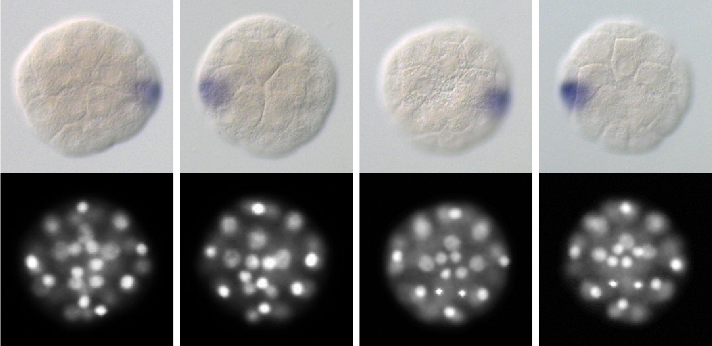
RK We have decisively identified that Lsdia1 is the long-sought handedness-determining gene of Lymnaea stagnalis by developing CRISPR/Cas9 gene editing techniques. Biallelic frameshift mutations introduced into the gene produced sinistrally coiled offspring generation after generation, in the otherwise totally dextral genetic background. We could also show that the gene sets the chirality already at the one-cell stage, the earliest observed symmetry-breaking event linked directly to body-handedness in the animal kingdom. The early intracellular chirality is superseded by the intercellular chirality during the third cleavage, leading to asymmetric expression of nodal and Pitx (two genes known to regulate chirality in vertebrates) and then to organismal body handedness.
Gene-edited F1 snails (49-cell stage), with in situ hybridisation for nodal, DAPI staining and selected cell types labelled.
How do you think chirality is transmitted through development?
RK This is the project we are currently working on. It is interesting that intracellular chirality within a cell is superseded by the intercellular chirality during the third cleavage (as proven by the fate of mechanically manipulated embryos at this stage), and then to the 24-cell stage when the cell fates are determined. We hope to report the results in the near future.
Left-right (LR) asymmetry is a widespread feature of animals – how much conservation do you think there is at a genetic and cytoskeletal level?
RK Although diverse mechanisms have been proposed for different phyla, we think a unified mechanism and the involvement of cellular chirality are probable. We believe that there are conserved mechanisms, given that diaphanous is a protein present in many phyla. We plan to elucidate the mechanisms of chirality establishment by LsDia1 at the molecular level, which may give insight into the conservation of the genetic control of LR asymmetry.
Do you have any advice for people wanting to do CRISPR/Cas9 mutagenesis in snails?
RK Gene editing of snails by CRISPR/Cas9 is not particularly difficult, as the method itself has already been well established in model organisms, although one must check for possible off-target effects. The most difficult aspects are raising the gene-edited eggs to adult snails, and successfully breeding them to produce the next generation.
When doing the research, did you have any particular result or eureka moment that has stuck with you?
MA Since it was not guaranteed that Lsdia1 was a handedness-determining gene, I sometimes felt anxious that the gene-edited individuals created over a long period would be wasted. At that time, the F0 individuals were starting to lay eggs, so I decided to count the number of hatched juveniles, and then felt a strong sense of discomfort. At that moment, I realized that the direction of shell coiling had changed into sinistral! The juvenile snails did not show any abnormalities except the shell-coiling inversion and moved around in the Petri dish vigorously. Then I was finally convinced that Lsdia1 was the snail handedness-determining gene. I was also surprised when I found one-cell-stage chirality.
And what about the flipside: any moments of frustration or despair?
MA It can sometimes get frustrating spending a long time caring for snails. Containers for breeding individuals should be cleaned weekly, thus I can’t take a long vacation leaving the lab. In particular, we are doing experiments with eggs just laid, but the quality of the eggs varies greatly depending on the health and age of the parent snail. It can’t be avoided in order to obtain correct results with good reproducibility.
So what next for you after this paper?
MA It was found that LsDia1 and LsDia2 give LR asymmetry to the one-cell-stage egg. Next, I would like to investigate the origin of organismal LR asymmetry by delving into the molecular level, focusing on the analysis of cytoskeleton dynamics in snails. In addition, as we have established gene editing technique for this snail, which is often used in the study of perception, learning and memory, I hope that our work will help this snail become a model organism for human disease research.
Where will this work take the Kuroda lab?
RK When one question is solved, more questions arise. We have so many interesting themes to work on: the roles of LsDia1, LsDia2 and other proteins in explaining the Dia1-dependent chirality within one cell, how the intracellular chirality is superseded by intercellular chirality (cytoskeletal dynamics), and how information on the geometrical arrangement of blastomeres at the eight-cell stage is transferred to the 24-cell stage, and then to nodal/Pitx expression, at the molecular level. My long-term research objective is to link macroscopic and microscopic phenomena, i.e. across the biological hierarchy, using chirality.
When one question is solved, more questions arise
Finally, let’s move outside the lab – what do you like to do in your spare time in Tokyo?
MA I like taking a walk doing Pokemon Go as it is good exercise. There are many interesting spots in Tokyo, historic buildings and monuments, beautiful parks and cityscapes, etc. Also I enjoy making small discoveries and surprises even in the back alleys that I had never visited before.
RK We both moved this April from Tokyo to Chubu University, Kasugai, near Nagoya. When in Tokyo, I often went to special exhibitions at the National Science Museum (such as ‘Lascaux – Cave Paintings of the Ice Age’, ‘Hunters of the Ocean’, ‘Wine – the Exhibition’ and ‘Deep Sea’) or other museums and galleries in Tokyo (e.g. ‘Winne-the Pooh – the original drawings from the V&A Museum London’ and ‘Vermeer and Dutch Art’). Once we have settled down in Kasugai, I hope to explore the area.
 (No Ratings Yet)
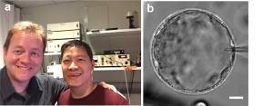
 (3 votes)
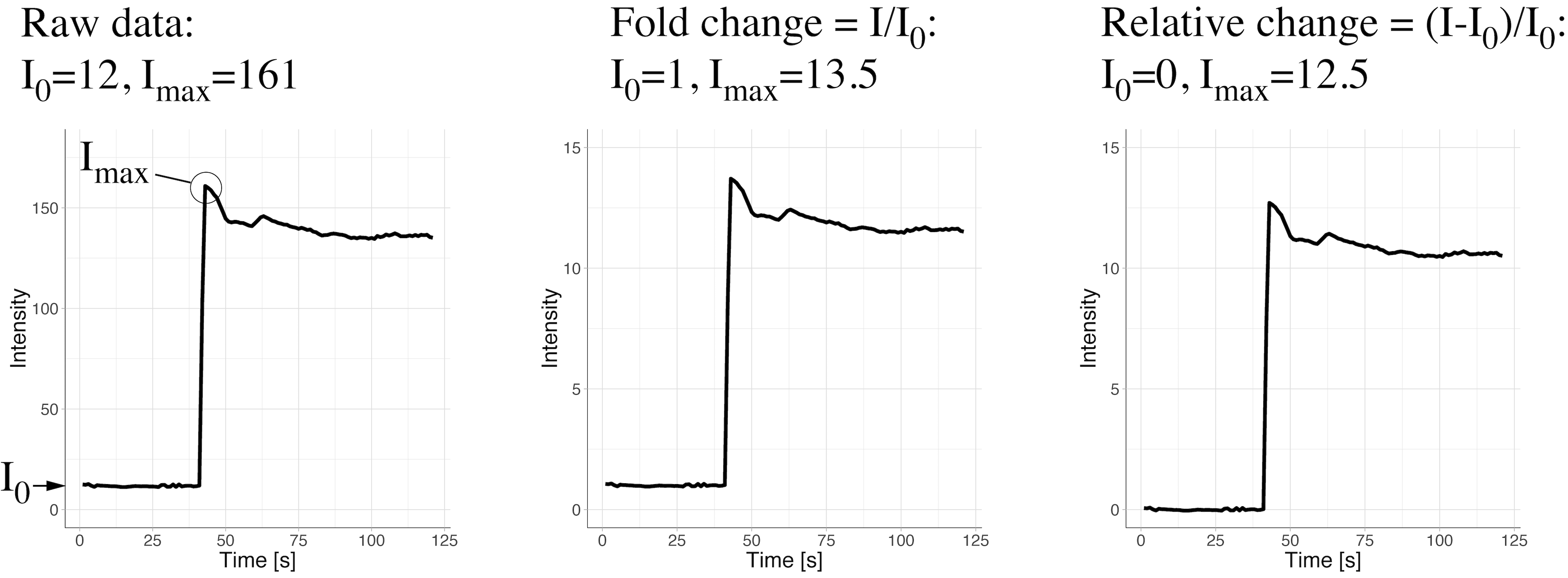

 (5 votes)


 What did Watson and Crick discover? Rosalind Franklin’s notes…