Closing Date: 15 March 2021
 The Faculty of Science and the Mathematical Institute invites applications for a
four-year
PhD Student on Mathematical Modeling of Cell-ECM Interactions During Angiogenesis (1.0 fte)
Project description
The opening is for a research position, within the field of mathematical or theoretical biology, computational physics, applied mathematics or computational science.
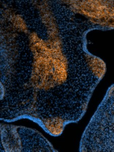
The outgrowth of new blood vessels from pre-existing vessels, called angiogenesis, is a crucial step in wound healing and tumor growth. Cell-based simulation models help to analyze how cells assemble into blood vessels and other tissue structures. Recently our group has developed novel techniques for modeling one of the key controlling factors of angiogenesis, the extracellular matrix (ECM). The ECM is a diverse class of jelly or hard materials providing structural support to the tissue and that acts as a medium for cell-cell communication.
The work will be embedded in the Multiscale Mathematical Biology team at the Mathematical Institute and the Institute of Biology Leiden. The team carries out mathematical biology research in close collaboration with experimental researchers. The team focuses on modelling pattern formation and morphogenesis in multicellular organisms and bacterial ecosystems. More information about the group can be found on biomodel.project.cwi.nl.
Key responsibilities
The project will be part of an NWO-funded Vici-project that will unravel how the extracellular matrix (ECM) coordinates the interactions between endothelial cells and other cells contributing to angiogenesis, and how modifications of the ECM, as they can occur for example near tumors, can modify the structure of new blood vessel networks. In this project you will work closely together with experimental researchers, that will test the mathematical models in the wetlab by imaging of cell cultures and zebrafish experiments. Your task will be to develop novel mathematical models, which will be informed by literature data and the insights coming from the wetlab. Your models will be experimentally validated by your direct colleagues in the wetlab, based on which you will correct and update your models. Based on cycles of iterative model refinement and experimental validation, you will unravel aspects of angiogenic sprouting, and prepare your insights for publication in the biological, biophysical, and/or biomathematical literature.
The mathematical models will be based on hybrid cellular Potts model, in which stochastic models of cell motility interact with continuum and discrete models of the ECM. You will contribute to the development of novel and efficient numerical methods for simulating the biomechanics of the ECM, that will interface naturally with pre-existing, stochastic models of endothelial cell behavior. You will integrate the novel methodology into our lab’s modeling framework Tissue Simulation Toolkit, and prepare the new developments for public release alongside our scientific publications.
Selection criteria
- Master’s degree in applied mathematics, computational/theoretical physics, theoretical biology, computer science or a related discipline;
- Excellent written and oral proficiency in English;
- Ability to work independently in a multidisciplinary environment;
- Fluent interdisciplinary communication skills with scientists in cell biology and mathematics;
- Keen interest in cell and developmental biology;
- Experience in (numerical) mathematical modeling (e.g., Cellular Potts modeling, particle-based simulations, PDEs, FBA);
- Excellent programming skills in C++.
Research at our faculty
The Faculty of Science is a world-class faculty where staff and students work together in a dynamic international environment. It is a faculty where personal and academic development are top priorities. Our people are driven by curiosity to expand fundamental knowledge and to look beyond the borders of their own discipline; their aim is to benefit science, and to make a contribution to addressing the major societal challenges of the future.
The research carried out at the Faculty of Science is very diverse, ranging from mathematics, information science, astronomy, physics, chemistry and bio-pharmaceutical sciences to biology and environmental sciences. The research activities are organized in eight institutes. These institutes offer eight bachelor’s and twelve master’s programmes. The faculty has grown strongly in recent years and now has more than 2,200 staff and almost 4,200 students. We are located at the heart of Leiden’s Bio Science Park, one of Europe’s biggest science parks, where university and business life come together. For more information, see http://www.science.leidenuniv.nl.
The Mathematical Institute (MI) is responsible for the research and education in mathematics and statistics at Leiden University. The institute has a strong international orientation. Its mission is to perform high quality research at the frontiers of mathematical knowledge, and to educate future generations of mathematicians and statisticians in a friendly but challenging environment. For more information see https://www.universiteitleiden.nl/en/science/mathematics.
The Institute of Biology Leiden (IBL) is an internationally oriented institute for research and education in biology. IBL performs top quality innovative fundamental and strategic research that will lead to scientific progress, contribute to solutions for societal challenges, and generate industrial opportunities, reflected in the general theme ‘Healthy Lives in a Changing World’. The Institute is organized in three multidisciplinary clusters: Animal Sciences & Health, Plant Sciences & Natural Products and Microbial Biotechnology & Health. For more information see: https://www.universiteitleiden.nl/en/science/biology. There is a large research community in The Netherlands (in particular, in Leiden), including many PhD students. There is close collaborating nationwide.
Terms and conditions
We offer a full-time position for initially one year. After a positive evaluation of the progress of the thesis, personal capabilities and compatibility the appointment will be extended by a further three years. Salary range from € 2,325.- to € 2,972.- gross per month (pay scale P, in accordance with the Collective Labour Agreement for Dutch Universities).
Leiden University offers an attractive benefits package with additional holiday (8%) and end-of-year bonuses(8.3 %), training and career development and sabbatical leave. Our individual choices model gives you some freedom to assemble your own set of terms and conditions. Candidates from outside the Netherlands may be eligible for a substantial tax break. Additional budget allows for research visits abroad and attendance of international conferences. More at http://www.workingat.leiden.edu/.
All our PhD students are embedded in the Leiden University Graduate School of Science www.graduateschools.leidenuniv.nl. Our graduate school offers several PhD training courses at three levels: professional courses, skills training and personal effectiveness. In addition, advanced courses to deepen scientific knowledge are offered by the research school.
Diversity
Leiden University is strongly committed to diversity within its community and especially welcomes applications from members of underrepresented groups.
Information
Enquiries about the position can be made to Prof. dr. Roeland Merks, merksrmh (on server:) math.leidenuniv.nl. Also see the group website at http://biomodel.project.cwi.nl.
Applications
To apply for this vacancy, please send an email to merksrmh (on server: ) math.leidenuniv.nl. Please ensure that you join to your application the vacancy number and the following additional documents:
• A letter of motivation
• An updated CV
• The letters of recommendation by 1 or 2 former supervisors
• The transcripts of your MSc studies
• Examples of previously written code
Only applications received before April 15th, 2019 will be considered.
Enquiries from agencies are not appreciated.
 (No Ratings Yet)
 Loading...
 last year and who has also served as Director on the board of The Company of Biologists, is now James Briscoe FRS! James’ lab at The Crick in London works on the molecular and cellular mechanisms of embryonic development with a particular focus on the developing spinal cord. You can hear more about his life in science in Katherine Brown’s Development interview, and his plans for Development in his inaugural editorial. Congratulations James!
 (1 votes)