The Giraldez laboratory at Yale University is seeking to recruit a highly qualified Associate Research Scientist as a long-term scientist in the laboratory (www.giraldezlab.org). Prerequisites for appointment on the research scientist track include a doctoral degree and relevant postdoctoral experience.
The successful candidate will bea highly-motived scientist with excellent organizational, mentoring and leadership skills. They will be responsible for coordinating the overall scientific operations of the Giraldez lab and will provide critical training and mentoring to individual lab members. In addition, the successful candidate will have the opportunity to participate in multiple research projects and drive a scientific project aligned with the major interests of the laboratory. The successful candidate will have the following attributes:
• A doctoral degree and relevant postdoctoral experience
• Excellent interpersonal and communication skills
• Excellent organizational skills and attention to detail
• Solid publication record and the ability to drive long-term, successful research projects
• Expertise in one or more of the following: molecular biology, chromatin biology, developmental biology, genomics, and/or imaging
This appointment can be renewed indefinitely provided the need for the position continues, the funding for the position is available, and the expectations for performance are met.
To apply to this position please submit the following using the Interfolio Link: https://apply.interfolio.com/50482:
1) A one-page cover letter describing your motivation for the position, research experience, and relevant mentoring/organizational experience,
2) biosketch or CV,
3) three letters of references
4) PDFs of three publications .
For inquiries please contact hiba.codore@yale.edu. Please include “Associate Research Scientist” in the subject of the email.
Applications are now open and will be considered on a rolling basis. Salary will be commensurate with experience and the appointment includes an attractive benefits package in line with an appointment in the research track within the Yale School of Medicine
Yale University is an Affirmative Action/Equal Opportunity employer. Yale values diversity among its students, staff, and faculty and strongly welcomes applications from women, persons with disabilities, protected veterans, and underrepresented minorities.
At the end of 2014, a friend asked me “What is your story”? I had just started my postdoc in the laboratory of Karla Neugebauer and was a bit perplexed by the direct question. I started talking about some loose project ideas of mine involving words like development, metabolism, and RNA – after all, I joined an RNA lab after doing my PhD on fly development, growth control, and lipid metabolism. But the story behind our paper did not start for another few months.
Historically, metabolism and developmental biology are deeply connected. This was emphasized in Joseph Needham’s mammoth three-volume work “Chemical Embryology” published in 1931 on the subject of physico-chemical embryology. This research faded away since the molecularization of experimental embryology, but over the last few years has reemerged as a quantitative field of study in developmental biology. Two consecutive meetings, the 2016 Company of Biologist Workshop ‘Metabolism in Development and Disease’ and the 2017 EMBO Symposium ‘Metabolism in Time and Space’ (Krejci & Tennessen 2017), highlighted that developmental biologists are once again investigating the role of cellular metabolism in growth, differentiation, and maturation during development. This resurgence of metabolisms role in development sets the stage for our story.
Developing embryos, like all living systems, are open systems exchanging energy and matter with their environment. They function out of equilibrium and require a continuous supply of energy to remain alive. From a thermodynamic perspective, metabolism can be regarded as an energy converter that directs energy from nutrients through an interconnected web of chemical reactions to meet the energetic and biosynthetic demands of growth, proliferation, and development. An emerging view is that this bioenergetic function of metabolism is tightly regulated in time and space in order to fulfill the changing energetic and biosynthetic demands of animal development (See Miyazawa & Aulehla 2018 and Gándara & Wappner 2018 for reviews). One particular example is that highly proliferative cells exploit increased glycolytic activity even in the presence of oxygen – a metabolic state known as aerobic glycolysis or the Warburg effect (Ward & Thompson 2012). How and why cells adopt this metabolic state has been the focus of intense research in the last decade and is not entirely understood. It has been recognized that this metabolic wiring supports the increased energetic and biosynthetic demands necessary for rapid cell growth and proliferation during development and disease. Indeed, implanted mammalian embryos seem to utilize Warburg metabolism with high levels of glucose uptake, glycolytic activity, and lactate production. Interestingly, early embryos undergoing rapid reductive cleavage divisions do not rely on glucose but rather use pyruvate, lactate and amino acids as energy sources (Gardner 1998). This indicates that early embryos rely on respiration to proliferate and switch their metabolic state during the transition from the cleavage to the blastocyst stage. This metabolic switch seems to coincide with the transition from maternal to zygotic instructions of developmental control known as maternal-to-zygotic transition (MZT) when maternally deposited RNA is degraded and zygotic gene expression is initiated.
Karla Neugebauer’s laboratory at Yale University focuses on the regulation of transcription and splicing in a variety of biological contexts, including MZT. When I joined the lab, RNA-interactome studies had just identified many ‘classical’ metabolic enzymes of intermediary metabolism as RNA-binding proteins and I was intrigued by the idea of combining expertise with the Neugebauer lab to study the dynamic interplay between transcription, RNA-binding proteins and metabolism during early zebrafish embryogenesis, particularly during MZT. Although this was the original idea for my project in her lab if you’ve read our paper you know that we ended up elsewhere… using calorimetry to measure the heat flow between a developing embryo and its surroundings. How did the project change so drastically? Stay with me, as I will explain in the next few paragraphs.
The change in a metabolic state is usually associated with a change in the energetic and biosynthetic requirements of the system under investigation. In the case of embryonic cleavage stage development, cells lack growth phases and thus become smaller as they divide. One could hypothesize that they use oxidative phosphorylation because of the absence of volumetric growth. However, increasing embryonic cell number demands precursors (e.g. nucleotides, fatty acids, and amino acids) for DNA replication, an increase in plasma membranes, and protein synthesis. Thus, the embryo must produce and/or polymerize the precursors necessary for cleavage stage development. Each cell of the embryo must also expend energy to assemble and disassemble cellular machinery (e.g. chromatin, mitotic spindles), generate forces needed to segregate the chromosomes and divide the cell, and change the activity of signaling pathways that enforce cell cycle phasing, even in the absence of volumetric growth. As development proceeds and the embryonic cell cycle gains G1 and G2 phases, the embryo faces the additional energetic demands of volumetric growth and might switch to aerobic glycolysis as a metabolic strategy to fulfill those. But what are those energetic demands? I realized that we lack a quantitative understanding of how the metabolic energy converted by different metabolic states is partitioned among the complex array of cellular processes that take place during cell growth, proliferation, and development. What if we could put numbers on energetic requirements of making a new cell or an embryo? Maybe then we will be able to understand why metabolism functions the way it does. I became fascinated by the thought of studying the energy budget of early embryogenesis and my focus shifted away from the regulation and interplay of RNA biology and metabolism during MZT. This is where our story truly started – with the question of how to measure the energy required for embryonic development.
The amount of energy dissipated by an animal per unit of time is defined as metabolic rate and is reported in energy units per unit time in Watt (joule/second) or Watt per kg body mass (W/kg). An average human at rest consumes ~2000 kcal per day and dissipates energy in the form of heat at a rate of about 100 W or 1 W/kg (Joules/s*kg). These estimates are conceptually based on the first law of thermodynamics. The change in energy (2000 kcal /day) equals heat dissipated (100W) minus work. The average human is at rest – not growing or conserving any energy – and does not perform any net chemical synthesis or anabolic reactions. All the 2000 kcal/day is converted by catabolic reactions and immediately used by various cellular processes defined as maintenance reactions to keep the individual alive or away from thermodynamic equilibrium. Now let’s consider a growing organism which must perform anabolic reactions and invests energy into chemical synthesis of new biomass in order to grow. In this case, the organism conserves energy and performs net chemical synthesis. Measurements of heat dissipation now reflect the difference between energy consumption in the form of nutrients and energy conservation by biosynthesis, which is equal to the net change in enthalpy of all the reactions taking place in the system. This is how heat dissipation differs from respiratory metabolic rate measurements such as O2 consumption or CO2 production. They reflect rates of catabolic reactions and are inherently blind towards energy conservation in the system by anabolic reactions.
Heat dissipation is directly connected to the laws of thermodynamics. When approaching this question, I thought, “maybe we can measure the heat flow between developing embryos and their environment using calorimetry.” It’s also possible that I always wanted to stick a developing organism in a calorimeter to see what I could measure… Isothermal titration calorimetry (ITC) is traditionally used to measure the heat transfer associated with a biomolecular interaction to determine the binding constant, stoichiometry, enthalpy and entropy of the binding event in solution, without the need of labels. After spending a few weeks of optimizing a 17 years old ITC machine housed in the biophysical core facility into measuring heat flow for prolonged periods of time without titrations, we were ready to try to measure the heat flow associated with early development. It worked incredibly well – and I was thrilled when I saw the first heat flow data from early zebrafish embryos undergoing cleavage stage development (see picture).
Isothermal calorimetry set up used to measure the heat flow between zebrafish embryos undergoing cleavage stage development and their environment. Not the actual first experiment. #mymachinewithoutme.
The initial finding was that the heat flow between embryo’s undergoing cleavage stage development and their environment increased over time. Surprisingly, however, I discovered that there was a small (~2% of the mean) but reproducible heat flow oscillation superimposed on the steady increase. The period and number of oscillations matched the division cycles taking place during this phase of embryogenesis. The oscillations suggested the presence of cyclic energetic events associated with embryonic cell proliferation, leading us to wonder if the oscillations are associated with the embryonic cell cycle.
After the initial discovery, I started a collaboration with Joe Howard, a biophysicist, to tackle the theory and thermodynamic aspects of heat flow during embryonic development. In the summer of 2015, Joe was invited faculty for the physiology course at the Marine Biology Laboratory (MBL) in Woods Hole, Massachusetts. We decided to take the project to the MBL for two weeks and I had the pleasure to work with two amazing students on the energetic costs of embryonic development. Manuel Razo was interested in the temperature scaling of the heat flow and Mathijs Vleugel started to investigate the underlying metabolic state and mitochondrial biology of embryos undergoing cleavage stage development. The two weeks at the physiology course and the MBL deeply influenced my thinking about the energetics of development and science in general. The course brings together scientists from diverse backgrounds and provides a truly collaborative and open environment. We started to throw ideas around and discussed what these heat flow oscillations could be? As the early embryonic cell cycle is solely composed of DNA replication, mitosis and cytokinesis, a prediction was that these heat flow oscillations could represent the energy used by either of those processes. My personal bet was on mitosis – but it turned out we were all wrong.
The experiment which pointed us in a new direction was when I blocked both DNA replication and mitosis by inhibitors. To our surprise, the heat flow oscillations persisted with a similar period and amplitude as control treated embryos. What followed was a series of quantitative heat flow measurements combined with perturbation experiments, theoretical modeling, and order of magnitude estimates for energetic costs of oscillatory cellular processes to investigate the underlying principle of the heat flow oscillations. We were able to show that they were driven by the phosphorylation and dephosphorylation reactions catalyzed by the cell cycle oscillator, the biochemical network controlling mitotic entry and exit, and thus revealed the energetic costs of cell cycle signaling.
In summary, the story started with an unconventional idea and a discovery. It evolved to show that quantitative heat flow measurements combined with perturbation experiments, theoretical modeling, and order-of-magnitude estimates can be a powerful approach to dissect the energetic costs of various cellular processes driving embryogenesis. In our work, we postulated that the energetic cost of cell cycle signaling likely reflects the thermodynamic burden of imposing accurate and robust timing on cell proliferation during development, as predicted by a theoretical tradeoff between energy dissipation and precision of biomolecular oscillators. I am currently working on establishing systems to measure the accuracy of the embryonic cell cycle and modulate the oscillatory heat flow amplitude and phase to test these in-silico predictions in-vivo and in-vitro. Furthermore, we have been able to allocate the oscillatory component to the energetic cost of the cell cycle signaling representing 2% of embryos total energy expenditure. What about the other 98%? Why does it increase during cleavage stage development – even absence of volumetric growth?
Searching for a post-doc who is passionate about both teaching and research. We are studying the interplay between division and inductive signaling. In particular, we are exploring how signaling receptors are trafficked in dividing cells to generate asymmetric induction of a cardiac progenitor lineage. We are also using comparative genomics to explore the evolutionary constraints that shape gene regulatory networks. We study these questions in the invertebrate chordate, Ciona robusta. Ciona embryos consists of extremely low cell numbers allowing high resolution analysis of intra-cellular dynamics in intact embryos. The ease of generating transgenic Ciona embryos make this an excellent model organism for undergraduate research. We have recently initiated a collaboration with Danelle Devenport at Princeton focused on similar processes in cultured mouse epithelial cells. This is a great opportunity for a post-doc with an interest in undergraduate teaching and research. My former post-doc is moving on to a tenure-track position at a small liberal arts college and it was clear that the combination of a strong research record along with a demonstrated commitment to undergraduate mentoring and teaching made her a strong candidate for these very competitive positions.
Applicants should have a PhD (or be close to completing one) in a relevant subject area. Excellent communication skills and a commitment to undergraduate mentoring are essential.
To apply or if you have questions about the position – please send your CV and a cover letter describing your interest to Bradley Davidson at bdavids1@swarthmore.edu. I will be in touch with instructions for submitting a formal application.
The Young Embryologist Network (YEN), is an academic body aiming to bring together early career scientists within the wide field of developmental biology, in order to provide opportunities to present talks and posters, network and collaborate, and gain research or career advice.
YEN was set up in 2008 by graduate students in the prestigious Department of Cell and Developmental Biology at University College London. Every year, the YEN hosts an annual conference at a UK research institution with great success. The conference is entirely organised by graduate students and junior post-doctoral scientists, and has remained free to attend since 2008, due to the generosity of sponsors and grants.
The 2019 conference is being held on the 13th of May 2019 at the Francis Crick Institute in London.
The deadline for Registration is the 19th April. Register here:
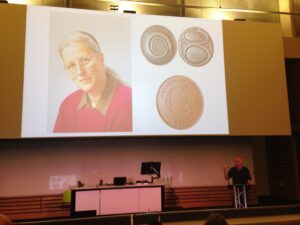
The Beddington Medal is the BSDB’s major commendation to promising young biologists, awarded for the best PhD thesis in Developmental Biology defended in the year previous to the award. Rosa Beddington was one of the greatest talents and inspirational leaders in the field of developmental biology. Rosa made an enormous contribution to the field in general and to the BSDB in particular, so it seemed entirely appropriate that the Society should establish a lasting memorial to her. The design of the medal, mice on a stylised DNA helix, is from artwork by Rosa herself. We would like to congratulate the 2019 winner of the Beddington Medal, David Munro, and would like to take this opportunity to give a brief overview of his career and the PhD project that was awarded the Beddington medal.
Jim Smith introduced the Beddington medal with heartfelt memories of Rosa Beddington and her time at the NIMR. Please read more of his thoughts here.
Some complicated selfies were taken as the medal was passed over before David went on to present the work that has deserved him this award.
In the words of his PhD supervisor:
“The really impressive thing about David’s work is that he did not come to my lab to fit in with an existing line of research but created one of his own”. Jamie Davies, University of Edinburgh.
David received his undergraduate degree in Sport and Exercise Science at the University of Stirling (2010-2014). With this, he achieved a first-class honours degree and the prize for the best overall performance throughout a physiology related degree (British Physiological Society Undergraduate Prize). His dissertation project investigated associations between ADRB2 mutations (an adrenaline receptor gene in humans) and athlete status/athletic ability measurements. Subsequently, he was awarded a University of Stirling Head of School Summer Bursary Award to remain in Stirling during the summer of 2014 and investigate the relationship between transcribed ultra-conserved regions of RNA (T-UCRs) and the development of diet-induced insulin resistance in humans (Summer 2014). He then moved to the University of Edinburgh for his MSc by Research in Biomedical Sciences (2014-2015). Again, he received a distinction and was awarded the Class Prize for best student. During this time, he studied the physiology of S-acylation the regulation of skeletal muscle energy expenditure by an obesity-associated phospholipase as part of two research placements.
David has been awarded the Beddington medal for his exceptional work performed during his 3-year MRC-funded PhD at the University of Edinburgh with Prof Jamie Davies and Dr Peter Hohenstein (2015-2018): The thesis is titled ‘Mechanisms of kidney vascularisation and the roles of macrophages in renal organogenesis’. During his PhD, he gave several oral and poster presentations at national and international conferences, supervised students (including a Gurdon Summer Studentship Awardee), established numerous international collaborations, was awarded travel grants (including a BSDB Conference Grant), and reviewed manuscripts for leading journals (including Cell Reports, Angiogenesis, and Scientific Reports). He is now a post-doctoral fellow at the UK Dementia Research Institute (University of Edinburgh; 2019- present), continuing research in macrophage biology under the supervision of Prof Josef Priller. His current focus in on brain macrophages (microglia) in development, neurodegeneration, and aging.
Thesis description
Kidneys are specialised organs that clean the blood, removing waste while retaining what is useful. This requires a complex vasculature, and its formation as a foetus develops is poorly understood. I started my PhD research by using advanced microscopy techniques to visualise how blood vessels form in three-dimensions in the mouse kidney. In doing so, I identified when and from where the first blood vessels enter the kidney, and how blood vessels pattern at the edge of the kidney throughout development.
Blood vessels can form through angiogenesis (branching of new vessels from pre-existing ones) and/or vasculogenesis (assembly of new vessels from the coalescence of endothelial precursor cells). It has long been thought that a combination of both processes occurs during kidney vascularisation; however, my thesis work indicates that this concept may not be correct. My data instead suggest that kidney vascularization relies on growth and remodelling of pre-existing vessels (angiogenesis) and does not depend on vasculogenesis at any point (Publications 1 and 5 in CV). When assessing the entire 3D vascular tree of the kidney, isolated endothelial cells were never observed at any developmental age. Instead, all vessels, including the newly forming ones, were connected to pre-existing vessels that could be traced to the major circulatory vessels.
I then focused on the blood vessels at the edge of the kidney, which I found to consistently and accurately pattern around a special collection of cells – the cap mesenchyme. The cap mesenchyme contains cells that eventually become the cleaning tubes of the kidney, the nephrons. This cell population undergoes rounds of splitting at the kidney’s periphery. As this happens, I demonstrated that blood vessels migrate through the newly opened regions between the separating cap mesenchymal populations (Publication 1 in CV). This occurs in cycles throughout development and is likely to be vital for the oxygenation of the kidney’s outer region, the site where important processes such as nephron formation take place.
I determined that a signalling molecule, semaphorin-3f, and its receptor, neuropilin-2, were expressed in a pattern consistent with them having roles in this cyclical patterning of blood vessels; however, using mouse models where the genes for these molecules were deleted, I established that they were not vital for this process (Publication 2 in CV).
I next shifted my research focus towards a specialised cell type known as the macrophage (macro = big; phage = eater) in the developing kidney (Publication 3 in CV). Macrophages are immune cells best known for clearing foreign and damaged cells. These cells have vital roles during animal development, but little is known about their specific functions during kidney development.
Macrophages arrived in the mouse kidney early during its development, where they were required to clear away misplaced cells to ‘set-the-stage’ for early kidney development (Publication 6 in CV, under review). Throughout later development, most macrophages wrapped around blood vessels and I demonstrated their ability to eat endothelial cells (which usually line the blood vessels) and red blood cells (which are carried within them) within the kidney. I also established that kidney macrophages produced many molecules linked to blood vessel development, and so I examined the consequences of macrophage-loss on blood vessel formation. Blood vessels normally form continuous networks in the kidney; however, when macrophages were depleted (by blocking a macrophage-survival signalling pathway), connections between renal blood vessels were reduced (Publication 6 in CV).
Publications
Munro DAD, Hohenstein P, Davies JA. 2017. Cycles of vascular plexus formation within the nephrogenic zone of the developing kidney. Scientific Reports. 7: 3273.
Munro DAD, Hohenstein P, Coate TM, Davies JA. 2017. Refuting the hypothesis that semaphorin-3f/neuropilin-2 guide endothelial patterning around the cap mesenchyme in the developing kidney. Developmental Dynamics. 246:1047-1056.
Munro DAD, Hughes J. 2017. The Origins and Functions of Tissue-Resident Macrophages in Kidney Development. Frontiers in Physiology. 8:837. (Review)
Mills CG, Lawrence ML, Munro DAD, El-Hendawi M, Mullins JJ, Davies JA. 2017. Asymmetric BMP4 signalling improves the realism of kidney organoids. Scientific Reports. 7:14824.
Munro DAD, Davies JA. 2018. Vascularizing the kidney in the embryo and organoid: questioning assumptions about renal vasculogenesis. Journal of the American Society of Nephrology. (Perspectives article).
Munro DAD, et al. Macrophages restrict the nephrogenic field and promote endothelial connections during kidney development. eLife 2019;8:e43271 DOI: 10.7554/eLife.43271
A postdoctoral position is available in the laboratory of Dr. Jessica Mark Welch in the Bay Paul Center to study the spatial organization of microbial communities in the human mouth. The successful candidate will use fluorescence in situ hybridization, spectral imaging microscopy, and computational image analysis to investigate microbial community structure and will interact closely with collaborators at other institutions as well as with the vibrant and collegial MBL scientific community.
The position is for 2 years, and may be extended beyond this period contingent on securing additional funding.
Physical requirements: This position requires fine motor skills and willingness to work with potentially biohazardous materials and standard laboratory chemicals including fixatives and solvents.
Basic qualifications: A Ph.D. in biological sciences or a related field is required.
Preferred qualifications: Experience with confocal microscopy, computational image analysis, bioinformatics, and/or microbiology is desirable.
Instructions: To apply, please visit the MBL Employment Opportunities website: https://www.mbl.edu/hr/employment/. The following documents are required: (1) a cover letter describing your interests, skills, and prior research experience, including any specific experience with the job responsibilities listed above; (2) a curriculum vitae/resume; and (3) the names and contact numbers of three persons who can be contacted for letters of reference, at least one of whom must have acted as your supervisor in a previous research position.
The Rohner Lab at the Stowers Institute for Medical Research has an opening for a Postdoctoral Researcher to develop an independent project investigating the molecular, genetic, and developmental mechanisms of how cavefish maintain health under diabetes-like phenotypes. The lab has previously found that the cavefish Astyanax mexicanus develop high-blood sugar and insulin resistance as part of their natural strategy to survive in the caves but without the usually associated health problems (Riddle et al. Nature. 2018 Mar 29;555(7698):647-651). Visit http://research.stowers.org/rohnerlab/ for more information.
The selected candidate will investigate the molecular mechanism underlying these impressive adaptations. The candidate will closely work with the core facilities at the institute to perform single-cell RNA sequencing, proteomics, and functional validation in vitro and in vivo. The candidate will receive strong support from the core facilities that provide advice, training and service to enhance the Institute’s interdisciplinary and collaborative research programs. Current core facilities are staffed by over 100 scientists with expertise in bioinformatics, cytometry, histology, imaging, microarray, next generation sequencing, transgenic and ES cell technologies, proteomics and molecular biology. The Stowers Institute offers a highly competitive compensation and benefits package.
The position is funded for two years through a grant by the Juvenile Diabetes Research Foundation and can be renewed for up to five years in order to allow enough time to develop a research program/publication record that makes the postdoc a strong candidate for an independent position. The Rohner Lab has a strong commitment for mutual success and is dedicated to providing support for all lab members.
Minimum requirements include a doctoral degree in the life sciences, chemistry, or biomedical engineering. Experience in one or more of the following areas is desirable: molecular biology, developmental biology, genetics, genomics, evodevo, physiology.
In addition to excellent verbal and written communication skills, successful candidates must be dynamic and highly motivated, work independently and creatively, able to work in a team-oriented environment, and proficient at problem solving.
Application Instructions: To apply, please submit (1) a brief cover letter, (2) a current CV, and (3) contact information for two professional references to Dr. Nicolas Rohner at nro@stowers.org cc: careers@stowers.org.
About the Stowers Institute for Medical Research
The Stowers Institute for Medical Research is a world-class basic biomedical research organization focused on improving our understanding of fundamental mechanisms of biology and using this knowledge to guide the development of innovative treatments to improve human health.
Our dedicated scientists collaborate across a variety of disciplines, studying many different aspects of health and disease. A primary goal of our research is to understand the principles that guide the function and behavior of living organisms and individual cells. Discoveries resulting from this kind of research often prove to be major milestones along the path toward novel therapies and cures (visit www.stowers.org). Jim Stowers, founder of American Century Investments, and his wife, Virginia, opened the Institute in 2000. Since then, the Institute has spent over 900 million dollars in pursuit of its mission.
Currently, the Institute is home to almost 550 researchers and support personnel; over 20 independent research programs; and more than a dozen technology-development and core facilities. The Institute has been ranked 3rd place by the Scientist for best places to work in the world: https://www.the-scientist.com/features/best-places-to-work-academia-2012-40676
Kansas City is an emerging metropolitan city in the Midwest with a high quality of living and affordability. Visit https://www.visitkc.com for information about living and working in Kansas City.
We are looking for an intellectually curious and motivated postdoctoral fellow to join in the fun exploring how glia regulate neurogenesis and neuronal fate-specification. Our approaches are interdisciplinary and involve genetics, live imaging, computational modelling, single cell RNA sequencing, etc. Available projects include: (1) Understanding how signals from glia impart unique neuronal identities (a follow up to Fernandes et al., Science, 2017). (2) Characterising glial diversity in the visual system. (3) Exploring glial involvement in neuroepithelial proliferation.
Applicants should have a PhD in a relevant subject area (or be close to completing their degree), excellent communication skills, a collaborative spirit and a kind heart. The ideal candidate will have a strong background in molecular biology, cell and/or developmental biology as well as experience with imaging. Knowledge of signal transduction, Drosophila genetics and bioinformatics are a plus but not essential.
Formal applications will be accepted online through UCL’s job portal till May 3rd, 2019. If you cannot meet this deadline but would like to apply, please contact Dr. Fernandes as soon as possible.
Proposed start date: August 1st, 2019 (Flexible).
For more details please contact Dr. Fernandes at vilaiwan.fernandes@ucl.ac.uk (along with a CV and cover letter describing research interests).
Note: Our lab and UCL value and support diversity. Funding is guaranteed beyond Brexit.
The Bressan Laboratory (www.bressanlab.com) at the University of North Carolina Chapel Hill is inviting applications for a postdoctoral fellow interested in developmental Cell Biology and Physiology research. The focus of the position will be to explore the genetic and molecular events that control cellular diversity during cardiovascular development. Specifically, candidates will conduct direct in vivo over expression, live imaging, cell sorting, primary culture, and next generation sequencing to explore how alterations in transcriptional activity and cellular mechanics influence physiological fate in the embryonic heart. The applicant is expected to manage an independent research project and to train students and other fellows in the laboratory.
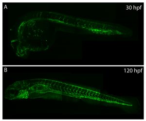
The correct patterning of embryonic tissues is essential for normal development. Aberrant patterning can lead to developmental abnormalities and pathogenic defects. Therefore, studying developmental patterning is important to better understand disease. The zebrafish embryo is a fantastic model for studying patterning during development owing to its optical clarity, small size and large clutch number. When coupled with dynamic transgenic lines, the picture of what occurs in the cell during these processes is starting to emerge.
The vasculature of zebrafish expands throughout development, providing developing tissues with oxygen and nutrients.
Understanding how the ubiquitous second messenger, the calcium ion, regulates cellular physiology during development has become an important question in biology. This is where my story begins. I arrived in Sheffield after obtaining a PhD position, eager for what lay ahead. The project was exciting; using new transgenic lines and cutting-edge microscopy to study the function of a poorly understood gene, tmem33, which we hypothesised would regulate calcium signalling within the developing vasculature. The preliminary morpholino knockdown data generated in my host lab before I started my PhD hinted at a vast cache of riches waiting to be uncovered. This data suggested a role for tmem33 in both vascular and kidney development. I was given some new toys to play with, a light sheet microscope and an endothelial-specific calcium reporter line. My first experiment in the lab was to analyse the mutant zebrafish line generated by my supervisor around which my PhD was supposed to focus. It was, of course, completely normal. No vascular defects here. It looked like my project was dead in the water before it had begun. My next few experiments yielded additional dead ends – analysing 34 TRP channels by in situ hybridisation, looking for specific vascular enrichment (spoiler alert – there wasn’t any).
Thankfully, science moves very quickly. My original project went from dead, to very dead as several papers at the time heavily criticised morpholino-based approaches (Kok et al, 2014; Schulte-Merker and Stainier, 2014; Stainier 2017; Robu, 2007) to back alive within the space of about six months. A single paper (Rossi et al, 2015) brought hopes for a revived project. In a series of elegant experiments, the authors described a situation where knockdown of the egfl7 gene by morpholino induced a robust vascular phenotype, but when this gene was mutated using genome editing the phenotype was absent. Interestingly, the authors showed that the egfl7 mutant displayed nonsense mediated decay of egfl7 transcripts and that when egfl7 morpholinos were injected into egfl7 mutants, the mutants were protected against the effects of the morpholino. The synthesis of these data was that there existed genetic compensation in the mutants but not in the morphants.
Finally, the stroke of luck I needed, our tmem33 mutants also showed nonsense mediated decay, so I set about injecting tmem33 morpholinos into our tmem33 mutants and I found strikingly similar results. This suggested that the reason the tmem33 mutants displayed no phenotype was because they displayed a kind of genetic protection, likely via a genetic compensation mechanism. I now had the beginnings of a successful project, a year in to my PhD. Rossi et. al used a new technology to address their issues, CRISPR interference (CRISPRi) – a modified version of the CRISPR/Cas9 system using an inactivated form of Cas9 (dCas9). The authors were able to reproduce the same phenotype they observed via morpholino knockdown using CRISPRi. I applied CRISPRi to knock down tmem33 and was able to reproduce our morpholino knockdown data of tmem33.
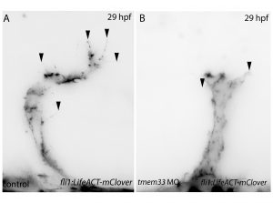
Filopodia are essential for cellular migration. Tmem33 morphants display reduced filopodia and delayed vascular migration.
Next, inspired by the conditional CRISPR approaches described in Ablain et al (2015), I sought to conditionally knock down tmem33 in endothelial cells by driving dCas9 expression under the control of the endothelial-specific fli1a promoter. Preliminary experiments using transient conditional knockdown were promising – only endothelial cells showed a phenotype! The next step was to generate a transgenic line which stably expressed dCas9 in endothelial cells.
Throughout this process, I had been studying tmem33 function with regards to endothelial cell physiology during angiogenesis. Since it was known that tmem33 functioned within the endoplasmic reticulum, using a calcium reporter line I began to test whether tmem33 knockdown altered endothelial calcium signalling. I found that tmem33 knockdown reduced observable endothelial calcium oscillations, which coincided with reduced endothelial cell migration. This was validated by both morpholinos and CRISPRi. Furthermore, I began using both knockdown approaches to position the tmem33 within the hierarchy of developmental angiogenic signalling. I found that tmem33 functions downstream of VEGF signalling but upstream of Notch and ERK signalling, identifying an essential function for calcium oscillations in mediating the response to Vascular Endothelial Growth Factor (VEGF) and inducing downstream signalling pathways essential during angiogenesis.
Calcium signalling during endothelial development.
Throughout my PhD, I’ve learned how the zebrafish can be a powerful tool for understanding basic biology and how a PhD is not a linear endeavour. You have to follow the research (and the results of others) and see where it takes you. I’ve also found that phenotypes (or rather, the lack of them) can be hard to interpret. If you think about it, nothing would be alive today if there weren’t contingency plans built into the genome. Unexpected negative results (I’m looking at you again, non-phenotypic mutants) are not worthless. In fact, they’re probably more interesting.
References
Kok, Fatma O., et al. “Reverse genetic screening reveals poor correlation between morpholino-induced and mutant phenotypes in zebrafish.” Developmental cell 32.1 (2015): 97-108.
Robu, Mara E., et al. “p53 activation by knockdown technologies.” PLoS genetics 3.5 (2007): e78.
Rossi, Andrea, et al. “Genetic compensation induced by deleterious mutations but not gene knockdowns.” Nature 524.7564 (2015): 230.
Savage, Aaron M., et al. “tmem33 is essential for VEGF-mediated endothelial calcium oscillations and angiogenesis.” Nature communications 10.1 (2019): 732.
Schulte-Merker, Stefan, and Didier YR Stainier. “Out with the old, in with the new: reassessing morpholino knockdowns in light of genome editing technology.” Development 141.16 (2014): 3103-3104.
Stainier, Didier YR, et al. “Guidelines for morpholino use in zebrafish.” PLoS genetics 13.10 (2017): e1007000.
 (No Ratings Yet)
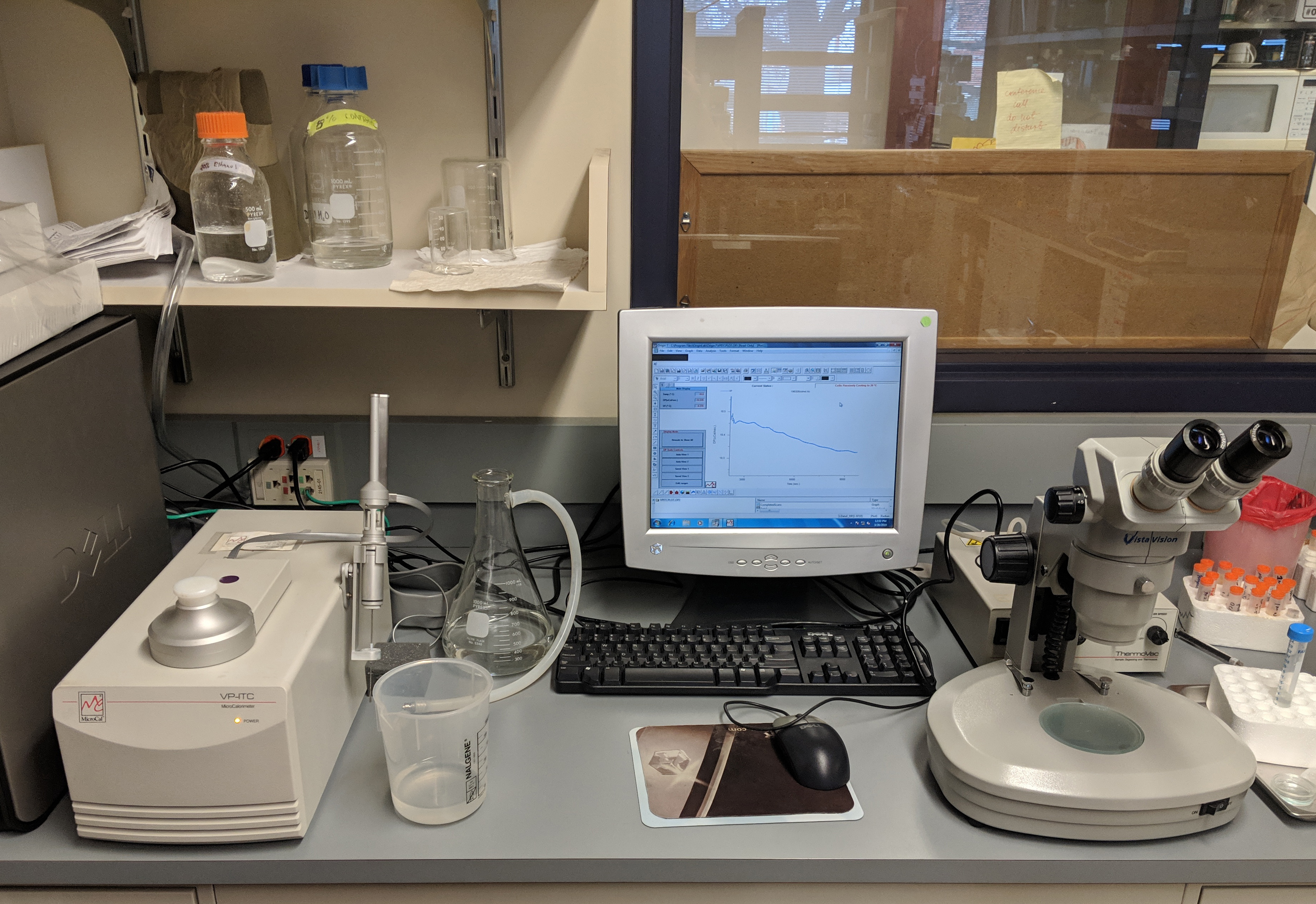
 (4 votes)
 (1 votes)
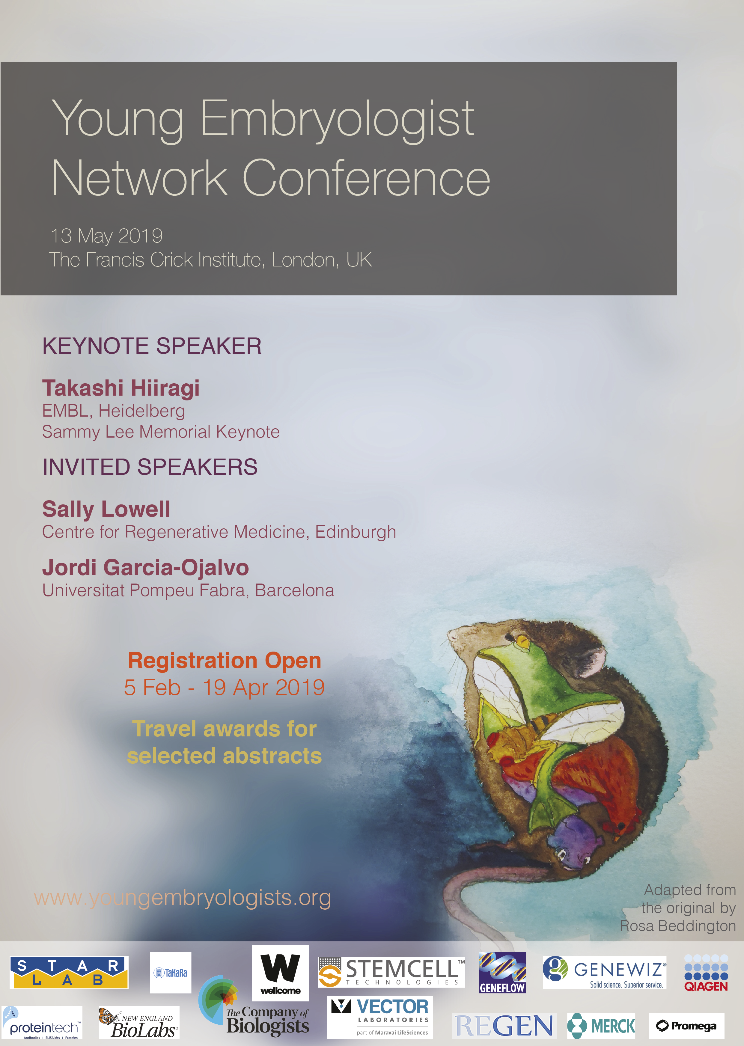
 Jim Smith introduced the Beddington medal with heartfelt memories of Rosa Beddington and her time at the NIMR. Please read more of his thoughts
  “The really impressive thing about David’s work is that he did not come to my lab to fit in with an existing line of research but created one of his own”. Jamie Davies, University of Edinburgh.
David received his undergraduate degree in Sport and Exercise Science at the University of Stirling (2010-2014). With this, he achieved a first-class honours degree and the prize for the best overall performance throughout a physiology related degree (British Physiological Society Undergraduate Prize). His dissertation project investigated associations between ADRB2 mutations (an adrenaline receptor gene in humans) and athlete status/athletic ability measurements. Subsequently, he was awarded a University of Stirling Head of School Summer Bursary Award to remain in Stirling during the summer of 2014 and investigate the relationship between transcribed ultra-conserved regions of RNA (T-UCRs) and the development of diet-induced insulin resistance in humans (Summer 2014). He then moved to the University of Edinburgh for his MSc by Research in Biomedical Sciences (2014-2015). Again, he received a distinction and was awarded the Class Prize for best student. During this time, he studied the physiology of S-acylation the regulation of skeletal muscle energy expenditure by an obesity-associated phospholipase as part of two research placements.
David has been awarded the Beddington medal for his exceptional work performed during his 3-year MRC-funded PhD at the University of Edinburgh with Prof Jamie Davies and Dr Peter Hohenstein (2015-2018): The thesis is titled ‘Mechanisms of kidney vascularisation and the roles of macrophages in renal organogenesis’. During his PhD, he gave several oral and poster presentations at national and international conferences, supervised students (including a Gurdon Summer Studentship Awardee), established numerous international collaborations, was awarded travel grants (including a BSDB Conference Grant), and reviewed manuscripts for leading journals (including Cell Reports, Angiogenesis, and Scientific Reports). He is now a post-doctoral fellow at the UK Dementia Research Institute (University of Edinburgh; 2019- present), continuing research in macrophage biology under the supervision of Prof Josef Priller. His current focus in on brain macrophages (microglia) in development, neurodegeneration, and aging.
Thesis description
Kidneys are specialised organs that clean the blood, removing waste while retaining what is useful. This requires a complex vasculature, and its formation as a foetus develops is poorly understood. I started my PhD research by using advanced microscopy techniques to visualise how blood vessels form in three-dimensions in the mouse kidney. In doing so, I identified when and from where the first blood vessels enter the kidney, and how blood vessels pattern at the edge of the kidney throughout development.
Blood vessels can form through angiogenesis (branching of new vessels from pre-existing ones) and/or vasculogenesis (assembly of new vessels from the coalescence of endothelial precursor cells). It has long been thought that a combination of both processes occurs during kidney vascularisation; however, my thesis work indicates that this concept may not be correct. My data instead suggest that kidney vascularization relies on growth and remodelling of pre-existing vessels (angiogenesis) and does not depend on vasculogenesis at any point (Publications 1 and 5 in CV). When assessing the entire 3D vascular tree of the kidney, isolated endothelial cells were never observed at any developmental age. Instead, all vessels, including the newly forming ones, were connected to pre-existing vessels that could be traced to the major circulatory vessels.
I then focused on the blood vessels at the edge of the kidney, which I found to consistently and accurately pattern around a special collection of cells – the cap mesenchyme. The cap mesenchyme contains cells that eventually become the cleaning tubes of the kidney, the nephrons. This cell population undergoes rounds of splitting at the kidney’s periphery. As this happens, I demonstrated that blood vessels migrate through the newly opened regions between the separating cap mesenchymal populations (Publication 1 in CV). This occurs in cycles throughout development and is likely to be vital for the oxygenation of the kidney’s outer region, the site where important processes such as nephron formation take place.
I determined that a signalling molecule, semaphorin-3f, and its receptor, neuropilin-2, were expressed in a pattern consistent with them having roles in this cyclical patterning of blood vessels; however, using mouse models where the genes for these molecules were deleted, I established that they were not vital for this process (Publication 2 in CV).
I next shifted my research focus towards a specialised cell type known as the macrophage (macro = big; phage = eater) in the developing kidney (Publication 3 in CV). Macrophages are immune cells best known for clearing foreign and damaged cells. These cells have vital roles during animal development, but little is known about their specific functions during kidney development.
Macrophages arrived in the mouse kidney early during its development, where they were required to clear away misplaced cells to ‘set-the-stage’ for early kidney development (Publication 6 in CV, under review). Throughout later development, most macrophages wrapped around blood vessels and I demonstrated their ability to eat endothelial cells (which usually line the blood vessels) and red blood cells (which are carried within them) within the kidney. I also established that kidney macrophages produced many molecules linked to blood vessel development, and so I examined the consequences of macrophage-loss on blood vessel formation. Blood vessels normally form continuous networks in the kidney; however, when macrophages were depleted (by blocking a macrophage-survival signalling pathway), connections between renal blood vessels were reduced (Publication 6 in CV).
Publications
“The really impressive thing about David’s work is that he did not come to my lab to fit in with an existing line of research but created one of his own”. Jamie Davies, University of Edinburgh.
David received his undergraduate degree in Sport and Exercise Science at the University of Stirling (2010-2014). With this, he achieved a first-class honours degree and the prize for the best overall performance throughout a physiology related degree (British Physiological Society Undergraduate Prize). His dissertation project investigated associations between ADRB2 mutations (an adrenaline receptor gene in humans) and athlete status/athletic ability measurements. Subsequently, he was awarded a University of Stirling Head of School Summer Bursary Award to remain in Stirling during the summer of 2014 and investigate the relationship between transcribed ultra-conserved regions of RNA (T-UCRs) and the development of diet-induced insulin resistance in humans (Summer 2014). He then moved to the University of Edinburgh for his MSc by Research in Biomedical Sciences (2014-2015). Again, he received a distinction and was awarded the Class Prize for best student. During this time, he studied the physiology of S-acylation the regulation of skeletal muscle energy expenditure by an obesity-associated phospholipase as part of two research placements.
David has been awarded the Beddington medal for his exceptional work performed during his 3-year MRC-funded PhD at the University of Edinburgh with Prof Jamie Davies and Dr Peter Hohenstein (2015-2018): The thesis is titled ‘Mechanisms of kidney vascularisation and the roles of macrophages in renal organogenesis’. During his PhD, he gave several oral and poster presentations at national and international conferences, supervised students (including a Gurdon Summer Studentship Awardee), established numerous international collaborations, was awarded travel grants (including a BSDB Conference Grant), and reviewed manuscripts for leading journals (including Cell Reports, Angiogenesis, and Scientific Reports). He is now a post-doctoral fellow at the UK Dementia Research Institute (University of Edinburgh; 2019- present), continuing research in macrophage biology under the supervision of Prof Josef Priller. His current focus in on brain macrophages (microglia) in development, neurodegeneration, and aging.
Thesis description
Kidneys are specialised organs that clean the blood, removing waste while retaining what is useful. This requires a complex vasculature, and its formation as a foetus develops is poorly understood. I started my PhD research by using advanced microscopy techniques to visualise how blood vessels form in three-dimensions in the mouse kidney. In doing so, I identified when and from where the first blood vessels enter the kidney, and how blood vessels pattern at the edge of the kidney throughout development.
Blood vessels can form through angiogenesis (branching of new vessels from pre-existing ones) and/or vasculogenesis (assembly of new vessels from the coalescence of endothelial precursor cells). It has long been thought that a combination of both processes occurs during kidney vascularisation; however, my thesis work indicates that this concept may not be correct. My data instead suggest that kidney vascularization relies on growth and remodelling of pre-existing vessels (angiogenesis) and does not depend on vasculogenesis at any point (Publications 1 and 5 in CV). When assessing the entire 3D vascular tree of the kidney, isolated endothelial cells were never observed at any developmental age. Instead, all vessels, including the newly forming ones, were connected to pre-existing vessels that could be traced to the major circulatory vessels.
I then focused on the blood vessels at the edge of the kidney, which I found to consistently and accurately pattern around a special collection of cells – the cap mesenchyme. The cap mesenchyme contains cells that eventually become the cleaning tubes of the kidney, the nephrons. This cell population undergoes rounds of splitting at the kidney’s periphery. As this happens, I demonstrated that blood vessels migrate through the newly opened regions between the separating cap mesenchymal populations (Publication 1 in CV). This occurs in cycles throughout development and is likely to be vital for the oxygenation of the kidney’s outer region, the site where important processes such as nephron formation take place.
I determined that a signalling molecule, semaphorin-3f, and its receptor, neuropilin-2, were expressed in a pattern consistent with them having roles in this cyclical patterning of blood vessels; however, using mouse models where the genes for these molecules were deleted, I established that they were not vital for this process (Publication 2 in CV).
I next shifted my research focus towards a specialised cell type known as the macrophage (macro = big; phage = eater) in the developing kidney (Publication 3 in CV). Macrophages are immune cells best known for clearing foreign and damaged cells. These cells have vital roles during animal development, but little is known about their specific functions during kidney development.
Macrophages arrived in the mouse kidney early during its development, where they were required to clear away misplaced cells to ‘set-the-stage’ for early kidney development (Publication 6 in CV, under review). Throughout later development, most macrophages wrapped around blood vessels and I demonstrated their ability to eat endothelial cells (which usually line the blood vessels) and red blood cells (which are carried within them) within the kidney. I also established that kidney macrophages produced many molecules linked to blood vessel development, and so I examined the consequences of macrophage-loss on blood vessel formation. Blood vessels normally form continuous networks in the kidney; however, when macrophages were depleted (by blocking a macrophage-survival signalling pathway), connections between renal blood vessels were reduced (Publication 6 in CV).
Publications