Macrophages are usually associated with immunity, but have increasingly appreciated functions in development and homeostasis. This week we meet the authors of a recent Science paper that identified a role for macrophages in zebrafish stripe patterning, revealing a remarkable ‘relay’ mechanism whereby macrophages help one type of cell signal to another via cytoplasmic extensions. Postdoc Dae Seok Eom and his supervisor David Parichy, recently appointed Pratt-Ivy Foundation Distinguished Professor of Morphogenesis at the University of Virginia, told us more.
David, can you give us your scientific biography and the questions your lab is interested in?
DP I started out in ecology and evolution as an undergrad at Reed College, studying maternal effects on tadpole growth and survivorship for four years with Bob Kaplan. With that experience I applied to E&E Ph.D. programs and ended up accepting an offer from Population Biology at UC Davis, with Brad Shaffer, a systematist and organismal biologist studying biogeography of salamander populations. But between when I applied and when I got there, I became more and more interested in developmental mechanisms and how they evolve. So I was really lucky that Brad has diverse interests and that Carol Erickson—a developmental biologist working on neural crest—was willing to serve as a co-advisor. For my dissertation I focused on the cellular bases for salamander pigment pattern development and evolution. Those are great animals, but the molecular biology and genetics were difficult at the time. So I switched to zebrafish and its relatives for my postdoc with Steve Johnson at Wash U Medical School in St. Louis. In Steve’s lab, we identified some of the mechanisms underlying the development of stripes and other patterns, and I carried this program into my independent career.
My lab has broad interests but an organizing theme has always been to understand the genes and cell behaviors underlying adult phenotypes, and how changes in developmental genetic mechanisms contribute to variation within and among species. We continue to work on pigment patterns, but we also want to know what regulates the stem cells that give rise to adult pigment cells and how “local” cellular mechanisms intersect with “global” endocrine control during development, homeostasis and regeneration. But our interests are wide-ranging so we’ve also worked on topics including skeletal development, zebrafish natural history, and the behavioural significance and cognitive processing of pigmentation. Right now we are doing a lot of work to understand scale morphogenesis and patterning in fish, and we’ve even started working on salamanders again, both pigment and regeneration.
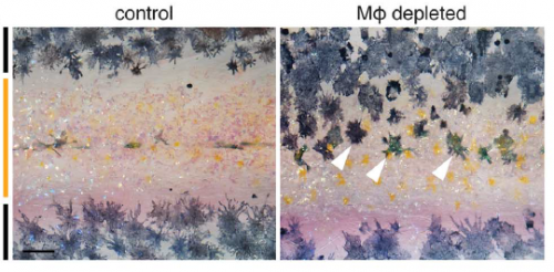
The macrophage depletion phenotype, from Figure 1, Eom & Parichy, 2017.
And Dae Seok, how did you come to join David’s lab?
DSE Actually, when I was at PhD training at the University of Texas at Austin, Dave was a faculty of my department, and I was interested in his work, but by then he was then planning to move to the University of Washington. While I was finishing my PhD training, my wife started her PhD at UW, so I had to find a postdoc position in Seattle. I realised Dave was there, and it was obviously a great second chance to work with him – and he accepted.
Aside from aesthetics, what makes zebrafish stripe development an attractive developmental model?
DP When I was a grad student, I wanted to find a system that could be studied from many different angles—molecular through organismal. And this was why I ended up focusing on pigmentation. For anyone with broad interests it’s just a natural: there’s a deep literature on pigment cells and pattern going back to the turn of the last century, the cells are visible even in the living organism as the phenotype is developing, the patterns themselves often have profound ecological significance, and there’s tremendous diversity of pattern across even closely related species. Of course there are also a variety of pigmentary disorders, including melanoma, and pigment cells develop and regenerate from stem cells—so there is obvious biomedical utility in studying pigment cells, especially with a system like zebrafish in which the genetic and cellular mechanisms are so accessible.
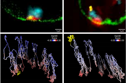
A macrophage drags an airineme to a melanocyte. Eom & Parichy, 2017.
Can you give us the key results of the paper in a paragraph?
DP We showed previously that consolidation of melanophores into adult stripes depends on interactions between these cells and xanthoblasts, the precursors of yellow xanthophores. During a specific stage of stripe development, xanthoblasts that happen to be present in future stripe regions extend very long, thin and meandering filopodia-like processes with membraneous vesicles at their tips. We called these projections “airinemes” after Iris—messenger of the gods—and Sir George Airy, who described limits on optical resolution, because the projections are such a pain to visualize. We saw airinemes “dock” with melanophores, and we found that blocking airineme extension prevented melanophores from consolidating properly into stripes, at least in part because of a defect in Delta-Notch signalling.
“Weird biology”
In Dae Seok’s new paper, we show that airineme extension and vesicle delivery depend on macrophages that are cruising around the local tissue environment. The macrophages recognize surface blebs on xanthoblasts and try to engulf them but continue to wander, pulling a membrane filament from the xanthoblast as they go. Eventually they wander across a melanophore and the vesicle and filament are deposited on the melanophore surface. If we get rid of macrophages, we prevent airineme extension and melanophores remain dispersed. Weird biology.
What convinced you to deplete macrophages in the first place? It might not seem like the most obvious place to look for patterning regulators…
DSE The idea initially came from the question of what would happen to unbound airineme vesicles. When xanthoblasts don’t meet the target melanophores, airineme vesicles detach from the airineme filaments and wander away. We knew the vesicles carry DeltaC and possibly other signalling molecules. Thus, these unbound vesicles should be somehow eliminated. The question then was what cell types can do that? One of the answers was the macrophage.
DP Plus we could constantly see macrophages wandering around in Dae Seok’s movies. How could we not try getting rid of them?
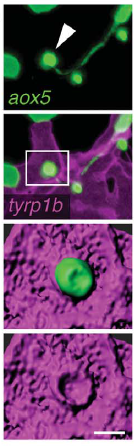
Airineme vesicle associated with melanophore membrane, from Fig. 4, Eom & Parichy, 2017.
Is there anything in the macrophage-depleted fish to suggest that other patterning or morphogenetic events may be affected, or is this just tissue specific?
DP We’re only starting to investigate roles for macrophages as well as airinemes in other tissues. Because macrophages wander all over, you could imagine a whole variety of possibilities for diffusion-like dissemination—or regulated attenuation—of signals in other contexts. The more people look, the more interesting things macrophages seem to do.
Can you hazard a guess as to whether you think the macrophages actively select signalling targets or just wander around randomly?
DSE We have no idea at this time but my guess is macrophages constantly probe the environment, and signals from the airineme vesicles instruct macrophages to detect the targets and drop the vesicles while they are randomly wandering around.
DP Yes, because Dae Seok showed that targeting is specific to a subset of melanophores, there has to be some sort of airineme vesicle–melanophore recognition system. It will be interesting to see whether this is somehow instructive to the macrophage itself or whether the macrophage is simply trying to eat the vesicle and can’t manage to do so, and therefore the vesicle gets displayed at the cell surface often enough that it can stick to a melanophore as its carrier macrophage wanders along.
When doing the research, was there a particularly exciting result or eureka moment that has stayed with you?
DSE I do not have good explanation for it, but I had very strong gut feeling that there are something going on between macrophages and airinemes. So of course, my most exciting moment was when I saw the macrophages interacting with extending airinemes.
Xanthoblast airinemes. Eom and Parichy, 2017.
And what about the flipside: any moments of frustration or despair?
DSE Our original plan was to add additional mechanisms of airineme vesicle-macrophage interactions beside phosphatidylserine, as we have several additional candidates. We ran into technical problems for this first paper but are now working on it again.
What next for you following this work?
DSE We have many questions about airineme-macrophage interactions, for example, what other signalling molecules carried in the airineme vesicles and how macrophages know what the targets are. I’m very excited about using our new super-resolution microscopes – they will open up many new possibilities.
“Our new super-resolution microscopes will open up many new possibilities.”
And where will this discovery take the Parichy lab?
DP Of course we’ll continue to pursue the mechanisms underlying this signalling relay in zebrafish pigment pattern formation. And we’d really like to know how these and other interactions have evolved to generate the very different patterns we see in Danio and beyond. But you know, a lot depends on the interests of the grads and postdocs who come to my lab. I just try to foster an intellectual environment and provide the resources to let people explore and go where the science leads. When we decided eight years ago to invest in live imaging we didn’t know what we’d find and we certainly didn’t expect this. So I’d be hesitant to make firm predictions.
Finally – what do you get up to outside of the lab when you are not playing with fish?
DSE One of my favourite things to do is visiting local breweries. My wife and I have a Saturday routine of having a lunch at a brewery, and then heading to the lab.
DP Legos. I like to spend as much time as I can with my four year old and my wife. So we do a lot of legos, trains and construction. Helicopters are big, too.
Our latest monthly trawl for developmental biology (and other cool) preprints. See June’s introductory post for background, and let us know if we missed anything
March was (yet) another bumper month for life sciences preprints. A glance at some of the names of last authors – Daniel St. Johnston, Denise Montell, Dennis Duboule, Roberto Mayor, Fiona Watt – shows that preprints are being embraced by established as well as early career scientists. The content itself covers every base in developmental biology, as well as a lot of exciting cell biology and modelling relevant for development, and an acronym-rich list of tools and resources.
The Molecular Link Between Auxin And ROS-Mediated Polar Growth. Silvina Mangano, Silvina Paola Denita-Juarez, Hee-Seung Choi, Eliana Marzol, Youra Hwang, Philippe Ranocha, Silvia M Velasquez, Jorge P Muschietti, Christophe Dunand, Hyung-Taeg Cho, Jose M Estevez
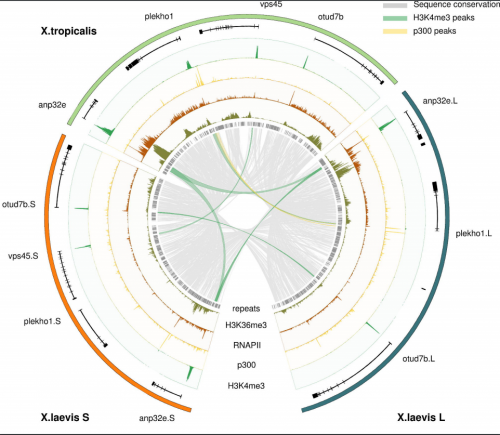
Alignment of X. tropicalis and the X. laevis L and S subgenomes, from Elurbe, et al.
Regulatory remodeling in the allo-tetraploid frog Xenopus laevis. Dei M. Elurbe, Sarita S. Paranjpe, Georgios Georgiou, Ila van Kruijsbergen, Ozren Bogdanovic, Romain Gibeaux, Rebecca Heald, Ryan Lister, Martijn A. Huynen, Simon J. van Heeringen, Gert Jan C. Veenstra
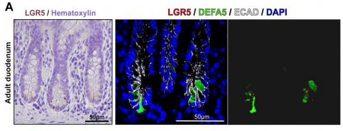
Looking for LGR5+ cells in the adult duodenum, from Dame, et al.
Identification, Isolation, and Characterization of Human LGR5-positive Colon Adenoma Cells. Michael K Dame, Durga Attili, Shannon D McClintock, Priya H Dedhia, Peter Ouilette, Olaf Hardt, Alana M Chin, Xiang Xue, Julie Laliberte, Erica L Katz, Gina M Newsome, David Hill, Alyssa Miller, David Agorku, Christopher H Altheim, Andreas Bosio, Becky Simon, Linda C Samuelson, Jay A Stoerker, Henry D Appelman, James Varani, Max S Wicha, Dean E Brenner, Yatrik M Shah, Jason R Spence, Justin A Colacino
An Algorithm for Cellular Reprogramming. Scott Ronquist, Geoff Patterson, Markus Brown, Haiming Chen, Anthony Bloch, Lindsey Muir, Roger Brockett, Indika Rajapakse
Rapid Sequential In Situ Multiplexing With DNA-Exchange-Imaging. Yu Wang, Johannes B Woehrstein, Noah Donoghue, Mingjie Dai, Maier S Avendano, Ron C.J. Schackmann, Shan Shan Wang, Paul W Tillberg, Demian Park, Sylvain W Lapan, Edward S Boyden, Joan S Brugge, Pascal S Kaeser, George M Church, Sarit S Agasti, Ralf Jungmann, Peng Yin
A toolbox of immunoprecipitation-grade monoclonal antibodies against human transcription factors. Anand Venkataraman, Kun Yang, Jose Irrizary, Mark Mackiewicz, Paolo Mita, Zheng Kuang, Lin Xue, Devlina Ghosh, Shuang Liu, Pedro Ramos, Shaohui Hu, Diane Bayron, Sarah Keegan, Richard Saul, Simona Colantonio, Hongyan Zhang, Florencia Pauli Behn, Guang Song, Edisa Albino, Lillyan Asencio, Leonardo Ramos, Luvir Lugo, Gloriner Morell, Javier Rivera, Kimberly Ruiz, Ruth Almodovar, Luis Nazario, Keven Murphy, Ivan Vargas, Zully Rivera-Pacheco, Christian Rosa, Moises Vargas, Jessica McDade, Brian S Clark, Sooyeon Yoo, Seva G Khambadkone, Jimmy de Melo, Milanka Stevanovic, Lizhi Jiang, Yana Li, Wendy Y Yap, Brittany Jones, Atul Tandon, Elliot Campbell, Stephen Anderson, Richard M Myers, Jef D Boeke, David Fenyo, Gordon Whiteley, Joel S Bader, Daniel J Eichinger, Ignacio Pino, Heng Zhu, Seth Blackshaw
Have you ever wondered what makes the shapes in the animal and plant kingdom so different? We take for granted the diversity of natural shapes that surround us, from a simple pine leaf to complex orchid flower. However, they pose one of the most beautiful scientific challenges. For centuries, scientists have been fascinated by how a certain shape is generated and what drives such diversity of shapes. I am no different! From my university days, I always wanted to study evolutionary development, to go into the depth of why and what lies behind the diversity of shapes in nature.
After my PhD days using genetics, molecular biology and comparative development as tools to study the evolution of shape in plants, I was keen to explore the upcoming computational modeling field as a tool to tackle complex development problems. This is where this article’s journey started, when I joined the lab of Enrico (Rico) Coen at the John Innes Centre to study the evolution and development of the complex 3D Snapdragon flower shape. I still remember my first contact with modeling. I was immediately hooked by the colorful shapes in the computer screen which looked so much like a snapdragon flower. However, nothing could have prepared me for the thousand lines of computational code that sustained the generation of the virtual Snapdragon model. At that moment I thought to myself, maybe I have bitten more than I can chew by wanting to do both the biology and the computational modeling.
The personal story that goes with the published article is a roller coaster of wrong hypothesis, failed and impossible experiments, and frustrating models but also glimpses of successes. You might be thinking, oh no this is going to be a tale of woe but let me disclose the end already – this is no sad story, this is the actual necessary evolution of thought, knowledge and personal growth that sustains any challenging scientific problem. For my feeling the story behind this paper feels more like a Tolkien novel with lots of downs, that you almost don’t expect to overcome, but also amazing ups.
Our approach at the start of my post-doc was to look into the working hypothesis generated by a previous in-house paper where the growth of the Snapdragon petals was analyzed and a virtual flower corolla was produced. The main assumptions underlying this model related to the spatiotemporal changes in the growth pattern due a local cell polarity inversion event combined with a local boost in growth rate.
To visualize the inversion of polarity in cells we decide to use an accepted marker of plant cell polarity that generally shows an asymmetric cellular localization – the PIN1 protein. For two long years, I struggled with PIN1 antibodies that didn’t work or produced slight promising subcellular signals that held us in the false hope of results. After changing the antibody company and spreading the risks by designing several different epitopes to the different PIN1s, I finally had a good working antibody that produced the expected pattern of cellular polarity in emerging organs. Armed with this tool, I initiated a marathon of immunolocalization sections across the petals at different stages of development to show the local inversion of cellular polarity. From the beginning it was obvious that something special was happening to the expression of the PIN1 in the predicted region at the predicted time by the model. However, after a year of thorough sectioning experiments and analysis, I was struggling to reconstruct the 3D PIN1 pattern from the 2D sections. At that point we wanted to start writing the paper to show the peculiar upregulation of PIN1 in the predicted region by the model but unfortunately the precise pattern of the PIN1 was still undetermined and led to a lot of team discussions and wild ideas on how to distil the result. Emotions were running high, as we were failing to grasp the final proof of the inversion of polarity. After we decided to put the paper on hold and re-think our approach, I got desperate! In other models system and exposed organs such as roots, the first solution would be to do whole-mount immunolocalization, where the whole organ is hybridized and the 3D patterns can be imaged. However, our tissue of interested was the inner surface of tiny petals, in buds as small as 500 microns. Desperate times require desperate measures, and after a bad night of sleep and probably a delirious morning, I started dissecting incredibly tiny petals and sticking them to a glass slide using prosthetic glue. Then, to make sure the antibody could penetrate the tissue, I boiled these fragile tissues cringing at every little water bubble brushing through the stuck petals. To check the cell/tissue integrity and in anticipation of no PIN1 signal, I also stained the petal tissues with a cellulose fluorescent stain called calcofluor. After three agonizing days of hoping that the tissue was not turned into a pulp, I descended the stairs that separate our lab from the confocal microscopy room with a heavy heart; this is our last crazy chance! If this doesn’t work, I am out of ideas. As I started up the confocal and focused on the first set of 5 petals, I couldn’t believe what I was seeing, a beautiful ladder of PIN1 signal in the regions of interest -I couldn’t contain my joy.
I picked up the phone and called some lab mates to be sure they could see what I was seeing (I couldn’t exclude delirious mirages at this point with my nerves on the fringe). After they confirmed what I was seeing and shared a happy dance with me, I went to get Rico from his office luring him with promises of something special without giving too much away. We still have photos of this moment that we shared with the four of us in a confocal room. There are moments that you don’t forget in your life and this one is engrained in my memory.
I finally could extract full epidermal patterns of PIN1 in wild-type and mutant petals at different stages of development, and guess what? I never saw an inversion or convergence of polarity in the region predicted by the model. I did see an upregulation of PIN1 at the predicted time and space but the pattern of PIN1 was mainly proximodistal (along one axis). This might feel like a bitter sweet result as it contradicted our model but actually this was quite an unexpected and exciting result. In my opinion, one of the beautiful givens in science is that it will surprise you. You make a hypothesis, your results prove it wrong and you need to re-evaluate your thinking. This was indeed what happened to us but as a result we realized that we didn’t need an inversion of polarity, as the key information required for growth lay in the orientation of the arrows (axiality), not in the direction in which they are pointing. So we came up with an alternative model that still involved a switch in growth orientations but without a polarity inversion (I can’t imagine I just summarized months of modeling with a sentence but there you have it!).
We then tested this model by correlating cell wall patterns with growth orientations and showing the switch in growth orientation at the predicted time and space.
Detail of Snapdragon petal stained with cell wall marker in order to visualize the orientation of cell division and growth (red lines indicate proximodistal growth while yellow lines indicate mediolateral growth with respective zoomed in areas). Adapted from Rebocho et al, 2017, eLife.
We were finally ready to write our results, publish our new model and make a contribution to the scientific understanding of 3D morphogenesis. However, one of our biggest challenges was still to surface – conveying the ideas developed over the years in words, while connecting the static 2D images of cell polarity and growth orientations with the 3D flower shape. When you read the paper now, the logic seems to fall into place, with the increasing complexity of the models and the experimental data driving a new model hypothesis. But the reality was a long winding road, with several versions of the paper; many rejected by ourselves, others by our critical colleagues, and others by peer-reviewed journals. This was a very frustrating time for the team and I am not really sure when we started to see the light, as this was an incremental and slow process, but at a certain point we felt that we were finally are on track. By deconstructing the model complexity (from square piece of tissue to wedge model to full corolla model and to mutant models) and conveying the idea of how differential growth behaviors can lead to tissue conflict resolutions, we were finally bringing the disjointed paper together. Our paper distilled three tissue conflict resolution behaviors leading to an out-of-plane deformation: surface conflict (differential growth/contraction rates between two surfaces), areal conflict (differential growth/contraction rates across a tissue) and directional conflict (differential orientation growth/contraction across a tissue). These three behaviors are the basis of many of the described morphogenetic events in plants and animals, and can be combined to produce the panoply of 3D shape complexity that surrounds us.
For me this journey made me a more complete scientist. I have explored new disciplines and learned to think in a different way. I have developed extra respect for the power of modeling and became humbler when it comes to tackling complex biological problems. Lastly, I realized that the essence of a piece of research lies in the evolution of thought of the researchers, their failures and frustrations, their stubbornness and their hunches, their commitment and their successes, and that is what makes science so amazing.
The Wellcome-Warwick Quantitative Biomedicine Programme Wellcome-Warwick Quantitative Biomedicine Programme was established to enhance the world-class interdisciplinary research environment at the University of Warwick by driving further development of our existing centres of excellence, including the Centre for Mechanochemical Cell Biology (www.mechanochemistry.org) and the Zeeman Institute for Systems Biology& Infectious Disease Epidemiology Research.
Warwick Medical School is seeking to appoint three outstanding early career scientists as Assistant Professors in Quantitative Biomedicine, aiming to expand our research on cell dynamics at the molecular, cellular and/or tissue scales. We are eager to recruit candidates using quantitative and/or interdisciplinary approaches and with an exemplary track record in cell biology, developmental biology, structural biology, computational biology or biophysics.
Successful candidates must have a strong track record with first class publications,together with the enthusiasm and expertise to contribute to our innovative undergraduate taught programme in Interdisciplinary Science. Evidence of being able to attract funding and/or fellowships would be a further advantage. You will also contribute to the Public Engagement interface of the QBP.
Successful candidates will receive a start-up package, laboratory space in the brand new extension to our mechanochemical cell biology building, access to state-of-the-art infrastructure, including light and electron microscopy, advanced proteomics, and support from our thriving and dynamic research community.
The posts will be in the Division of Biomedical Sciences, Warwick Medical School and the successful candidates will be expected to play an active role in advancing the mission of the QBP. The posts will be subject to a five year probation period and once successfully completed, promotion to Associate Professor will follow, subject to criteria set out by the University of Warwick being met.
Potential candidates are encouraged to make informal contact with the Directors of the Wellcome Warwick Quantitative Biomedicine Programme, Profs. Mohan Balasubramanian (M.K.Balasubramanian@warwick.ac.uk) and Andrew McAinsh (A.D.McAinsh@wawick.ac.uk).
To be considered, please fill out an online application, including a CV, names of three expert referees who are able to comment on your readiness to embark on an independent career, a one-page cover letter and a two-page research proposal describing an exciting research program in cell dynamics.
A postdoctoral position is available in the laboratory of Dr. Sophie Astrof to study roles of cell-extracellular matrix interactions in cardiovascular development and disease using cell biological approaches and mouse model system. The project will involve investigation of signaling by extracellular matrix in development and differentiation, utilizing state-of-the art imaging and genetic approaches. In our lab, we use genetics, conditional mutagenesis, and transgenic approaches to explore roles of tissue microenvironment during organogenesis and disease. Experience with genetic manipulation, embryology and cell biology is desirable. My laboratory is a part of the Center for Translational Medicine at Jefferson Medical College (http://www.tju.edu/jmc/medicine/translational_medicine/faculty/astrof.cfm?detail=0) located in the heart of Philadelphia. To apply, send a letter of interest, CV and names and contact information of three references to sophie.astrof@gmail.com…
The Woods Hole Embryology Course, which will celebrate its 124th birthday this year, is a continual source of beautiful images (and videos) of development. Since 2011 the Node has run a competition for the community to pick the best images from a given year – the winning pictures become immortalised as Development covers!
Below you will find 4 images from the 2015 course, Round 1. Choose the one you would like to see on the cover of Development by voting on the poll at the end of the post (you can see full size versions by clicking on the images).
The poll is set up to allow only one vote per person, and closes at 12.00 GMT, Friday 14th April. Results will be announced Tuesday, 18th April.
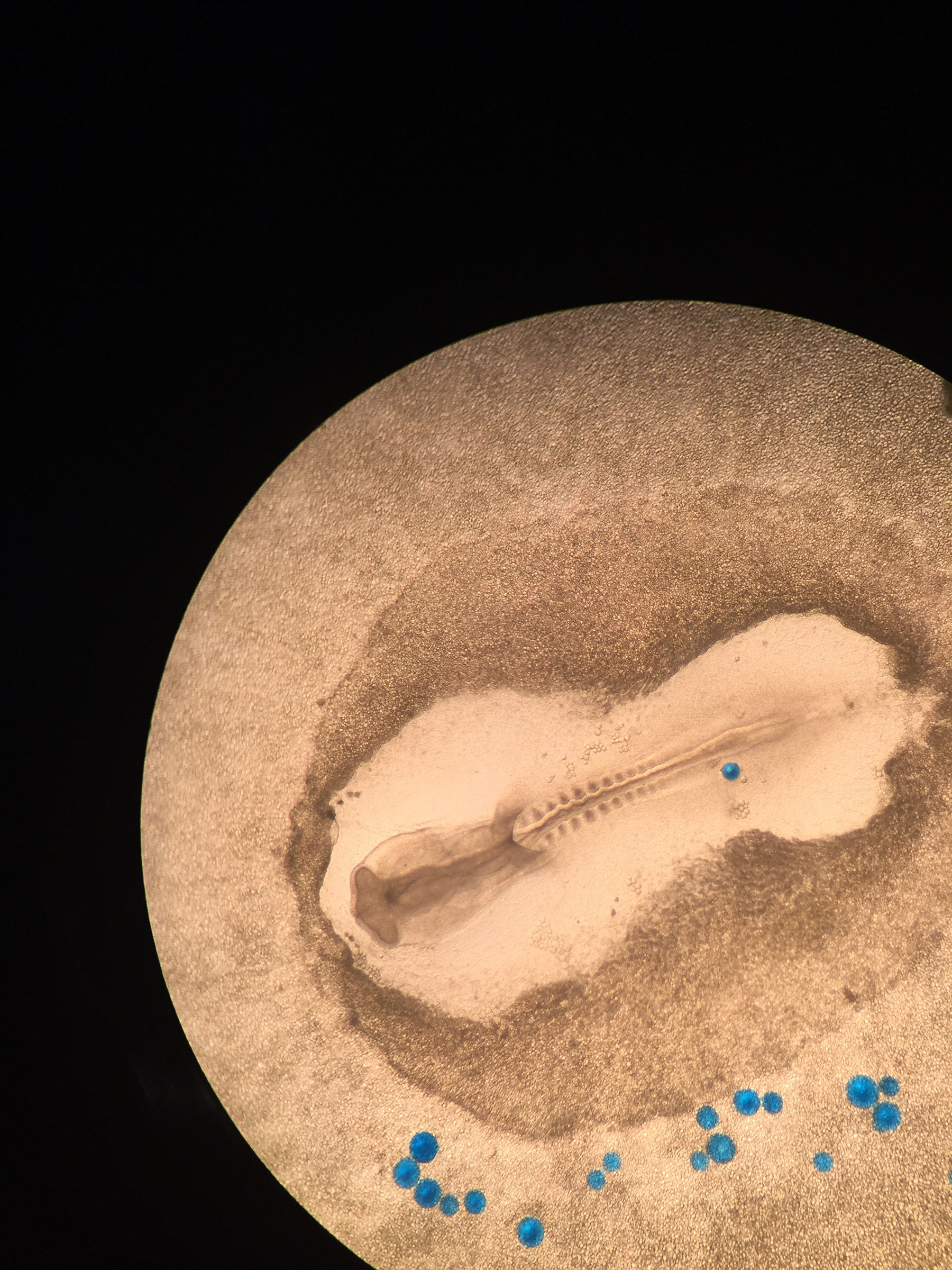
Chicken
Stage 10 chick embryo with noggin coated beads (blue). Imaged with cell phone.
Theodora Koromila
CalTech, USA
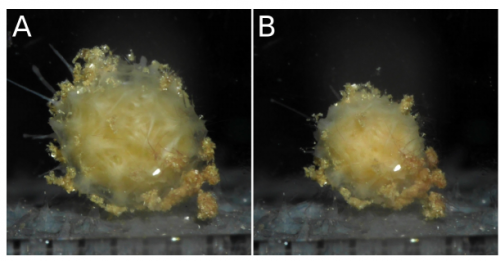
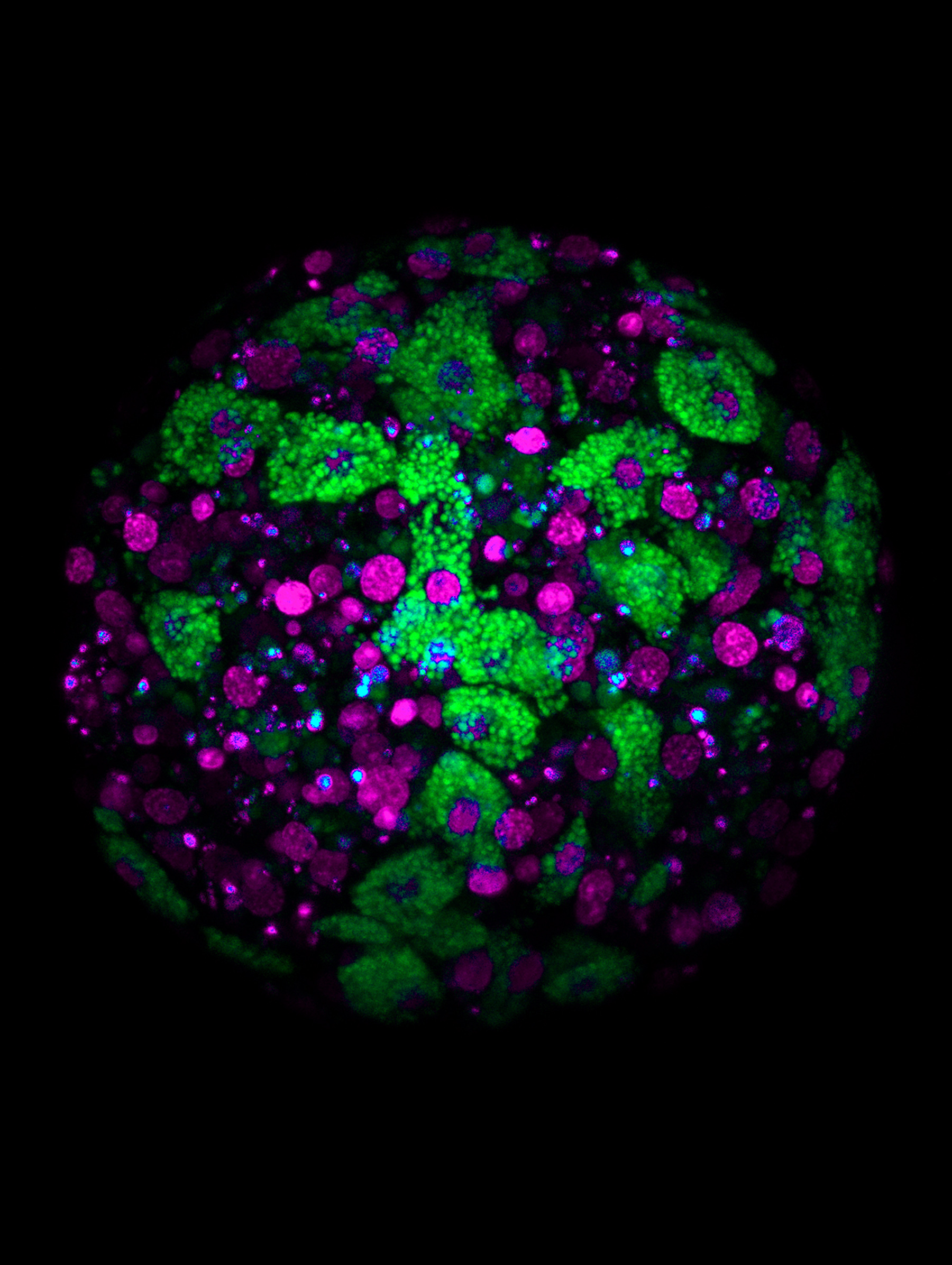
Clathria
10 hours post-dissociation aggregate of cells of the marine sponge, Clathria prolifera. A subset of the cells contains autofluorescent vesicles excited by a 405nm laser (shown in green). The nuclei are labeled with Hoechst staining (shown in purple), and the two signals were separated by spectral imaging and linear unmixing on a Zeiss LSM 780 confocal.
Shun Sogabe
The University of Queensland, Australia
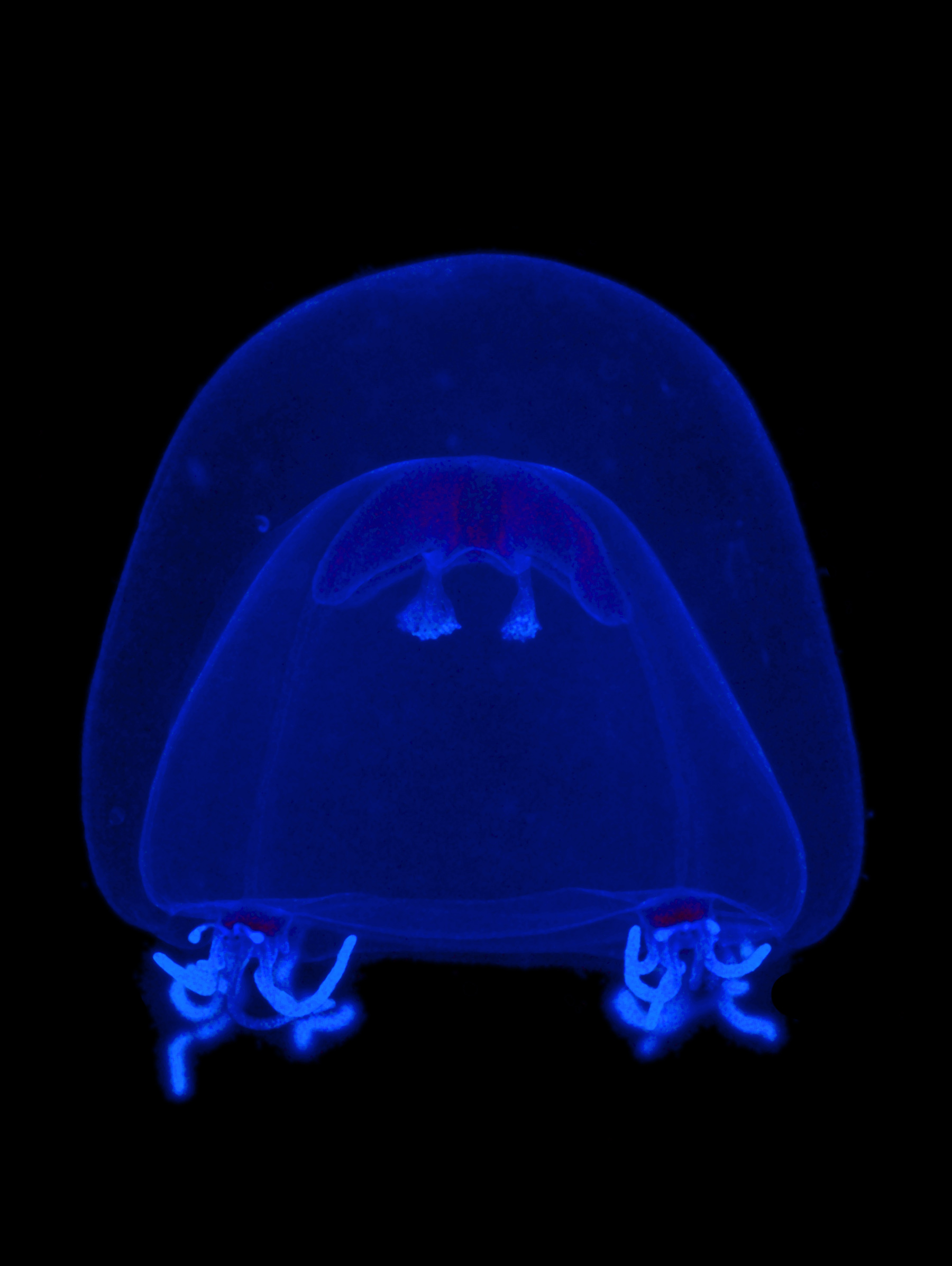
Jellyfish
Juvenile jellyfish Nemopsis bachei collected in a plankton tow. Stained with DAPI (nuclei). Imaged with a Zeiss AxioImager and processed with Photoshop.
Chiara Sinigaglia
Observatoire Océanologique de Villefranche sur Mer/ CNRS, France.
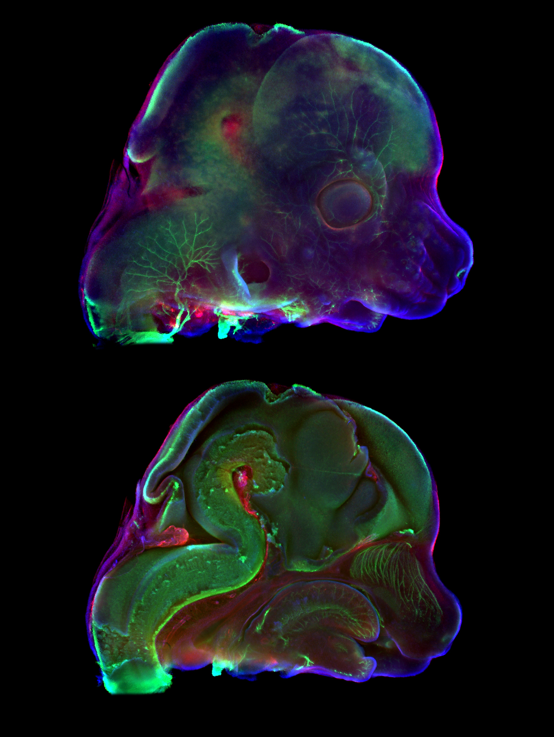
Mice
Midsagittal section through E12.5 mouse embryo head showing muscle fibers (MF20, red), nerve fibers (TUJ1, green) and nuclei (DAPI, blue). Head surface on the top, surface of the section plane on the bottom. Captured using Zeiss Axio Zoom V16 stereomicroscope. The final image was composed in GIMP.
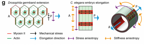
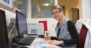
This year marks the centenary of D’Arcy Thompson’s On Growth and Form, an attempt to outline the physical and mathematical principles underpinning the generation of biological form. Modern day developmental biologists, bolstered by new technologies, have taken up Thompson’s cause to try to understand the mechanics of development, particularly with regard to morphogenesis. While the generation of forces by the actomyosin cytoskeleton has received a lot of attention, how the material properties of developing tissues influence morphogenesis is less well understood. Today’s paper was recently published in eLife, and investigates the relationship between forces and tissue stiffness in the elongation of the C. elegans embryo. We caught up with lead author Thanh Vuong-Brender and hersupervisor Michel Labouesse of the Institut de Biologie Paris-Seine, to hear the story behind the work.
The Labouesse lab, with Thanh second from left and Michel third from left.
Michel, can you tell us your scientific biography and the questions that your lab is trying to answer?
ML I did my undergrad in Maths/Physics, but chose to do a PhD in Genetics, which appealed to the mathematical neuron I had. I fell in love with C. elegans through a series of seminars by Sydney Brenner – I like the concept of the lineage – and I went to get worm training with Bob Horvitz at MIT. Initially interested by cell fate specification, I rapidly moved to analyse epithelial morphogenesis, and progressively realized that I could thereby feed my second physically-oriented neuron.
Broadly speaking, in the lab we want to understand how mechanical forces impact on cellular processes. Indeed, a cellular phenotype corresponds to its global fate and its 3D organisation; so the challenge is to understand how mechanical forces can modify gene expression programs and/or cell shape determinants, which are defined by junction and cytoskeleton organisation, plus trafficking. Addressing these issues is not as trivial as it may look. At the molecular level, one can think that identifying the structure that senses the force, and the signal transduction that can next modify cell fate or shape should be enough. But it is unlikely to be so. First, the effect or forces is rarely an isolated one-time event, but is often repeated such that the question of timing/periodicity becomes more central than for a chemical signal. Second, understanding how a force can have an effect generally requires thinking, not (only) in biochemical terms, but chiefly in physical terms. Entities to be considered should be energy, entropy, elasticity.
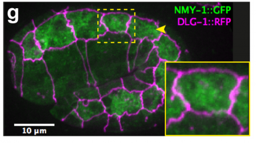
Myosin distribution in embryonic epidermal cells, from Figure 1, Vuong-Brender et al., 2017, eLife
And Thanh, how did you come to work in Michel’s lab?
TV-B I was trained as a physicist. My PhD was about the use of automated imaging and fluorescent markers for diagnosis of cervical cancer. After that, I went to work for a small company that developed automated imaging systems. I realised that I was more interested in academic research to explore and understand natural phenomena. During my PhD, I learnt some biology but not as much as I wanted. So I looked for a postdoc during which I could learn more about biology and came across Michel lab’s papers on mechanical problems of C. elegans embryonic elongation. I found the subject attractive, maybe because it presented to me a mechanical problem to solve. I did not understand all the biology but I thought it was really interesting to learn and to work on it. I sent my postdoc application to Michel and was really lucky to be accepted.
In an interview with Current Biology in 2005, you said you were excited by the challenge of understanding the mechanics of development. I wonder what you think of the progress the field has made in the 12 years since then?
ML The field has evolved tremendously, in part due to progress in imaging and data processing, and in part because a new generation of scientists with strong background in physics has entered the area. In my field, papers that brought key paradigmatic changes, which in retrospect seem quite common sense, include the demonstration that a morphogenetic event requires small increments that progressively modify the cell (Martin et al, Nature; Rauzi et al, Nature; Solon et al, Cell). Another one is that two apposed tissues with distinct mechanical properties will twist (Savin et al, Nature).
You write that the material properties of developing tissues have received less attention than the forces that act on them. Why do you think this is?
ML There are two probable reasons: in vertebrate embryos, the field has more frequently focused often on global movements (although the issue of stiffness has been pointed out more than 30 years ago in Xenopus), whereas in fly embryos, the field has focused on processes dependent on cells having a homogenous behaviour.
TV-B Deformation depends both on forces and material properties, so in theory, one can have as many regulatory pathways of shape formation through the regulation of material properties as through forces. The role of forces has been intensively investigated through the studies of non-muscle myosin and its regulatory pathways. Many studies have suggested the role of anisotropic material properties, like the elongation of Drosophila developing eggs or trachea. The role of material properties has received more attention in plants, but studies and mechanical measurements of material properties in animal morphogenesis are scarce. Our aim was to bring attention to this important parameter.
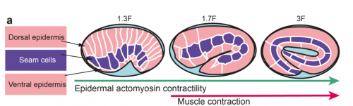
Overview of C. elegans embryo elongation, from Figure 1, Vuong-Brender et al., 2017, eLife
What makes the C. elegans embryo a good model for developmental mechanics?
ML The worm is very simple and many of its past successes have been linked to cell biology (PAR proteins, netrin, centrosome assembly, apoptosis, EGF/Ras signalling, to name a few). Its only drawback is that the embryo is quite small, very fragile and not easily amenable to approaches available in other species.
TV-BC. elegans embryonic elongation is very simple and different from other animal models, since it does not involve cell division or cell migration, but is mostly driven by cell shape changes. The number of cells is limited so that one can investigate at the cell resolution and the whole embryo. Other advantages are the easy genetics and worm cultivation.
Can you give us the key results of the paper in a paragraph?
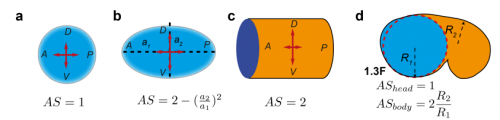
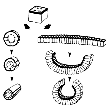
TV-B In the last step of embryonic development, the embryos of C. elegans transform from a ball of cells into the characteristic cylindrical shape of a worm. This process is powered by the association of the molecular motor myosin II, and the actin cytoskeleton in the embryonic epidermis. The epidermis is made up of six strips of cells running along the head-to-tail axis. Myosin II is mostly active in two strips of cells on the two sides of the embryo (lateral cells), but has low activity in the upper and lower strips of cells (dorsally and ventrally to the lateral cells). It is unclear how this distribution of myosin causes embryos to elongate only along the head-to-tail axis. Using laser nano-ablation, we have probed the forces exerted and the material properties in the embryonic epidermal cells. The results show that myosin’s activity in the lateral cells induced constriction around the embryo, sort of similar to the effect of a boa constrictor tightening around its prey. At the same time, the actin filaments in the dorsal and ventral strips form rigid bundles oriented along the circumference. They form a “belt” preventing the constriction from causing the cells at the dorsal and ventral strips embryo to expand. Finally, the only direction the embryo can elongate is along the head-to-tail axis.
Modelling anisotropic stresses in different shapes, from Figure 4, Vuong-Brender et al., 2017, eLife
How important was the modelling to complement and reinforce your experimental data?
ML It was critical in two ways. First to guide the interpretation of experimental data; second as suggested in the answer to the first question, because understanding a process involving a mechanical force should include a physical dimension whenever possible.
TV-B The mathematical modelling helps us to understand mechanistically what we observed. It can also predict the behaviours of the system in another situation or the role of another component in the process. So it can be your initial hypothesis or feedback, which combines with the experiment data to make the work evolve.
Are the anisotropies of stress and stiffness established by particular signals in the cell, and could this be this linked to cell fate choice in the embryo?
ML These are key mostly unsolved questions for the future.
TV-B I think that the anisotropies of stress and stiffness are linked to the cell specific myosin/actin regulator distribution, which have been shown to be different between lateral and dorso-ventral epidermal cells. It is likely to be linked to the lateral/dorso-ventral cell fate, but remains to be proved.
Comparing models of fly and worm elongation, from Vuong-Brender et al., 2017, eLife
When doing the research, was there a particularly exciting result or eureka moment that has stayed with you?
TV-B I did not really have a “eureka”, but for me, everyday is like an adventure. There were problems (technical or theoretical) to solve and challenges to overcome because we always tried new things. For more routine stuff, I tried to make it better or quicker. There are small victories like “yes ! it (laser ablation) works”, “I got my CRISPR knock-in strain”, “ the experiment data matches (the theoretical one))”… which led me through the days and frustrations sometime. Well, like every scientist, I hope to have a “bigger eureka” in the future.
And what about the flipside: any moments of frustration or despair?
TV-B Yes, I was trying desperately to make optogenetically-controlled gene expression work in worms. I have tried 3 different systems, none of these worked. Finally I tried a different optogenetic method controlling protein aggregation, but did not have the time to finish it. One consolation is that my preliminary results were used by someone else. I told myself that the failures are part of the quest and became now more resilient to them.
Where next for you following this work?
TV-B I am trying to shift (again) to another domain of research. During my postdoc, my interest for microbiology, symbiosis between microbes and the origin of eukaryotes has grown. So I decided to go to study the microbial diversity. It will be totally different but surely I will learn a lot of things.
30 year old foresight about embryo mechanics – from Jim Priess and David Hirsch’s Developmental Biology paper.
And what is the next step for the Labouesse lab?
ML I am quite proud of the work we did with Thanh and our two other co-authors, as I think it accounts in physical terms for a big part of the early phase of elongation. Incidentally, I want to pay tribute to a visionary landmark paper of our field written in 1986 by Jim Priess, a PhD student at the time, who had foreseen almost everything in early worm elongation. Now, I want to account for the second phase of C. elegans elongation that requires the mechanical input of muscle contractions (which Jim had not touched upon), and want to reach a similar level of understanding. We are nearing this phase.
STEM Graduates is a graduate recruitment agency and jobs board. We offer permanent salaried roles to students and graduates from Science, Technology, Engineering and Mathematics disciplines. We believe these candidates have a unique set of career needs that can only be met by a specialist within this field. We launched STEM Women in 2016 to provide a specific place for female careers advice, profiles of women in STEM and a dedicated job board.
We are always looking to expand what we can offer STEM students to make them more employable in their highly competitive markets. That is why we are excited to launch our recent partnership with the Science Council. This partnership will include a variety of activities and exciting content.
The Science Council will host a careers advice blog topic each month written by STEM Graduates (most recent blog post here) and we will be focusing on educating our candidates on the benefits on offer from joining the Science Council. We will also be listing the numerous specific professional bodies under the science umbrella including the Royal Society of Biology, the Institute of Science and Technology and the Institution of Environmental Sciences. We will achieve this through our social media channels, a new dedicated section on our website and with weekly articles.
We are proud to endorse the ‘working towards registered scientist’ (Registered Scientist (RSci)) initiative that has been launched by the Science Council. This will focus on the conduct, competence and professional development of early years’ scientists. For many graduates this initiative will be the first step towards becoming a chartered scientist.
The Science Council is a membership organisation for professional bodies and learned societies across the disciplines of science. They are in a unique position, bringing together a range of disciplines and sectors to reflect the multi-disciplinary practice of science in today’s society.
We are looking to expand this section further with other societies, associations and communities so please let us know if you have any ideas.
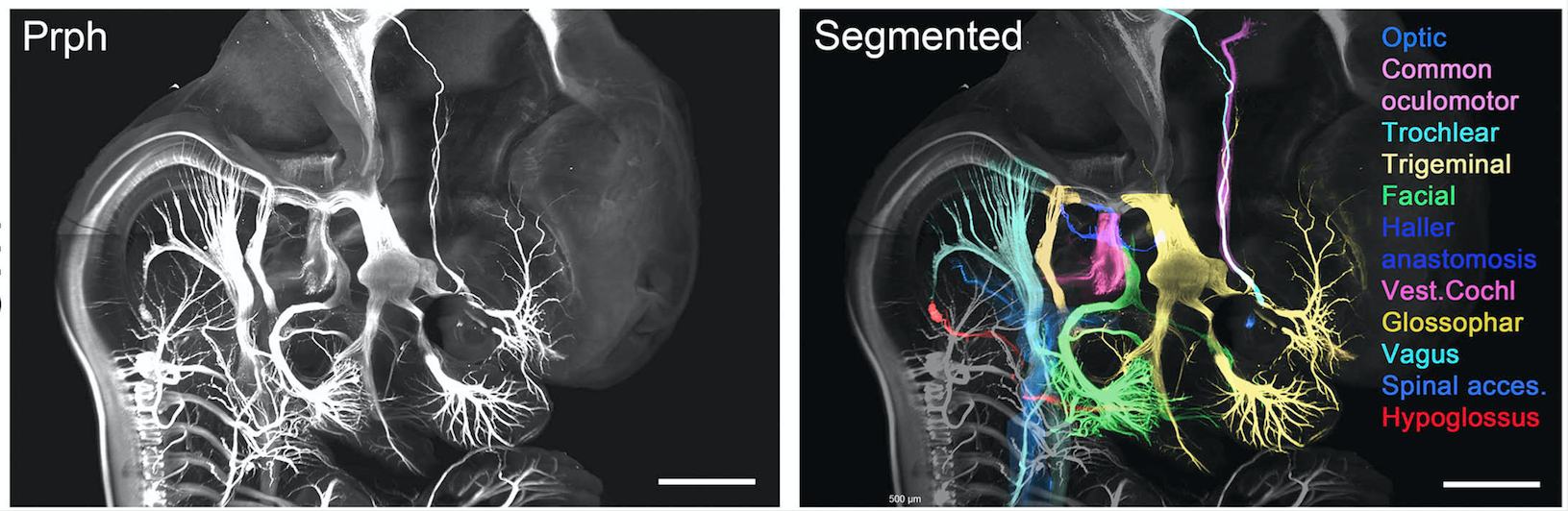
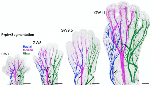
View of the head of a transparent 7 week embryo labelled with Peripherin. Cranial nerves have been pseudocoloured. From Fig1. Belle et al., Cell, 2017
Throughout history, the desire of scientists to understand physiology and disease by thoroughly studying anatomical features, has always faced an intractable limitation: they cannot simply see through the tissue! Dissection has therefore been the modus operandi of anatomists: from Galen’s pioneering studies, to modern day biologists who routinely section tissues to label structures for microscopic analysis.
Whilst these methods have informed a wealth of knowledge linking anatomical form to function, they are inherently flawed due to a 3-Dimensional appreciation of structures being lost. This has been especially problematic for the study of human development, where structures are continually evolving, and therefore a precise visualisation has been impossible to achieve through traditional methods and anatomical atlases. Coupled with the difficulties in tissue access, our understanding of human development has progressed perhaps the slowest of any biological process since the 1930’s; whilst in some cases observations of lower vertebrates have subsequently been erroneously applied to humans.
“Birth defects of structural or functional origin currently affect more than 3% of births”
Without a good understanding of physiological development, we lack the fundamental knowledge required for clinicians and researchers to tackle a healthcare issue that inflicts a severe healthcare and emotional burden. Recent advances in non-invasive, in vivo imaging techniques, have shown great promise in detecting congenital abnormalities as well as providing information on gross topological features of fetal development; however, they lack sufficient resolution in order to inform developmental biologists of currently unknown features of organogenesis. This has recently been most strikingly highlighted by the surge in Zika virus infections and reports of its detrimental effects on cephalic development.
The developing innervation of the human hand from 7-11 weeks gestation. From Fig2. Belle et al., Cell, 2017
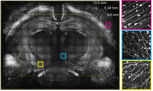
In order to begin rapidly addressing the gaping holes in our knowledge, we have begun to form a cutting edge research consortium of developmental biologists, to build a detailed human cell atlas. Here we have expanded the use of clearing techniques to human tissues and immunolabeled with over 40 antibodies, intact human embryos during the first trimester of gestation (from 6 to 14 gestational weeks).
In recent years, several techniques have been developed for tissue clearing, in which whole organs are rendered macromolecule permeable and optically transparent. Among tissue-clearing techniques, the process called 3D imaging of solvent-cleared organs, or 3DISCO has been proved to be a simple, robust and inexpensive method for 3D analysis of immunolabeled transparent organs in embryonic and postnatal mice.
Currently organised along seven organ systems, the project aims to expand and evolve as more data is added. Current data in the atlas examines molecular organogenesis based on over 40 samples comprising 1,500,000 optical sections – making it the most complete 3D analysis of early human development currently available.
Constructed by imaging intact tissues and whole embryos, the atlas will be an invaluable tool for researchers with the ability to explore cell distributions, count proliferating cells in each organ etc. whilst also being useful for didactic purposes, with the ability to 3D print models to inform health science teaching programs.
This has long been the dream of developmental biologists, which has finally been realised by the use of organic solvent based clearing techniques (3DISCO/iDISCO) combined with light sheet imaging. Together, these powerful approaches allow inexpensive labelling of any cell population of all developing organ systems during development and imaging at cellular resolution whilst fully maintaining structural relationships. To further highlight the robustness and power of this technique to analyse human development, it should be noted that these data were generated in just over one year by a small team of researchers.
And this is just the beginning, as the online resources are open access – available for researchers all over the world to analyse and contribute to, along with new data we will acquire. The goal of the project is to make a continuously updated 3D molecular reference atlas of human cells during development, paramount to a better understanding of human development during health and disease.
The online video series of immunolabeled tissues along with the original data sets is available at https://transparent-human-embryo.com/ . The 3D database was developed with Keen eye technologies with support from the “fondation voir et entrendre”
University of Oregon biologists have figured out how zebrafish perfectly regenerate amputated fins with a precisely organized skeleton.
Adult zebrafish fins, including their complex skeleton, regenerate exactly to their original form within two weeks after an amputation. The process, they found, is driven by clusters of specialized skin cells that migrate over reforming bones, known as rays, and escort bone cells into the right positions to form individual bones of a branched skeleton.
These skin cells produce a protein called Sonic hedgehog, which interacts with bone-building cells called osteoblasts to promote bone patterning during fin regeneration.
“The orderly reconstruction of zebrafish fins is amazing to see,” said Kryn Stankunas, a professor in the Department of Biology and member of the Institute of Molecular Biology. “Zebrafish fins, which are akin to our limbs, regenerate perfectly. The zebrafish bony rays re-branch just like the original structure. This would be like losing your arm and watching it progressively regenerate complete with a hand and fingers — all the bones restored in their original configuration.”
The findings will not lead to humans re-growing lost limbs, Stankunas said, but such advances in understanding the fundamental processes of regeneration in related vertebrate organisms will inform innovative and targeted therapeutic strategies to improve the repair of broken bones.
“The mechanism — how the skin and bone cells dynamically move and interact using the signaling pathway — is elegant and unexpected, broadening the project’s impact on regenerative medicine,” Stankunas said.
Hedgehog signaling, he added, is also linked to several human cancers.
“The zebrafish fin provides a tractable and simple model to decipher mechanisms of regenerative skeletal patterning,” the researchers wrote in their paper in the March 28 issue of the journal Development, a publication of the non-profit Company of Biologists in the United Kingdom.
Benjamin E. Armstrong, who earned a doctorate in biochemistry in 2016, was the study’s lead author. Scott Stewart, a research professor in the Institute of Molecular Biology, co-directed the project.
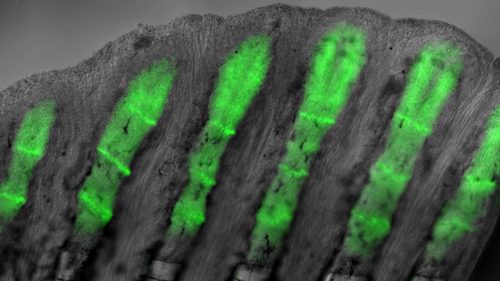
Green fluorescent proteins show where bone-building is occurring in the regeneration of a zebrafish caudal fin that had been amputated. Complete repairs begin at the tail’s base and gradually proceed to the tip, a process that is completed within two weeks. Courtesy of Kryn Stankunas
The research team used genetically modified zebrafish that produces a fluorescent protein that helps identify the subset of skin and bone cells that respond to Hedgehog signals. The fluorescent marker appears green under the microscope until illuminated with ultraviolet light to photo-convert the green protein to red.
This photo-conversion method revealed that repairing skin cells collectively move towards the tip of the regenerating fin. At particular times, Sonic hedgehog is induced in skin cell clusters that then split into two pools. Simultaneously, the skin cells activate a Hedgehog response in adjacent osteoblasts. That drives them to associate with the skin cells and co-migrate into split groups. The now separated bone cells continue to regenerate replacement bone, but now forming two rays instead of one – a branched skeleton.
“We could see that the bone cells responding to the skin-produced Sonic hedgehog become physically attached to the migrating skin cells”
“We could see that the bone cells responding to the skin-produced Sonic hedgehog become physically attached to the migrating skin cells,” Stewart said. “The pathway is quickly turned off but the now split groups of bone cells will then form two separated mature bony rays connected at a branch point.”
To define the functions of the Hedgehog signaling pathway, the researchers used a new chemical inhibitor, BMS-833923, to turn off Hedgehog signaling in their experimental fish. With Hedgehog blocked, the skin and bone cells failed to interact, and the fin regenerated with stick-like rays rather than forming a branched skeleton.
The inhibitor used in the study is in clinical trials against some forms of human cancers, but it had not been used in zebrafish. The Hedgehog pathway is most associated with basal cell carcinoma and medulloblastoma, Stankunas said.
“The Hedgehog response is absolutely required for branching and not essential for any other aspect of regeneration,” Stankunas said. “Instructions that drive the branching come from the skin cells moving into two groups and likewise dividing the osteoblasts. This is new information. It is the traffic pattern generated by the signaling that regenerates the fin. It is skin and bone working together.”
###
Astra Henner, lab manager and research assistant, was the fourth co-author of the paper.
The National Institutes of Health funded the project through a training grant to Armstrong and research grants to Stankunas and Stewart.
Source: Kryn Stankunas, associate professor of biology, 541-346-7416, kryn@uoregon.edu
Note: The UO is equipped with an on-campus television studio with a point-of-origin Vyvx connection, which provides broadcast-quality video to networks worldwide via fiber optic network. There also is video access to satellite uplink and audio access to an ISDN codec for broadcast-quality radio interviews.

 (1 votes)
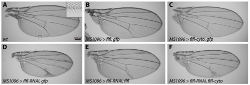
 (No Ratings Yet)
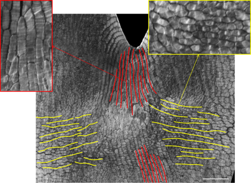
 (8 votes)