the Node in 2016
Posted by the Node, on 23 December 2016
2016 will live in infamy for many reasons, but perhaps we can seek a little catharsis in science, and in some of the wonderful developmental biology that the Node has promoted this year.
This end of year roundup comes in two sections – the top 20 most read posts, and then some of my personal favourites that may have been posted late in the year or gone somewhat underneath the radar. I joined the Node half way through the year and one of the real highlights of the job so far (aside from making GIFs) has been helping out with – and reading – a diverse and interesting suite of posts.
Skate development from the Gillis lab, who reached #4 in our top 20.
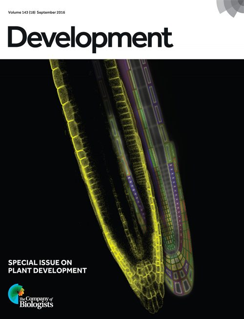
2016 Most Read Top 20
- In memory of Marcos Vidal – Ross Cagan and Eyal Gottlieb
- BarBarPlots – Helena Jambor
- Woods hole cover competition – Round 1
- Gills, fins and the evolution of vertebrate paired appendages – Andrew Gillis
- The Doctor of Delayed Publications – the remarkable life of George Streisinger – Máté Varga
- Raw Data: a cautionary tale – Katherine Brown
- Sweetening with a pinch of salt: maximized Cas9 efficiency in zebrafish – Alexa Burger
- MBL Embryology Course 2016 – Joaquín Navajas Acedo, Aleisha Symon and Tsai-Ming Lu
- Woods Hole cover competition – Movie round
- A day in the life of a spider lab – Anna Schönauer, Daniel Leite and Christian Bonatto
- An interview with Peter Lawrence
- Preprints in publishing – Katherine Brown
- The people behind the papers #1
- A day in the life of an embryonic stem cell lab – Helena Pérez Valle
- drosophila.me – manage your fly stocks and crosses – Mario Metzler
- Drawing Embryos, Seeing Development – Beatrice Steinert
- Revisiting the classics: coupling embryology with genomics to alter cell fate – Marcos Simoes-Costa and Marianne Bronner
- An interview with Melina Schuh
- A Tale of Trunks or Zen and the art of doing a PhD – Rita Aires
- Time-Lapse Recording of Pre-Implantation Mouse Development – Katie McDole
As you can see this represents quite an accurate cross section of typical Node content – from interviews to resources, and research to personal histories.

My personal favourite from the top 20 is Máté Varga’s magisterial history of George Streisinger. I knew nothing of Streisinger before reading this piece, one of the few ‘longread’ types we featured. I urge you to sit down with a coffee and take the time to read it; his really was a remarkable life, and developmental biology owes him a lot for initiating the zebrafish field. As a fan of the history of science, I also loved Beatrice Steinert’s piece on Edwin Conklin and drawing in early embryology. The introductory paragraph sets the scene beautifully:
“Today, when we want to capture an image given by the microscope we can either snap a photograph of it or obtain a computer-generated image. But prior to when photographic methods began making their way into biology labs and journals, this meant you had to draw it. For embryologists, this meant creating accurate, detailed drawings of either live or fixed embryos. Because developing embryos are three-dimensional, complex, and constantly changing, being able to render them by hand, let alone to see and make sense of them, was no simple feat. The task required meticulous observation of both the form and movement of cells, tissues, and structures. Pencil and paper weren’t used only as a recording device to create figures for publication, they also served as a form of note taking and played a central role in guiding embryologists’ observations of specimens placed under the microscope.”
Sticking with the MBL, I enjoyed Joaquín Navajas Acedo, Aleisha Symon and Tsai-Ming Lu’s account of the legendary Woods Hole Embryology course – their piece captures the intensity and slight sense of madness of those weeks by the shore. Of the posts about research, my pick is Marcos Simoes-Costa and Marianne Bronner’s account of their beautiful work on the neural crest, which blended personal and scientific stories brilliantly:
“Marianne had been interested in this question for a long time, and for more personal reasons – while recovering from a bicycle accident (face plant resulting in a broken nose and deviated septum), she learned from the craniofacial surgeon that facial cartilage is very difficult to replace”
Outside of this Top 20 (which is obviously a little biased towards the front end of the year), I’ll highlight three pieces that intersect art and science. Mark Hintze, a developmental biologist, and Diana Gradinaru, an illustrator, talked us through their animation ‘Developmental Biology: Life’s symphony.’ It really is worth a watch:
We also heard from two scientists-cum-artists: Mia Buehr, who uses machine embroidery to create wonderful versions of embryonic and fully formed model organisms, and Beata Edyta Mierzwa, whose drawings capture the magic of cell biology and the travails of research.


I always enjoy our Day in the Life posts, and as well as the two that made the top 20, I found Martin Minařík’s cross-continental account of life working on gar (fishes related to sturgeons) fascinating and funny. It was also a pleasure to get to know a bunch of scientists through our People Behind the Papers feature and will be continuing this throughout 2017. The piece that I am perhaps personally most proud of comes from our Forgotten Classics series, where I had the privilege of discovering Rosa Beddington’s work for the first time.
Thanks to all our authors and thanks to all our readers!
See you in 2017
 (1 votes)
 (No Ratings Yet)
 (6 votes)