The community site for and by
developmental and stem cell biologists

Regeneration thwarts ageing in newts

Posted by , on 17 January 2016

Konstantinos Sousounis and Panagiotis A. Tsonis


The human eye is built to deliver the sense of vision. The eye lens is one of the organs playing role in focusing the light to the retina. Lens injury or disease leads to blurriness or even blindness in human patients. This is not the case for newts. These small amphibian urodeles can have their entire lens removed, and yet they can regenerate a brand new one! In fact, regeneration in newts is not limited to the lens, but extends to many organs from brain, heart, spinal cord to limbs and tails. Ever since the discovery that newts can regenerate numerous body parts, many scientists have focused on deciphering the mechanism underlying regeneration (Sanchez Alvarado and Tsonis, 2006; Tsonis and Fox, 2009). The mechanism as a whole is still unclear, despite the numerous findings of the various molecular interactions underlying regeneration. In addition to the mechanism of regeneration, two important questions remained unanswered:

(1) Knowing that body parts can be regenerated, how many times can an organ be regenerated?

(2) Does age affect/limit the regeneration capacity of an organism?

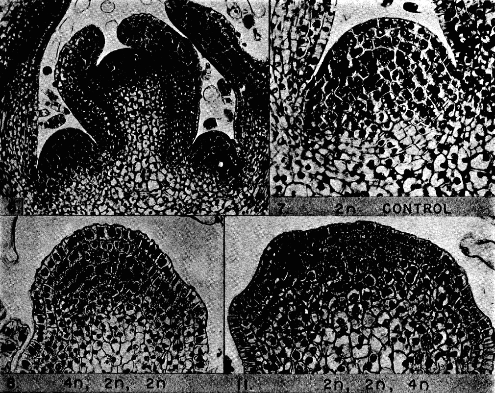
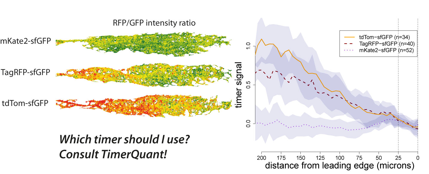
These two questions cannot be answered experimentally by the commonly used “injury induction-time point-collection-analysis” paradigm. These experiments need to include repetitive insults to the same animals over a prolonged period of time and will, thus, require a commitment from the researcher to take extremely good care of the animals while maintaining these experimental colonies for a long time. Now, this is not an easy task. To put things into perspective, keep in mind that newts are kept in water, and water is an environment that without prompt cleaning can get pretty dirty quite fast. Diseases can also spread, so that one infected newt can easily wipe out the rest of a group in a tank. So, rule number one: Extreme super animal care! Something you get used to after the first couple of weeks in an amphibian lab. Many other crucial issues arise. For how long do you need to be super vigilant for newts being used in an ageing study? When does a newt age? How many years can a newt live [keeping in mind that a mouse is considered aged after its first birthday]? Well, according to Goin et al, 1978, newts can live as long as 25 years. That is almost a three-decade-long newt ageing study, which would also mean that an actual study designed to investigate the effect(s) of ageing on regeneration in newts would span way beyond the time typically needed for students to obtain their Ph.D. or post-doctoral researchers to complete their fellowship. That is not all; surviving surgery is challenging and requires a lot of post-surgical care on the animals that will be operated twice every year since 6 months is sufficient period of time for an organ to be fully regenerated and for a newt to recover well before the next surgery. Taken together, the requirements for the newt ageing study make it seem unimaginable that a person might actually carry such a study and investigate how repetitive regeneration is affected throughout the lifespan of a newt (not to mention receiving funding). Unimaginable but not impossible for Goro Eguchi who in 1994 at the National Institute for Basic Biology at Okazaki, Japan took on this task and put a start to this project. The model chosen was that of lens regeneration in the Japanese newt. One of the main advantages of this model is the fact that the lens is in an enclosed environment (the eye), and when removed through a small slit in the cornea the injury is local and the wound closure heals very fast. The lens is regenerated from the iris via a process called transdifferentiation, whereby the uniformly pigmented cells of the dorsal iris undergo a complete morphological, transcriptomic and molecular transformation to regenerate the lost lens (Henry and Tsonis, 2010). The process is considered complete after 6 months and the newly regenerated lens is identical to the original. In our study, the first lens removal was performed on 14-year-old newts. These newts were 30-year-old at the end of the study, and they had their lenses removed 18 times over a period of 16 years. Then, at the end of the study, our team (based at the University of Dayton and the Sanford Burnham Prebys Medical Discovery Institute) had these repeatedly regenerated lenses analyzed for the first time. The conclusion was clear: repeated regeneration or ageing did not affect regenerative capacity in newts (Eguchi et al, 2011). Fiber structure and expression of crystallin and lens-specific genes were similar to never-regenerated lenses from young newts. Even though that study was groundbreaking, many questions were left unanswered. What we wanted to investigate next was whether the regenerated lenses were aged and if the process of regeneration had affected the underlying genetic profile. To get the most out of the tested samples, we decided to use RNA-sequencing. We collected the repeatedly regenerated lenses (now 19 times regenerated; 18 years after the initiation of the project), the dorsal iris (the source of regeneration) and the tails (to serve as controls since this tissue was never removed or regenerated in these animals). We also collected the same tissues from young newts that never regenerated the lens. Altogether the samples totaled 30, with n = 5 animals per each group. In the absence of a genome, de novo assembly of the newt transcriptome was required. With 30 samples at hand, multiplied by 150 million reads for each sample, we were left with a complex task that even super computers found challenging to execute this de novo assembly. With suggestions from Tritiny while using their program, the task was completed and the final gene expression data was emailed to us. Holding our breath, we checked the data and BINGO: zero genes differentially expressed in Experimental Lenses versus Control Lenses! After an overwhelming feeling of joy we went onto data analysis. The tail samples were found to have the most differentially regulated genes, followed by the iris and then the lenses with none. At first glance, it was obvious that there was a gradient of age-dependent regulation based on the degree of regeneration (or degree of cell replacement in the case of iris if you prefer) that each tissue had experienced. To clarify how we came about knowing that the tail and iris samples were indeed aged, we used previous studies on other vertebrates and even flies that had identified, for example, that down-regulation of mitochondrial genes is a common signature of ageing. We identified this pattern and other gene patterns too in our samples. That told us that there was a robust transcriptional program that is utilized with every single lens removal, which guarantees the completion of the process with the outcome of a perfectly identical lens (Sousounis et al, 2015). Another interesting finding was that the iris, the tissue that regenerates the lens, had shown signs of ageing but the regenerate, the lens, did not! That showed us that the transdifferentiation process can be really compared to an “extreme makeover – cell edition.” Considering that human lenses are vastly affected by ageing and that their composition is really not that different from newt lenses, our findings hold great translational promise for future studies. The newt is not only capable of reprogramming adult cells to dedifferentiate and regenerate, but also has inherent mechanisms to deactivate pathways that lead to ageing. These findings could pave the way to avenues of experimentation towards the prevention of the process of ageing.

References

Eguchi G, Eguchi Y, Nakamura K, Yadav MC, Millán JL, Tsonis PA. Regenerative capacity in newts is not altered by repeated regeneration and ageing. Nat Commun. 2011; 2:384. doi: 10.1038/ncomms1389.


Goin, C. J., Goin, O. B. & Zug, G. R. Introduction to Herpetology 3rd edn (W.H. Freeman, 1978).


Henry JJ, Tsonis PA. Molecular and cellular aspects of amphibian lens regeneration. Prog Retin Eye Res. 2010; 29(6):543-55. doi: 10.1016/j.preteyeres.2010.07.002.


Sánchez Alvarado A, Tsonis PA. Bridging the regeneration gap: genetic insights from diverse animal models. Nat Rev Genet. 2006; 7(11):873-84.


Sousounis K, Qi F, Yadav MC, Millán JL, Toyama F, Chiba C, Eguchi Y, Eguchi G, Tsonis PA.  A robust transcriptional program in newts undergoing multiple events of lens regeneration throughout their lifespan. eLife. 2015; 4. pii: e09594. doi: 10.7554/eLife.09594

Tsonis PA, Fox TP. Regeneration according to Spallanzani. Dev Dyn. 2009; 238(9):2357-63. doi: 10.1002/dvdy.22057

Thumbs up (17 votes)
Loading...

Tags: , ,
Categories: Research

Navigate the archive

Use our Advanced Search tool to search and filter posts by date, category, tags and authors.

FlyBase Biocurator (Cambridge, UK)

Posted by , on 15 January 2016

Closing Date: 15 March 2021

We are seeking a full-time biocurator to join the FlyBase group at the University of Cambridge, UK. If you are looking for a fulfilling, fly-related career away from the bench, and enjoy the challenge of organizing complex data and presenting it clearly and concisely, then this is the job for you!

The primary responsibility of the FlyBase-Cambridge site is to identify genetic data from scientific articles concerning Drosophila and to record these data in a systematic manner. The data we capture include all genetic objects (genes, alleles, transgenic constructs, etc.) plus associated information such as phenotypes, genetic interactions, gene product functions and models of human disease. Curated data are subsequently integrated into the FlyBase database and made freely available via our website.

Established staff also:

  • research strategies to improve data curation;
  • work with other FlyBase sites to improve data display/querying on the website;
  • interact with our user community by answering HelpMail and giving presentations/workshops at research conferences;
  • have the opportunity to develop programming skills;
  • contribute to FlyBase publications.

Informal enquiries to Dr Steven Marygold (sjm41@cam.ac.uk).

Further details and a link to the application form are here.

Closing date for applications is 14th February 2016.

Thumbs up (No Ratings Yet)
Loading...

Categories: Jobs

Two Postdoctoral Fellow Positions (skeletal muscle biology or neurological disorders, at Johns Hopkins University)

Posted by , on 14 January 2016

Closing Date: 15 March 2021

Two Postdoctoral Fellow positions are available in Gabsang Lee laboratory at Johns Hopkins University. These positions are NOT for stem cell expert, rather for who has OUTSTANDING expertise on either biochemistry, skeletal muscle biology, optogenetics OR single-cell studies. Previous experience on human pluripotent stem cells is not necessarily required, and we value more on the ‘non-stem cell’ experience. Research project will be developed based on the applicants’ talent and interest, in a conjunction with our research topics.


The Lee lab has been establishing novel methodologies to specify human induced pluripotent cells (hiPSCs), and to direct convert somatic cells, into multiple lineages, including induced neural crest (Kim et al., Cell Stem Cell, 2014), peripheral neurons (Oh et al., in revision), Schwann cells (Mukherjee-Clavin et al., in revision) and skeletal muscle cells (Choi et al., in revision) using multiple genetic reporter systems. We continue to study human developmental and degenerative disorders (pain disorders, demyelination disorders, and muscular dystrophies) to unravel the underlying cellular/molecular mechanism toward realistic therapeutic approaches.


Areas of research topics of these positions are below.
– Neural cell fate determinations
– Pain disorders
– Demyelination disorders
– Insulin resistance
– Muscular dystrophies
– Satellite cell biology


Applicants can send (by Feb 10th, 2016) a CV and the names of three references to the address listed below:

Gabsang Lee DVM, PhD

Johns Hopkins University

School of Medicine

Institute for Cell Engineering

Departments of Neurology and Neuroscience

leelabjob@gmail.com

Thumbs up (No Ratings Yet)
Loading...

Categories: Jobs

An interview with Melina Schuh

Posted by , on 14 January 2016

This interview first featured in the Journal of Cell Science and is part of their interview series Cell Scientists to Watch.


Melina Schuh received her diploma degree in biochemistry from the University of Bayreuth, Germany, where she completed her Diploma thesis with Stefan Heidmann and Christian Lehner. She went on to do her PhD with Jan Ellenberg at the European Molecular Biology Laboratory in Heidelberg, Germany. In 2009, after a bridging postdoc with Jan, Melina started her own group at the MRC Laboratory of Molecular Biology in Cambridge, UK. Since January 2016, she is a Director at the Max Planck Institute for Biophysical Chemistry in Göttingen, Germany, and will establish a new department focussing on meiosis. She is an EMBO Young Investigator and a recipient of the 2014 Lister Institute Research Prize, the 2014 Biochemical Society Early Career Award and the 2015 John Kendrew Young Scientist Award. Her lab is studying meiosis in mammalian oocytes, including human oocytes.


Figure1

What motivated you to become a scientist?

To some degree I became a scientist by chance. I had many different interests in school, and I was struggling to decide whether I wanted to become a journalist, an architect or a scientist. My friends and family encouraged me to study biochemistry because they thought that it suited me most. But I don’t think I knew at that stage what it would be like to work as a scientist. Studying biochemistry was a very good choice, though, and I never had any regrets. I loved the way science was taught at university.


In your PhD you developed methods for studying meiosis in mouse oocytes. What problems did you have to overcome, how did you devise your system?

There were very few high-resolution studies of mouse oocytes when I started my PhD in Jan Ellenberg’s lab at EMBL in Heidelberg. Jan’s lab had worked with starfish oocytes but had no experience with mammalian oocytes. I wanted to develop a system that would allow us to image meiosis in oocytes at high resolution by using a confocal microscope. This was not trivial because oocytes normally develop inside the body and they are very sensitive cells. I had to find out how to culture the oocytes outside the body on a confocal microscope, how much light they could tolerate, and how to label and follow structures – like the chromosomes – over the entire course of meiosis, which takes more than 12 h. Much of it was learning by doing, but I also got very valuable advice from various people around me.


You continue to work on meiosis in mouse oocytes. What are the particular questions that your group is currently trying to answer?

Our main aim is to understand how defects at the interface between chromosomes and the cytoskeleton lead to aneuploid eggs and pregnancy loss in mammals. We have also recently started to study meiosis and chromosome segregation directly in live human oocytes, which has not been possible before. This is a new direction of research in my lab that I am very excited about, as it should allow us to investigate why human eggs are so likely to be aneuploid.


What parts of the cytoskeleton are you specifically looking at?

We have evidence that, in human oocytes, the way that the spindle assembles is different from how it assembles in mitotic cells. Spindle assembly takes more than half a day in human oocytes. In mitosis, it takes just 30 minutes. So the question is: why are human oocytes so slow in assembling a spindle? Oocytes from many species assemble the spindle without centrosomes. Instead, they use acentriolar microtubule organising centres (aMTOCs), which functionally replace the centrosomes. We could not find these aMTOCs in human oocytes. We discovered that human oocytes assemble the spindle by a mechanism that is chromosome-dependent and mediated by the small GTPase Ran. The spindles need to be extensively reorganized while the chromosomes become aligned in the spindle centre. This could be one of the reasons why spindle assembly takes so long. These are, at least to our knowledge, the first studies of chromosome segregation in live human oocytes.


Fig. 2.
Mouse oocyte during polar body extrusion. Chromosomes are shown in cyan, actin in green, microtubules in magenta. (©MRC Laboratory of Molecular Biology, M. Pasternak and M. Schuh).


Have we been looking at an incomplete system when studying meiosis in mouse oocytes?

Mouse oocytes still resemble human oocytes in many aspects. We will need to study human oocytes in much more detail to evaluate how closely they resemble mouse oocytes.


How did your collaborations influence your research? Do you have any advice on collaborating?

It is helpful to collaborate with scientists that you enjoy interacting with and where the communication is working well. Which is crucial to a successful collaboration.


Everyone makes mistakes. How do you deal with them in the lab?

It is unavoidable that you make mistakes at some point in your career. And you should, of course, try to learn from your mistakes. But it is equally important that you don’t get hung up on these mistakes. You need to accept that you cannot be perfect. Try to always remind yourself of all the things that are working, the things you are doing well and to simply enjoy the science.


What advice on how to establish a successful academic career would you give?

I would recommend that you try to do something new and exciting, even if it is difficult. Be creative, ambitious and fearless in your approaches. Do not think that something cannot be done just because nobody has done it yet. It is good if you try to develop something new, because this helps you to stand out from the crowd, become known for what you are doing, advance your career and get tenure.


What do you think about the feasibility of being both a good parent and a good scientist?

I think this is a challenge for many parents. Pregnancy, maternity or paternity leave, the many sleepless nights that come with a baby, and the time involved in looking after children will all have an impact on your productivity. There is not much that can be done about this. It does help to have excellent day care, ideally subsidised by institutes. It is also important that line managers and institutions in general are understanding that there will be a transient period of time when progress is going to be a bit slower. Having this support will help you to stay focussed on your work and to get through this busy time with confidence.


You left your home country, Germany, to start your own research group here in Cambridge. Do you think of returning to Germany one day?

I have been offered a great position at the Max Planck Institute for Biophysical Chemistry in Göttingen. I very much enjoy working at the LMB, but my husband is still working in Germany while I am in the UK with two kids. Moving back to Germany will allow us to live together as a family while doing research at a fantastic institute – it couldn’t be better.


Also watch this additional clip:

Thumbs up (1 votes)
Loading...

Tags: , ,
Categories: Interview

In Memory of Marcos Vidal (1974-2016)

Posted by , on 13 January 2016

This obituary was written by Ross Cagan and Eyal Gottlieb, and first appeared in Disease Models & Mechanisms.


With the untimely death of Marcos Vidal, we have lost a good friend and a creative, brilliant colleague who made important contributions to the field of cancer biology through fruit fly research. Marcos began his research into Drosophila at Ross Cagan’s laboratory in 2003 first at Washington University in St. Louis and later at Mount Sinai Hospital in New York. In 2009 Marcos was appointed as Research Group Leader at the Beatson Institute for Cancer Research in Glasgow.


I (Ross) had just finished giving a talk in the Renal Division at Washington University School of Medicine when a young scientist approached me. He was a biochemist but liked the idea of using fruit flies to study diseases. Marcos Vidal was an impressive young man and I was excited to have him join the laboratory. A few days later a woman approached with the same request. Julia Cordero assured me that her relationship with Marcos would not be a problem, that they were used to working together and that they never fought. I sat them on opposite sides of the laboratory.

Marcos VidalMarcos and Julia were a brilliant young couple who instantly made the laboratory smart and fun. Marcos took up cancer research, the second person to ever work on this disease in the laboratory. He loved everything about conducting science and he was ambitious. He worked crazy hours. With a laconic Argentinian accent, he kept up a running science conversation that lasted until now. Julia worked on separate topics and they lived balanced lives. Marcos loved board sports from snowboarding to kite- and wind-surfing and was superb at them. Marcos and Julia made friends easily. The Facebook postings and the emails we’ve received and the conversations we’ve had over the past few days are a reminder just how much they were loved. We all rooted for their success because they were really good people.

Marcos pushed the fly as a discovery platform further than almost anybody else. He was enthusiastic to exploit the power of drug screening. He had a feel for the organism and, perhaps most importantly, he had a feel for what constituted an important biological question. In 2005 he validated Vandetanib as a candidate therapeutic for medullary thyroid carcinoma. The drug was approved in 2011 for clinical use. He explored the role of Src in oncogenesis, providing evidence and mechanisms that position metastasis as an immediate early event, promoted at least in part by local cell interactions within the epithelium. Later on, as an independent Group Leader in Glasgow, Marcos focused on the cancer promoting role of the tumor microenvironment and the immune system. Using the fruit fly as a cancer model, Marcos demonstrated that the genetic composition of a tumor is a key determinant as to whether Tumor Necrosis Factor (TNF) acts as a tumor suppressing or tumor promoting factor. He further characterized the crosstalk between epithelial tumors and the innate immune response, demonstrating that the TNF-Toll non cell-autonomous signaling cascade dictates tumor cell fate. Marcos used the fruit fly cancer model creatively, and he did it with a style unmistakably his own.

As a couple, Marcos and Julia were a true science power: young and smart and handsome. In Glasgow, Marcos started out as a Junior Group Leader at the Beatson Institute, becoming my (Eyal) valued neighbor and peer. Julia helped him set up his laboratory, and later started her own. They lived the lives of young professionals, with an international twist. They worried about keeping two leadership research positions, worried about their two young children and whether Argentinians could adapt to the Scottish weather. But they soon became central to the Beatson Institute, got involved with the local community and established successful professional and personal lives in Scotland. Beyond his work in the lab, Marcos dedicated his energy and free time to his family. He spent long, happy hours passing his boarding and drawing skills to his son Lautaro, and riding his bike with his daughter Mara.

Marcos fell ill 18 months ago and those extremely difficult times were a testimony to the central role his family and friends played in his life. Marcos worked as hard as he could to make a recovery. He received tremendous support from his colleagues and friends and, most of all, from Julia. Despite struggling with his illness, Marcos sailed through his Tenure Review with flying colors and in July 2015 he became a Senior Research Group Leader at the Beatson Institute and a Professor at the University of Glasgow.

Marcos Vidal died on January 2, 2016. Julia, Lautaro and Mara lost a warm-hearted, dedicated and loving husband and father; the science community has lost a brilliant young scientist and a good friend.


Thumbs up (22 votes)
Loading...

Tags:
Categories: News, Research

Forgotten classics- Cell layers in the shoot meristem

Posted by , on 12 January 2016


Satina, S., Blakeslee, A.F., and Avery, A.G. (1940) Demonstration of the Three Germ Layers in the Shoot Apex of Datura by Means of Induced Polyploidy in Periclinal Chimeras. American Journal of Botany 27, 895-905

Recommended by Jane Langdale (University of Oxford)


If you read about plant development in textbooks you will be told that the shoot meristem, which gives rise to all the stems, leaves and flowers, is formed of distinct cell layers. In dicotyledonous flowering plants there are three layers: the outermost layer (L1) will give rise to the epidermis; the L2 layer will produce most of the subepidermal tissues of leaves and flowers; and the innermost L3 layer forms the stem and vasculature tissues. That these layers exist and that they make different contributions to the shoot does not seem so surprising now, but there was a time when even the number of the then called ‘germ’ layers was a topic of debate. There had been much interest in meristems since the mid 19th century, and scientists like Hanstein had already suggested that meristems might have 3 germ layers in some plants. There was the need to carefully determine whether that was the case in all higher plants.

In the late 1930s, Amos Avery and his colleagues were on the case. But how do you investigate cell lineages when you are 60 years too early to use ‘basic’ labelling techniques like GFP-tagged proteins? What was needed was a way to genetically modify some of the cells of the plant so that you could identify their descendants and hence follow their fate, but it didn’t necessarily have to be a precise change. Realising this, Avery and colleagues took a blunt approach. They soaked seeds of Datura plants in colchicine to induce chromosome duplications. As colchicine inhibits spindle formation, chromosome duplication occurs without cell division, generating cells of different ploidy within a single seed – a chimera. It is important to note that the shoot meristem is fully formed in the embryo and hence these treated seeds contained shoot meristems in which some cells were polyploid and others were not.


FC1- Fig 1

Reproduced with permission of the American Journal of Botany


The seeds were grown in fields over the summers of 1938 and 1939, and the plants were regularly pruned to encourage the formation and outgrowth of lateral shoot meristems (which were ultimately derived from subsets of cells in the primary embryonic shoot meristem). Such an experiment is of course a game of chance, and as a result Avery and his colleagues obtained a huge range of chimera types, which they then used to study different aspects of plant development. In this particular paper, they examined the shoot meristem. Using cell size as a broad readout of ploidy, they examined how the cells of the shoot meristem were affected by the procedure. They noticed that you could find plants in which all the cells of the first layer had a different ploidy to the rest of the meristem. Sometimes they found plants in which the change in ploidy happened only in the cells of the second layer, and sometimes only in the cells of the third layer. And very occasionally you could find a plant where all three layers had different ploidy, e.g. the first layer was 2n, the second layer was 4n and the third layer 8n. Since ‘the response of any one layer to colchicine is independent of the response of the other two’, the authors concluded that the shoot meristem of Datura must have three independent germ layers. They also observed another phenomenon that is now textbook fact, namely that the cells in the 1st and 2nd layer (L1 and L2) mainly undergo anticlinal cell divisions (where the division plane is perpendicular to the cell layer) while the cells of the third layer can undergo divisions in any orientation.

This paper essentially reported the results of a single experiment, and did not go on to give further details on the subsequent development of the germ layers and on which organs originated from which layer. This is perhaps something we would expect in a modern paper, but science was different in those days so the authors saved those experiments for a subsequent paper. However, one should not underestimate the importance of this work. As Jane Langdale (who recommended this paper) put it, this was the ‘first demonstration that shoot apical meristems of flowering plants operate in the context of functionally distinct layers. A concept that is now so embedded that students rarely wonder how it was first shown.’


Further thoughts from the field


This study is a prime example of an early clonal analysis in plants. It provided direct information about the cell division patterns in shoot apical meristems and laid the basis for the tracing of cell lineages and the origin of tissues. Most importantly, it established the existence of three germ layers in plants.

Kay Schneitz, TU Munich (Germany)


Though molecular genetics has shown the importance of communication among layers in shoot meristem organization, we are far away from understanding its complexity and how this communication orchestrates layer-specific gene expression and the identities of individual cell layers. In the data rich science that we practice today, this paper is a must read for every developmental biologist, as it conveys the power of a single and carefully thought about experiment.

Venugopala Gonehal, UC Riverside (USA)


The American Journal of Botany has kindly provided free access to this paper until the end of June 2016!


—————————————–
by Cat Vicente


Forgotten classics doodle squareThis post is part of a series on forgotten classics of developmental biology. You can read the introduction to the series here and read other posts in this series here.


Thumbs up (10 votes)
Loading...

Tags: , , ,
Categories: Education, Research

Forgotten classics of developmental biology- a new Node series

Posted by , on 12 January 2016

You just started your research career, or maybe you just moved fields. The first thing on your to-do list is to catch up with the literature. What has been the latest progress? What are the open questions in the field? Ideally, you would go all the way back to the beginnings of the field and acquire a broad perspective of how research has changed and how the earlier works have impacted on current research. But time is short. The literature is just too vast, and the bench is calling, urging you to get started on your experiments. So you read a couple of recent comprehensive reviews, a bunch of papers from the last few years and a handful of the most significant earlier studies and move on to start your research project.

There is nothing wrong with this approach- there really is a vast number of papers out there, and time is limited. Unfortunately, this means that many important papers are overlooked. Certain ‘facts’ are taken for granted, and how they were obtained is not questioned. That chance encounter with an old result that could change the course of your research never takes place.

Forgotten classics doodleAware of these issues, we have decided to launch a new series of posts called ‘Forgotten Classics of Developmental Biology’. We asked prominent researchers from all areas of developmental biology to suggest their favourite hidden gems and to explain why we should all revisit them. In this series we will provide you with a summary of each paper, alongside comments from the person who recommended it as well as from other researchers in the field. For those papers still not freely available online, we also aim to ask the publishers if they will provide free access to each paper for a limited period of time after the post is published. We hope this will give everyone a chance to benefit from re-reading these forgotten classics!

The list of papers that we plan to feature in this series is still growing, so if there is a ‘Forgotten Classic’ that you think we should cover, please get in touch. We would love to hear your suggestions.


Thumbs up (11 votes)
Loading...

Tags: ,
Categories: Education, Research

Mole- So now you have your own lab! Part VIII- Money Matters

Posted by , on 11 January 2016

This cartoon was first published in the Journal of Cell Science. Read other articles and cartoons of Mole & Friends here.


Embedded ImageEmbedded Image

Part I- ‘The imposter’

Part II- ‘The teaching monster’

Part III- ‘The Pact’

Part IV- ‘The fit’

Part V- ‘The plan’

Part VI- ‘FCTWAWKI’

Part VII- ‘Beaten and bruised’

Thumbs up (2 votes)
Loading...

Tags: ,
Categories: Discussion, Lab Life

Applying tandem timers to measure signalling and gene expression dynamics in developing embryos

Posted by , on 11 January 2016

figNode

The signalling systems that conduct the orchestra of embryonic development are fantastically complex and dynamic. We owe much of our knowledge of in vivo signalling dynamics to advances in microscopy and protein tagging with fluorescent reporters that have allowed visualization of signalling proteins. Looking forward, however, it is clear that simply analyzing the localization patterns of proteins is only the first step to fully understand signalling processes. Of course, a protein’s presence at a particular time or place in the embryo does not tell us if it is actively signalling or lying dormant until its activity is required. Therefore, generic methods to distinguish actively signalling proteins from passive proteins will be tremendously useful advances for researchers interested in developmental processes.

A few years ago, Darren Gilmour at the European Molecular Biology Laboratory (EMBL) in Heidelberg, Germany, realized that since many signalling events lead to the degradation, stabilization, or relocalization of participating proteins, methods for detecting changes in protein turnover could provide powerful proxies for in vivo signalling activity. Gilmour and his graduate student Erika Donà therefore took advantage of a dual-color genetically encoded fluorescent reporter called a “tandem timer”, newly developed by Michael Knop in the next-door lab at the EMBL. Tandem timers report on the age of protein populations through the relative fluorescence of adjacent slow- and fast-maturing fluorophores (Khmelinskii et al. Nat. Biotechnol 2012). In collaboration with Knop and his postdoc Anton Khmelinskii, Gilmour and Donà tagged the chemokine receptor Cxcr4b with a tandem timer with a view to observing Cxcl12a chemokine signalling activity across a migrating tissue, the zebrafish posterior lateral line primordium. They wagered that this strategy was likely to inform on chemokine signalling activity since Cxcr4b receptors undergo rapid internalization and degradation upon ligand-mediated activation. Higher Cxcl12a levels would increase Cxcr4b internalization rate and this should be reflected by a decrease in the receptor’s population age (increased protein turnover). Gilmour and Donà also understood the potential long-term implications of their attempt; a successful proof-of-principle study might lead to the adoption of the tandem timer as a generic tool for the detection of signalling activities.

At the time I was a postdoctoral researcher in Wolfgang Huber’s group at EMBL, and having collaborated with Knop and Khmelinskii on the original tandem timer paper, I was happy to get involved in this first project using tandem timers in embryos. Using image processing and analysis tools from R and Bioconductor software, I measured tandem timer signalling readouts from 3D images of migrating tissues, and used a mathematical model of timer behaviour to interpret the results. The model describes the maturation kinetics of the two fluorophores on the timer, as well as production and degradation rates of the tagged protein. What surprised and delighted me throughout my collaboration with Gilmour and Donà, was the extent to which quantitative analysis and modelling was able to feed back on subsequent experimental design, and positively influence later results. Ultimately our tandem timer observations, along with other data, allowed us to demonstrate that a self-generated chemokine gradient guides the migration of the zebrafish posterior lateral line primordium (Donà et al. Nature 2013).

Tandem timers can be constructed with virtually any combination of spectrally separated fluorescent proteins with different maturation rates. Therefore, the first question an experimentalist typically asks when using tandem timers is: which fluorophores should I choose? We have been contacted by many groups asking exactly this question. As our ability to design timers and interpret timer data has benefited greatly from the use of modelling, we decided to write a paper outlining how our modelling tools could be used by other researchers interested in tandem timer research. To make these tools easily accessible we developed an interactive web application, TimerQuant, and made all our code available through the open-source software platform Bioconductor. For our new paper, which is now published in the journal Development (Barry et al. Development 2016), we systematically investigated the effect of relevant experimental parameters on timer signal, a measure of how good a particular timer is at detecting differences between two protein half-lives. To validate the predictions of our model, we reinvestigated the Cxcr4b signalling gradient identified in our original study using three tandem timers that had the same fast-maturing fluorophore (sfGFP), but different slow-maturing fluorophores (mKate2, mCherry, TagRFP). We found that timer signal decreased as the maturation times of the slow- and fast-maturing fluorphores became more similar. Readouts became noisier as protein abundance decreased. Timer signal increased with the maturation time of the slow-maturing flurophore, albeit at the expense of noisier readouts. While these conclusions might already be expected from a more qualitative reasoning, the quantitative model also led to some unexpected and intriguing findings. For example, Förster resonance energy transfer (FRET) between the fast- and slow-maturing fluorophores actually increased timer signal, a prediction that was borne out in our experiments, and which may be an important consideration for future timer designs.

In the initial model protein production (expression) and degradation rates were kept constant. Given that in developmental contexts these are likely to change over time, we decided to explore the effect of dynamics on timer ratio profiles. We modelled constant increases and decreases of protein expression and degradation rates as well as sudden bursts of expression and degradation, and looked at model solutions over time. The simulation results showed timer ratio profiles over time producing clear, characteristic responses to expression and degradation dynamics. We were surprised by the extent to which expression and degradation responses were distinguishable from one another. These additions to the model further help in the interpretation of timer ratio results in non-steady state conditions, which is often the situation when studying dynamic developmental processes. Moreover, as degradation rate could be experimentally fixed, for example by using non-degradable (or slowly degradable) versions of tandem timers, these model predictions open up the exciting possibility of using tandem timers to observe gene expression dynamics with time-lapse microscopy.

Explore Further:
Interactive TimerQuant software applications including those showing timer responses to dynamically changing gene expression can be used online at http://chronos.embl.de/TimerQuant/ or offline through the TimerQuant software package on the Bioconductor website.

Papers Referenced:
1. Barry JD, Donà E, Gilmour D and Huber W, TimerQuant: A modelling approach to tandem fluorescent timer design and data interpretation for measuring protein turnover in embryos, Development 143(1), 2016
2. Donà E, Barry JD, Valentin G, Quirin C, Khmelinskii A, Kunze A, Durdu S, Newton LR, Fernandez-Minan A, Huber W, Knop M and Gilmour D, Directional tissue migration through a self-generated chemokine gradient, Nature 503(7475), 2013
3. Khmelinskii A, Keller PJ, Bartosik A, Meurer M, Barry JD, Mardin BR, Kaufmann A, Trautmann S, Wachsmuth M, Pereira G, Huber W, Schiebel E and Knop M, Tandem fluorescent protein timers for in vivo analysis of protein dynamics, Nature Biotechnology 30(7), 2012


Thumbs up (4 votes)
Loading...

Tags:
Categories: Research

New updates on Node emails

Posted by , on 11 January 2016

Here on the Node you can sign up for several mailing lists, but since they were implemented at different stages each sign up page was in a different place. To makes things simpler and easier, we have now created a single page where you can sign up to our three mailing lists:

  • Node Weekly emails– a list of all new posts published on the Node in the last week.
  • Occasional writing ideas– for those who want to post on the Node but need some inspiration.
  • Occasional news from The Company of Biologists– updates from our sister journals, workshops and grants.

If you haven’t sign up to these lists yet, or only signed up to some, you can do it now in our new Sign up page. We also added a new Sign up box on the right hand side column of the Node, so that this new page is easy to find. New users will also be given the option to sign up to these email lists upon registration.

You will also notice that that our daily update emails have now become weekly. In our recent survey several of you commented that our update emails were sent too frequently, so we hope that a weekly email will work better. Our first weekly email will arrive to your inbox tomorrow, also featuring a new look. As always, we welcome your feedback on these changes. Please feel free to leave a comment here or drop us an email!

Thumbs up (No Ratings Yet)
Loading...

Categories: News