9-21 April 2015 Cold Spring Harbor Lab Xenopus course:
Xenopus is an extraordinary in vivo model for cell and developmental biology. The ease of loss- and gain-of-function approaches allows rapid mechanistic analysis; this can be combined with classic embryological manipulations and state of the art imaging. As advances in human genomics rapidly expand our list of novel disease genes, Xenopus is emerging as a powerful, high-throughput system for studying unknown gene function.
For this year’s course, we are fortunate to have two fully funded fellowships for applicants who come from non-standard backgrounds (ie, physics, math, computer science, engineering), as well as several half scholarships for people entirely new to Xenopus.
Informal queries to Karen Liu (karen.liu@kcl.ac.uk) or Mustafa Khokha (mustafa.khokha@yale.edu).
In 2014, the British Society of Developmental Biology (BSDB) has initiated the Gurdon Summer Studentship program with the intention to provide highly motivated students with exceptional qualities and a strong interest in Developmental Biology an opportunity to engage in practical research. The 10 successful applicants spent 8 weeks in the research laboratories of their choices, and the feedback we received was outstanding. Please, read the student reports, kindly sent to us by George Hunt.
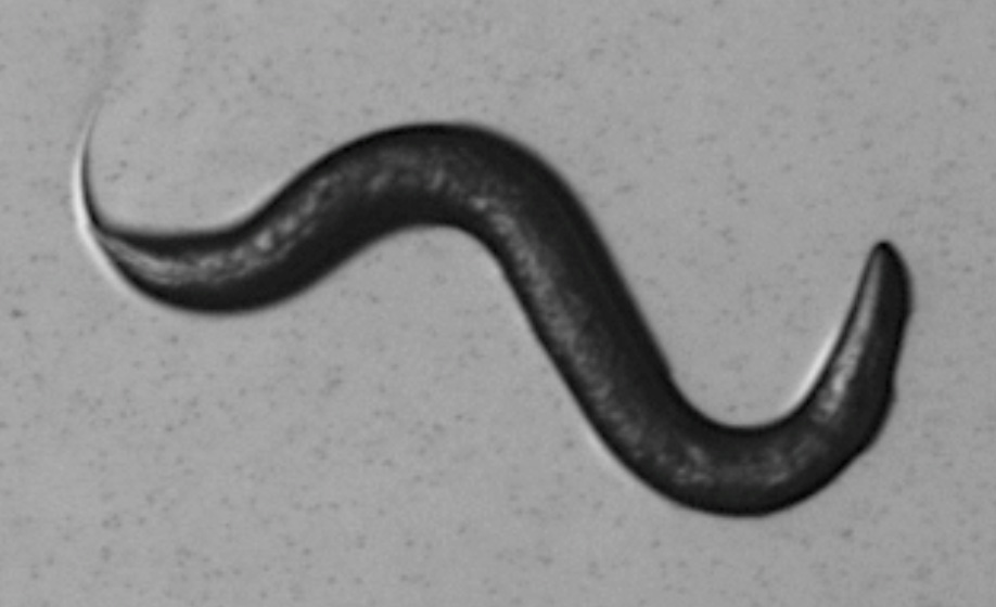
Modelling Developmental Neurological Disorders and Childhood-Onset Epilepsy in Caenorhabditis elegans
During the summer of 2014 I was a recipient of a Gurdon Summer Studentship awarded by the BSDB. The studentship provided the opportunity to undertake a research project in the Laboratory of Ian Hope at the University of Leeds where I study Biology.
Abnormalities in the development of the central nervous system have been implicated in the epilepsy group of chronic neurological disorders. These disorders affect fifty million people worldwide and are characterized by spontaneous recurrent seizures resulting from excessive, synchronous neuronal activity [1,2]. Improving our understanding of the biomolecular basis of epilepsy is of great importance in the search for new therapeutics. Caenorhabditis elegans provides a genetically amenable, and experimentally tractable system to model disease-relevant mutations and was the focus of this study.
Homo sapiens KCNT1 encodes a sodium-activated potassium channel that is widely expressed in the central nervous system and mutations of KCNT1 have been identified in patients with drug-resistant childhood-onset forms of epilepsy. The C. elegansKCNT1 orthologue slo-2 is widely expressed in both neurons and muscle cells [3] where it is activated during hypoxia-like physiological conditions by raised concentrations of chloride and intracellular free calcium [4]. Protein alignment identified KCNT1 amino acid residues implicated in childhood-onset epilepsy that are conserved from H. sapiens to C. elegans SLO-2. These KCNT1 variants, R474H and R928C, are associated with malignant migration partial seizures in infancy (MMPSI) [5] and autosomal dominant nocturnal frontal lobe epilepsy (ADNFLE) [6], respectively.
In order to generate R474H and R928C epilepsy-associated variants of C. elegans slo-2, equivalent mutations were introduced using the CRISPR-Cas9 system [7] to produce novel transgenic strains which can then be used to study the role of these mutations in the development of epilepsy.
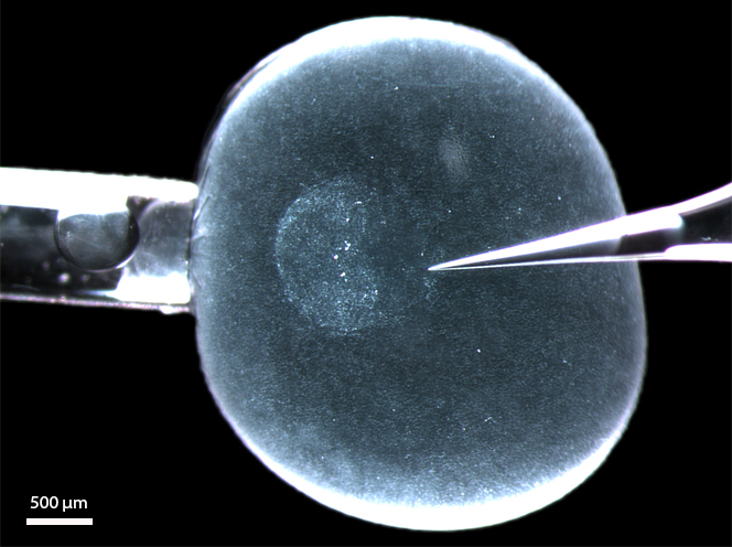
Synthetic guide RNAs (sgRNA) were used to direct Cas9 nuclease activity to regions of the slo-2 locus in close proximity to the target mutational sites. Cas9-mediated cleavage generates double-stranded DNA breaks (DSBs) that are repaired in vivo by either homology-directed repair (HDR), using a homologous template, or by non-homologous end joining (NHEJ), which generates short insertion/deletion (indel) mutations. To incorporate the epilepsy-linked point mutations into the C. elegans genome, worms were microinjected with plasmid vectors expressing the modified sgRNAs and Cas9 alongside slo-2 fragments that had been PCR mutagenized to include the desired point mutations. This generated DSBs at the slo-2 locus followed by HDR to mend the DSB and introduce the desired point mutations.
From fifteen F0 hermaphrodites injected, I obtained twenty-eight transgenic F1 individuals. Ten of these produced transgenic progeny and ten independent transgenic strains were established of which six had been targeted with the R474H equivalent alteration and four with the R928C equivalent. Microinjected DNA forms large extra-chromosomal arrays that are not transmitted to all progeny in a brood and the rate of successful CRISPR-Cas9 alterations in C. elegans varies depending on target loci [7]. To screen transgenics for alterations of slo-2, I performed single worm PCR on progeny of transgenic individuals to amplify slo-2 fragments containing the target mutation loci and assayed fragment length for the presence of indels by agarose gel electrophoresis. Following CRISPR-Cas9 generation of a DSB, activation of the NHEJ repair pathway can produce short indels at the repaired loci, providing a useful method for confirming successful CRISPR-Cas9 activity. Smaller than expected slo-2 fragments were detected in the progeny of transgenic individuals from three different strains; however, further extensive screening is required to determine whether these represent deletions within slo-2 or non-target amplification. Following confirmation of the efficacy of the CRISPR-Cas9 system the next step will be sequencing of slo-2 to identify mutants with the desired epilepsy-linked point mutations.
The transgenic and mutant strains will provide a useful resource in further studies of the biomolecular basis of drug-resistant childhood-onset epilepsies as they should allow production of the specific genomic modifications sought in slo-2. The effects of the slo-2 mutations on the functioning of the C. elegans neuromuscular system would then need to be characterized and findings could provide improvements in our understanding of how specific KCNT1 variants give rise to epilepsy. This research will contribute to an existing network of KCNT1 research currently being undertaken by Jonathan Lippiat and Steve Clapcote at the University of Leeds.
Overall, the BSDB Gurdon Summer Studentship provided a great opportunity to experience working in a professional research laboratory, and has strongly reinforced my desire to pursue a career in research.
References:
[1] Kwan, M.D. Schachter, S.C. and Brodie, M.J. (2012) Drug-Resistant Epilepsy. The New England Journal of Medicine 365, 919-926
[2] Engel, J.E.R. (2013) Seizures and Epilepsy (2nd Edition), New York: Oxford University Press
[3] WormBase web site (2014) Release: WS244 — [LINK]
[4] Santi, C. et al. (2003) Dissection of K+ currents in Caenorhabditis elegans muscle cells by genetics and RNA interference. PNAS 100, 14391-14396
[5] Barcia, G. et al. (2012) De novo gain of function KCNT1 channel mutations cause malignant migrating partial seizures of infancy. Nature Genetics 44, 1255-1259
[6] Heron, S.E. et al. (2012) Missense mutations in the sodium-gated potassium channel gene KCNT1 cause severe autosomal dominant nocturnal frontal lobe epilepsy. Nature Genetics 44, 1188-1190
[7] Friedland, A,E. et al. (2013) Heritable genome editing in C. elegans via a CRISPR-Cas9 system. Nature Methods 10, 741-743
Our jobs page has been busy this month, with several new postdoc positions advertised. Here are some of the other highlights:
Research:
– Christoph and his colleagues wrote about their recent Development paper, where they inhibited the Rho-kinase ROCK in the rabbit embryo, discussing the implications of their work in our understanding of the evolution of vertebrate gastrulation.
– and Chistele’s latest Stem Cell Beauty post focused on a Stem Cell Reports paper where Meinhardt and colleagues embed embryonic stem cells in a 3D matrix to generate 3D neuroepithelial cysts.
– and the newly created Pan-American Society for Evolutionary Developmental Biology is running their first meeting this August.
The Node survey:
The Node was launched almost 5 years ago, and it is now time to revise its design and functionalities. Help us develop the Node for the future by answering a few questions in our survey. To thank you for your time, at the end of the survey you can choose to enter a prize draw to win a bag of goodies from the Node and Development!
Also on the Node:
– Our latest model organism post comes all the way from Dunedin in New Zealand! Read Megan’s post ‘A day in the life a honeybee lab‘!
– There are many ways to get involved in science outreach. We have reposted a Development Spotlight article on this topic.
Maize has a rich history as a model organism for genetics; Rollins Emerson began describing mutants in the 1930’s. Large chromosomes, amenable to cytology, aided Barbara McClintock in her critical discovery of transposable elements for which she earned a Nobel Prize in 1983. As developmental biologists, we treasure maize for its large and varied meristems, which are totipotent, like animal stem cells. These are excellent for microscopy, particularly RNA and protein in situ hybridization. Now, GFP-tagged fusion protein lines are also available (http://maize.jcvi.org/cellgenomics/index.php).
Maize is a domesticated form of Teosinte and comparison to its ancestor has revealed loci important for grass domestication (see Doebley and Stec, 1993 and Doebley et al., 1997). Grasses include most staple starches, not only corn but wheat, rice, sorghum, millet, sugarcane and others. So, there are practical benefits to studying maize as well.
Field season sets apart the daily life of a maize developmental geneticist from that of colleagues working with laboratory organisms. Controlled pollinations are technically simple, requiring only bags, staples and markers, and yet are undeniably physical. We use waxed bags to protect the ear shoots from rogue, windborne pollen. A few days later, the tassel, which bears staminate flowers and pollen, emerges. Not many geneticists are dwarfed by their subjects and need to wrangle 40cm of male inflorescence into a brown paper bag. This is particularly true for those of us who are below average height.
Bagging a maize tassel in Nayarit, Mexico, January 2015. Photo by Angus Vajk.
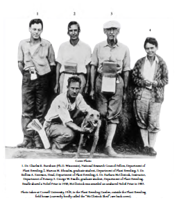
Of course, laboratory life does not end as soon as the kernel hits the soil. Summers are rife with mad-dash genotyping and tissue collection and the normal responsibilities of correspondence, teaching and writing. This is why the real joy is tending the winter nursery. Frequently, particularly off-season, we fly thousands of miles to attend our winter nurseries. East Coast-based maize geneticists typically send winter plantings to Puerto Rico whereas those of us on the West Coast winter in Mexico or Hawaii. Yes, these trips are romantic — in the travel sense — where you feel a connectedness with researchers that came before. These trips are coordinated reunions of former and current colleagues such that fond memories of potluck BBQs, scorpion sightings, torrential rains and of near-death driving experiences are commonly recalled and newly generated. I regularly picture McClintock and Emerson (referred to by Rhoades as “the spiritual father of maize genetics”) in their make-shift pollinating aprons and knickers and feel proud to carry on illuminating the wonders of maize.
Reprinted with permission of W. B.Provine, the Department of Plant Breeding and Genetics, and the publisher, from Kass, Lee B. (Ed.). 2013. Perspectives on Nobel Laureate Barbara McClintock’s publications (1926-1984): A Companion Volume. The Internet-First University Press. URI:http://hdl.handle.net/1813/34897
In preparation for winter nurseries, lab members from all over the country package seeds for individual experiments and these are carefully organized. Shipping is timed with colleagues. Field management companies receive our seeds (after phytosanitary inspection) and carefully plant them for us. Multiple, staggered plantings are prepared so that inbred (homozygous control) lines (think ecotype if you work with Arabidopsis) are shedding pollen for the largest window of time; these we often share – knowing a colleague will happily return it to us.
A few lucky researchers will perform the controlled pollinations for their co-workers. I have been fortunate enough to be a staff research associate in the developmental genetics lab of Dr. Sarah Hake at the Plant Gene Expression Center – a collaborative institute of the USDA Agricultural Research Service and the UC Berkeley Department of Plant and Microbial Biology – for fifteen years. I have pollinated on Molokai eight times and in Mexico four. Pollinating schedules are coordinated so that we break up the work, each going for 1-2 weeks in overlapping windows. We frequently mop up the last few crosses for our friends or help each other while the field is peaking and most plants are sexually mature. Sometimes, there are not enough hours in the day so we must help each other – pollinating by headlamp is not recommended!
Dr. Clint Whipple, Assistant Professor at Brigham Young University and Dr. Cliff Weil, Professor at Purdue University look for lodicule (a floral organ) phenotypes in maize spikelets over lunch at José’s taco stand.
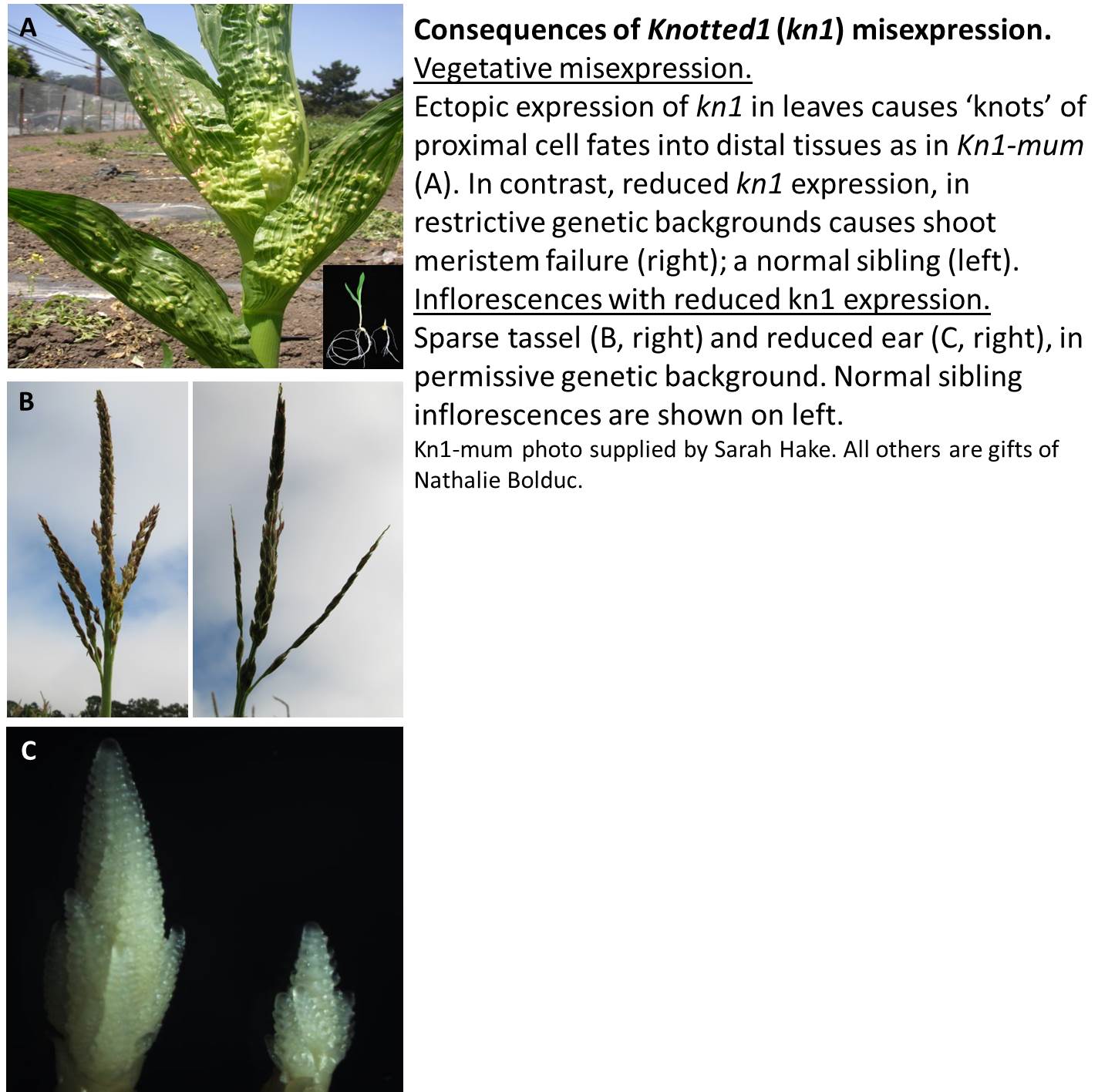
Most of us are mutant-lovers; perhaps including the reader? Nothing is better than traipsing about in a jungle of new and old “friends” as we frequently call them. People have studied maize for about a hundred years, so we even call some of our best friends “classic mutants.” These include such beauties as knotted1 (kn1), with its telling gain-of-function phenotype of protuberances of proximal tissue fates into distal organs and a satisfyingly opposite loss-of-function phenotype of small, early-terminating inflorescences. Knotted is required for meristem maintenance and causes ectopic growth when ectopically expressed (e.g. in leaves). Or, depending on background, loss of function mutants may be shootless, not unlike mutants of its related Arabidopsis gene, shootmeristemless.Kn1 was the first homeobox gene cloned in plants. To me, there is no clearer example of shared descent. If animal homeobox genes regulate proximal/distal patterning in limbs and plant homeobox genes regulate proximal/distal patterning in leaves, then we have all originated from the same lovely stew.
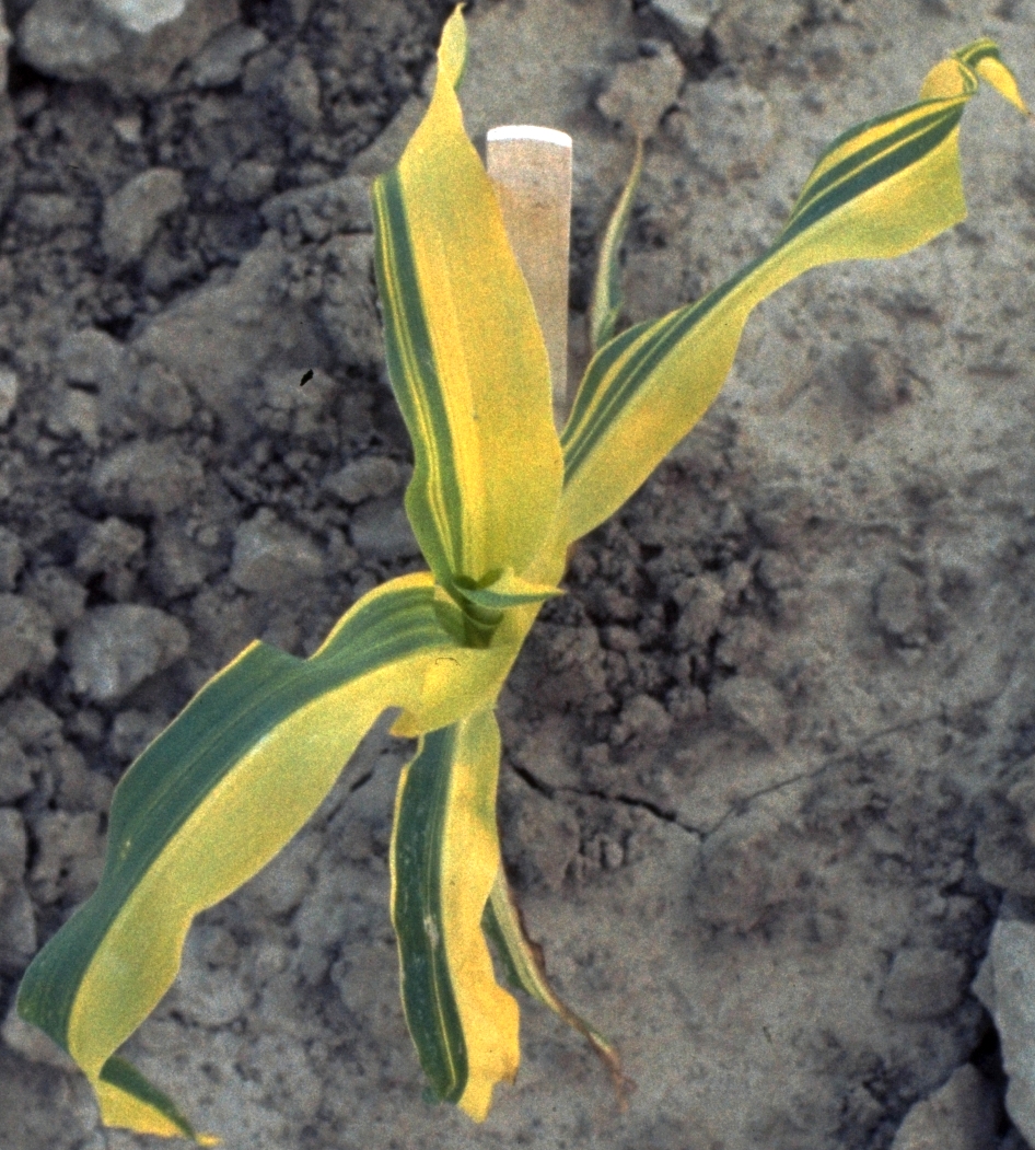
A real pleasure is to tour a field of families from a mutagenesis experiment. Ideally, this is done by the side of an expert. Perhaps, Gerry Neuffer will show you his favorite half-plant chimera, visible only as an M1 (+/-) plant because it would not survive without the healthy half of the plant coaxing it along.
Oil yellow Chimera: An immature half-plant chimeric greenish brilliant yellow, Oy1-N1459, M1 mutant origin plant , showing consequence of EMS-induced mutation in two-stranded sperm nucleus of treated pollen grain. Photo and caption courtesy of Gerald Neuffer, please see his wiki at http://mutants.maizegdb.org/doku.php.
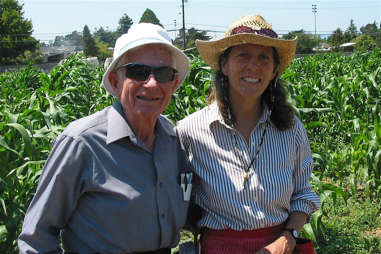
Gerry Neuffer and Sarah Hake at the Gill Tract Research Plot, near Berkeley, CA (2006). Photo courtesy of Sarah Hake.
He would even tell you that the frequency of certain mutants occurring in EMS populations is different than those arising from transposon populations such as Mutator or Ac/Ds. This would open the fascinating topic of his hypotheses regarding the mechanisms behind these long-term observations (for review see his chapter, Neuffer et al., ‘Mutagenesis – the Key to Genetic Analysis’ in the Handbook of Maize, 2009 pp 63-84), which is best had over a plate of lomo pork at the annual luau on Molokai, Hawaii.
Our pollinated ears are anxiously awaited and will land at their home research institutes in mid-March. This is also the timing of the annual Maize Genetics Conference. This year, and every other it is held in the Midwest of the US. If you are an aspiring or confirmed mutant lover, consider attending March 12-15, 2015 in St. Charles, Illinois or attending in the future. I hope to meet you there!
This post is part of a series on a day in the life of developmental biology labs working on different model organisms. You can read the introduction to the series here and read other posts in this series here.
Fully funded post-doctoral positions are available to investigate molecular, genetic and cell biological mechanisms of development in the sea anemone Nematostella vectensis in the Gibson lab at the Stowers Institute for Medical Research. Broad project goals are: 1) To utilize established genome engineering and advanced imaging methods to investigate the morphogenesis of polarized epithelia during early embryonic development and 2) To utilize established gain- and loss-of-function approaches to interrogate signal transduction, pattern formation, and growth control in an early-branching metazoan system. Specific research goals are flexible and can be fit to the interests of successful applicants.
Candidates should be dynamic and highly motivated with a demonstrated record of creativity and a recent PhD degree. Experience in genomics, cell and molecular biology and experimental embryology are highly desired, along with training in confocal microscopy and phylogenetic analysis. To apply, please send a single .pdf file containing your CV, email addresses for 3 references, publications and a statement of postdoctoral research interests to: mg2@stowers.org.
Our laboratory, at the EMBL/CRG Systems Biology Unit in Barcelona, is looking for an excellent and highly motivated postdoc to study the functions and evolutionary impact of neural-specific alternative splicing in vertebrates.
The major goal of the project – funded by the European Research Council (ERC) – is to understand the in vivo functions and evolutionary impact of a program of neural-specific protein isoforms that are conserved across all vertebrates. These isoforms, sometimes diverging by only one or two aminoacids from the onneural isoforms due to microexons (see Cell 2014, 159:1511-23), are expected to be crucial for terminal neurogenesis and synaptic function, and unique to vertebrate species. The applicant will mainly use zebrafish as a model organism to investigate these questions. In addition to these, the candidate will be encouraged to develop his/her own scientific ideas.
The applicant is expected to be passionate about evolution, neuroscience and/or developmental biology.
Strong experience on zebrafish research, particularly on nervous system development and/or in vivo neuronal differentiation, is required. Previous experience with the CRISPR-Cas9 system, and interest on transcriptomic analyses are an advantage, but not necessary. The applicant should be able to work rigorously, independently and flexibly. The candidate will be responsible for his/her own project within the research group, including carrying out experiments, data analysis and interpretation. Fluency in English (spoken and written) is expected.
The position has a fully covered, competitive salary for up to five years, but the applicant will also be encouraged to apply for independent funding.
The Institute
The Centre for Genomic Regulation (CRG), is an international biomedical research institute of excellence, based in Barcelona, Spain, whose mission is to discover and advance knowledge for the benefit of society, public health and economic prosperity.
The breadth of topics, approaches and technologies at the CRG permits a broad range of fundamental issues in life sciences and biomedicine to be addressed. Research at the CRG falls into four main areas: gene regulation, stem cells and cancer; cell and developmental biology; bioinformatics and genomics; and systems biology.
With more than 350 scientists from 41 countries, the CRG excellence is based on an interdisciplinary, motivated and creative scientific team that is supported by high-end and innovative technologies.
The centre’s other main strategic goals are: to translate basic scientific findings into benefits for health and economic value for society; to provide advanced and excellent training to our scientists; and to communicate and establish a bilateral dialogue with society.
Experience on zebrafish research, particularly on nervous system development and/or in vivo neuronal differentiation.
Additional beneficial skills:
Experience with CRISPR-Cas9 system.
Interest and experience on transcriptomic analysis.
Languages:
Fluent level of English
Soft skills:
Passion for evolutionary biology.
A highly motivated and organized candidate.
Capable of working in group, and with a high degree of work autonomy.
The Offer
Duration: 1 year renewable contract up to 5 years.
Estimated annual gross salary: A competitive salary will be provided, which will be well matched relative to the cost of living in Barcelona, and adjusted according to experience.
Starting date: as soon as possible from April 2015.
We offer work in a highly stimulating environment with state-of-the-art infrastructure, providing the successful applicant with unique opportunities to develop a strong technical portfolio.
Application Procedure
All applications must include:
A presentation letter addressed to Manuel Irimia
A full CV including contact details.
Two contacts for further references.
All applications must be addressed to Manuel Irimia and be submitted to the following email address: rrhh@crg.es. Please include as email subject the reference “Postdoc-NeuralAS”.
Deadline: Please submit your application by 13th February 2015
Centre de Regulació Genòmica (CRG)
Doctor Aiguader 88, 08003 Barcelona (Spain)
Growing organs in vitro is one of the ultimate dreams of any stem cell biologist. As such, it seems obvious that some of these organs will need to be grown in 3D. This is why stem cell 3D culture systems are very fashionable among scientists. They are increasingly successful and a fair amount of exciting scientific publications have blossomed in the recent years.
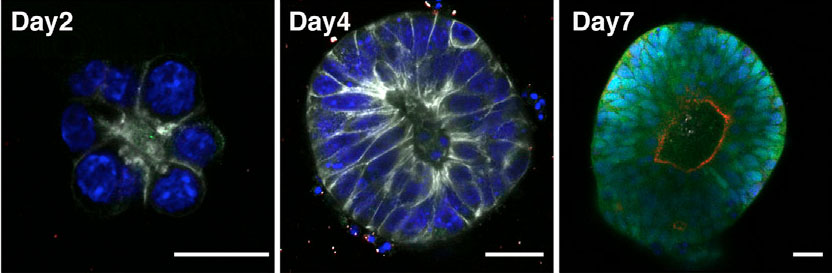
One of these was recently published in Stem Cell Reports by Meinhardt and colleagues. They show that individual embryonic stem cells (stem cells that can become any type of cell in the body), when embedded in a 3D matrix and grown with a medium that induces neural differentiation, can form structures (called neuroepithelial cysts) that mimic early neural development.
In this picture, you can observe mouse embryonic stem cells cultured in the 3D matrix (called Matrigel) for 2 days (left picture), 4 days (middle picture) and 7 days (right picture). Up until day 4 of culture, you can observe a fairly simple “flower” structure with the protein E-cadherin in white and cell nuclei containing DNA in blue. After 7 days in culture, you can observe a more organized structure with a lumen (inside space of a tubular structure) with the proteins Sox1 in green and N-cadherin in red, which are typical markers of ‘neuroepithelial’ cells. Also, the expression of N-cadherin is polarized. From these observations, the authors conclude that in their culture conditions, mouse embryonic stem cells grow into an organized structure of more specialized ‘neuroepithelial’ cells.
Further experiments presented in this study show that both the timing and the developmental steps that are observed during the differentiation of mouse embryonic stem cells into these neuroepithelial cysts are similar to those observed during mouse development. Additionally, they show that upon stimulation with various “morphogenetic” factors (substances governing tissue development and the positions of the various specialized cell types within a tissue) they can obtain structures resembling a patterned neural tube with a dorso/ventral axis.
This study nicely illustrates how 3D stem cell culture systems are an invaluable tool to study the action morphogenetic factors during the development of organs Also, these “mini organs” could also constitute a source of tissues (in this case, the neural tube), which hopefully, one day in the near future, will find their way into real 3D stem cell therapies!
Picture credit:
Meinhardt, A., Eberle, D., Tazaki, A., Ranga, A., Niesche, M., Wilsch-Bräuninger, M., Stec, A., Schackert, G., Lutolf, M., & Tanaka, E. (2014). 3D Reconstitution of the Patterned Neural Tube from Embryonic Stem Cells Stem Cell Reports, 3 (6), 987-999 DOI: 10.1016/j.stemcr.2014.09.020
The days of the solitary scientist toiling away within the isolation of the academic ivory tower are something of the past. This is not only true when actually doing science, as projects become more collaborative and global, but also when considering the relationship between scientists and the public. Science outreach is something that most of us may already be familiar with, and yet the perception that outreach is not for the committed scientist is still there. In many quarters, outreach is seen as an occupation for those who are transitioning to an ‘alternative’ career or for naturally gifted communicators. However, the economic crisis was a wake-up call to many. With governments all over the world cutting their budgets, and with public opinion playing a big role in influencing these decisions, it became clear that outreach is not just about helping the public gain some understanding of scientific concepts but it is also about ensuring that they appreciate why it is important and why governments (and hence the tax payer) should support it. In other words, we don’t just have a moral duty as scientists to inform the public but we have a very practical necessity to justify our activities to
those who fund our work.
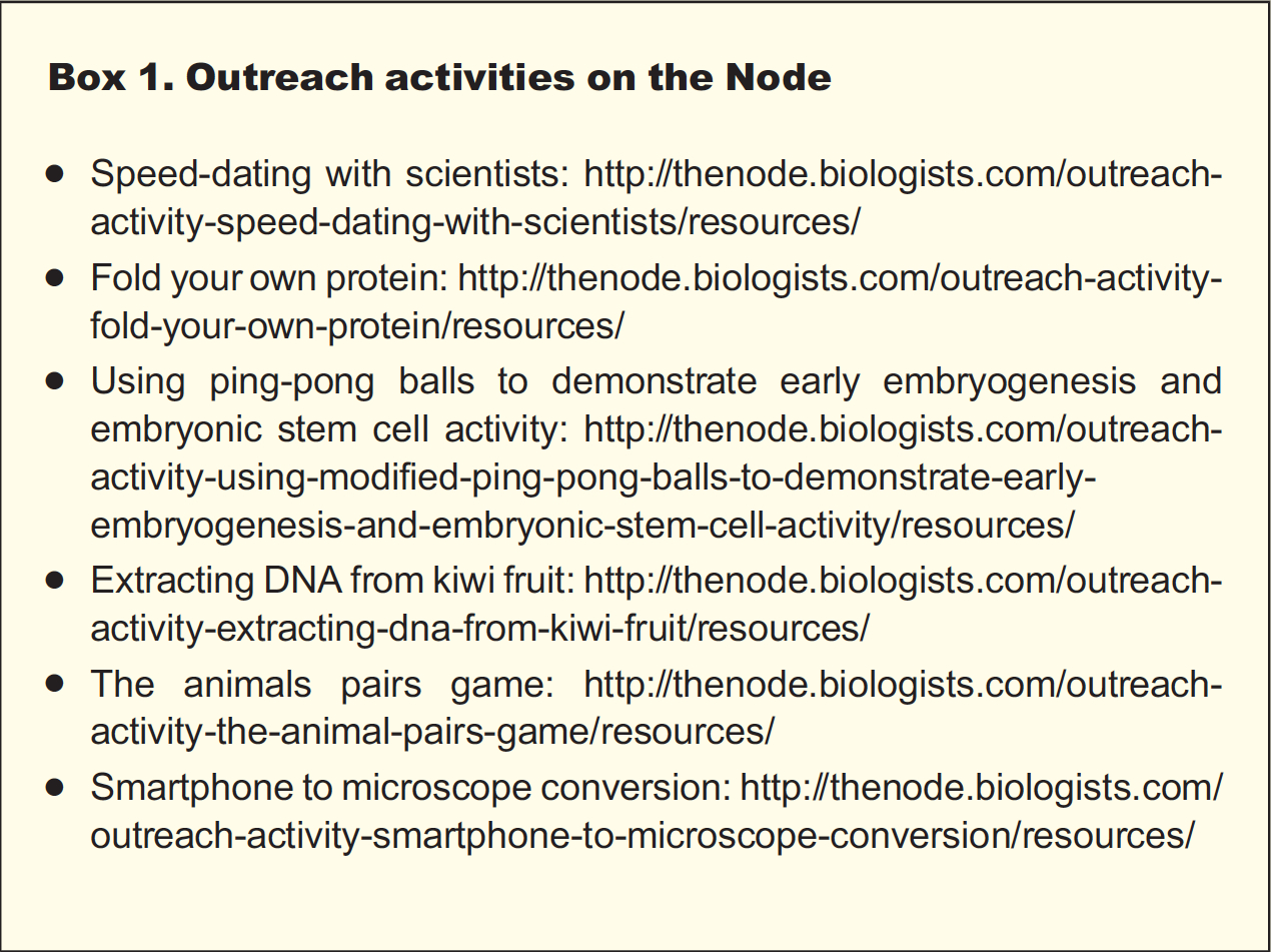
For the uninitiated, communicating science outside academic environments can be a daunting prospect. Where to start? Where do you find an audience? What should you be communicating – basic concepts of biology or your specific research project? In late 2013 we launched a series on the Node dedicated to science outreach. This was an auspicious time to launch this series, as developmental biology outreach received an enormous boost as the focus of the 2013 Royal Institution Christmas Lectures, which were widely broadcast on the BBC (https://thenode.biologists.com/of-worms-and-womenreflections-on-the-life-fantastic-christmas-lectures-2013/events/).We had two main objectives with this series. First, to inspire our readers by showcasing the variety of outreach projects available, including tips and insights from the scientists and communicators involved. Second, we also wanted to provide a set of easy, tried-and-tested outreach activities (with step-by-step instructions) to make that first outreach experience a little easier (see Box 1).
When first thinking about science outreach, table-top activities at departmental open days and science festivals are what many scientists have in mind. They are very good starting points. The department or festival organisers deal with the logistics, and these events attract a crowd of children and adults who are there because they want to learn – your audience is already interested and eager to be captivated. As a participant you only need to worry about your own stall, and our easy outreach activities (see Box 1) can be a great place to start. The post by the Biology Builders, a group of Nottingham scientists who organised a stall at the Royal Society Summer Science Exhibition, provides interesting insights into the challenges and rewards of outreach in this context (https://thenode.biologists.com/making-an-exhibition-of-your-science/resources/).
Another good way to get involved in outreach is by collaborating with local schools. There are many ways to bring science to school kids, and the key is to develop projects that take into account the social and economic context of the schools involved. Microscopes 4 Schools (https://thenode.biologists.com/microscopes4schoolshands-on-microscopy-for-children/education/) brings affordable microscopes to the already well-furnished Cambridge schools. Here the aim is to amaze children with the beauty of science and inspire a new generation of scientists. Ciencia al tiro (https://thenode.biologists.com/outreach-program-ciencia-al-tiro-scienceimmediately/education/) takes place on the other side of the globe, reaching out to some of the most deprived schools in Chile. The aim of this project is to reduce some of the inequalities of the education system and to provide extra support and attention to children who might not have many opportunities. Science is shown to be something very practical that can help daily life, by developing projects that teach concepts but also have practical applications, such as installing solar showers to provide hot showers to the children. The projects and activities help the children think outside their daily reality and hopefully inspire them to aim higher.
If a collaboration with a local school is not possible, there are other ways to reach school children. I’m a Scientist, Get me out of here (https://thenode.biologists.com/im-a-scientist-get-me-out-ofhere/resources/) is an online project in which scientists join online chats to answer questions from school children. You are faced with the usual challenges of outreach, such as the need to be clear without being patronising and the ability to deal with difficult questions. In addition, and in the fashion of the similarly named TV show, the scientist with fewest votes from the students is evicted at the end of each day. You must be fast as well as a good communicator!
Another way to influence how science is communicated in schools is by interacting with the teachers themselves. This is the mandate of the European Learning Laboratory for Life Sciences (ELLS), launched by the European Molecular Biology Laboratory (EMBL) in 2003 (https://thenode.biologists.com/embl-ells/resources/). This science education facility aims to train teachers, bringing them up to date with the latest discoveries, helping to bring molecular biology to the classroom and providing opportunities to interact and learn from research scientists.
Schools and science festivals are great ways to be involved in outreach, but the audience is already primed to learn and has an interest in science. How to reach those that are less likely to be inspired by what science has to offer? One way to reach a new audience is by choosing an object or topic dear to them. Mission Peluches (https://thenode.biologists.com/a-cuddly-science-outreachproject/education/) is an outreach project developed in France that is particularly popular with young children. The starting point is the soft toy, an object that every child has and is emotionally attached to. The children are encouraged to become naturalists of the toy world, classifying their soft toys as if discovering new species. Placing that soft toy in the context of others in the collection naturally develops into a discussion of the different mechanisms of evolution.
One notoriously difficult age group encompasses teenagers and young adults. The Gulbenkian Institute has found a novel way to engage with this audience by moving their outreach activities to where the young people are (https://thenode.biologists.com/sciencepowered-by-music-2/resources/). The institute is next door to the venue of one of the biggest music festivals in Portugal. By establishing a partnership with the festival’s organisers, the researchers at the Gulbenkian Institute organise a stall at the festival every year, reaching out to the unsuspecting music fans. The spirit of this project is similar to that of other outreach projects that are moving outreach to less formal settings, such as recent
efforts to bring science to pubs or bars (https://thenode.biologists.com/cosy-science-science-cafes-in-the-pub/events/).
One interesting activity developed by the Gulbenkian Institute as part of their activities at the music festival is the opportunity to ‘speed-date’ with scientists. This gives revellers the chance to discuss science one-on-one with a scientist. The most important aspect of this activity, though, is that it allows young people to realise that scientists are just normal people (especially when they have a drink in their hand, sun glasses on, and are as interested as you in the next act playing!). This highlights an important aspect of science outreach: it has wider objectives besides just teaching scientific concepts. Soapbox Science (https://thenode.biologists.com/soapbox-science-on-londons-south-bank-using-flies-to-fightcancer/education/) is another outreach project that aims to change the way that science is perceived, focussing specifically on women in science. Female scientists are invited to stand on top of a box in a busy pedestrian area and grab the attention of passers-by with an engaging presentation of their work. Part of the challenge is to get the attention of the audience, but this project is also unashamedly about showing that women have a role to play in science. Successful, enthusiastic women take the centre stage, showing young girls that science is for them too.
For many of us, the initial interest in science was not sparked by a specific concept but rather by a beautiful image or an unusual perspective. Bringing such moments to the public is also an important aspect of outreach. A research group in France (https://thenode.biologists.com/cellular-architectures-at-the-nuit-blanche-2013-in-paris/education/) aimed to do just that, and at quite a scale. They participated in Nuit Blanche, which is a big arts festival in Paris, by projecting videos of cells onto the façade of a 17th century building. This medium does not allow a complex explanation of concepts, but it is a fantastic way to share the beauty of science and spark the curiosity to investigate further. As the scientist behind this project stated: “curiosity and wanting to understand needs to be promoted as a value by itself”.
As you might imagine, adapting movies of cells into an excitingmusical show that is projected onto a building is not an easy task for a scientist, and close collaboration with an artistic company was an essential part of this project. The willingness to collaborate with experts in other areas can therefore be another important aspect of outreach (unless of course you have an academic background in both; for example, science and art – https://thenode.biologists.com/outreach-series-evo-devo-art/interview/). We must also be open to acquiring new skills. As scientists, we already have many of the skills required for successful outreach, such as the ability to speak in public, presentation and writing skills. However, these need to adapted and readjusted to a different audience (or several different audiences), and collaborations, in particular with professional science communicators, can help make that transition.
Improving and widening your skill set is just one of the many ways in which you can personally benefit from being involved in outreach. As the organisers of Cosy Science explained in their post, outreach “makes it easier to step back from day-to-day problems and see the bigger picture, question basic assumptions […] and think outside the box”. If you are lucky and a bit creative, your outreach project might even contribute to your research. Worms Watch Lab (https://thenode.biologists.com/worm-watch-lab-real-data-real-outreach/education/) is an example of a citizen science project (http://scistarter.com). Faced with the challenge of analysing hours and hours of C. elegans videos, a laboratory in Cambridge transformed this non technical but time consuming activity into a game that anyone can play. As a consequence, the public is entertained while learning about the science behind the game and contributing to a real research project.
As the posts already featured in this Node series attest, there are many opportunities to be involved in outreach, from joining an ongoing project to starting your own event, from organising a stall to broadcasting a radio programme (https://thenode.biologists.com/onthe-air-bringing-developmental-biology-to-the-radio/education/). Although it is true that efforts to communicate with the public remain mostly unrewarded in science, where publications are the only currency, there are signs of a new trend. In many countries, funding bodies are starting to require their grant applications to include a section on public communication, and the opportunities to apply for outreach funding are also increasing. There are even a few job positions in which outreach is not only encouraged but also an integral part of the job alongside traditional research (https://thenode.biologists.com/best-of-both-balancing-research-and-outreach/education/).
We hope, therefore, that this series, which is still ongoing, will be a good introduction to the exciting world of science outreach and that it may inspire and encourage those of you not already involved to step out of the ivory tower and give it a try.
Here are the highlights from the new issue of Development:
Invadosomes: aiding axonal invasion
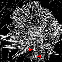
Invasive cells such as immune and metastatic cancer cells form protrusions known as invadosomes, which mediate adhesion to the underlying substrate and induce extracellular matrix degradation – thus promoting invasiveness. On p. 486, Timothy Gomez and colleagues demonstrate that invadosomes can also be found in axon growth cones, where they have not previously been characterised. Primarily using Xenopus spinal cord neurons as a model, the authors use high-resolution imaging techniques to identify and characterise these structures, both in culture and in vivo. Importantly, similar structures can be found in other Xenopus neuron populations, as well as neurons derived from human pluripotent stem cells. In functional assays, the authors find that axon growth in culture is unimpeded by disruption of the invadosome protein Tks5, but in vivo outgrowth of motoneurons into the periphery does require invadosome activity. Thus, these data not only demonstrate the presence of invadosomes in growth cones, but also provide evidence for a specific role for these structures during certain phases of axon outgrowth.
Taking the strain out of stem cell renewal
For many years it has been clear that embryonic cells from different mouse strains differ in their properties for generating embryonic stem cells (ESCs). Specifically, ESCs can be generated and maintained from some (‘permissive’) strains in the presence of serum and LIF, whereas these conditions are insufficient to support self-renewal of cells derived from other genetic backgrounds (‘non-permissive’ strains). Here (p. 431), Satoshi Ohtsuka and Hitoshi Niwa set out to understand the reasons behind the differing potential of ESCs from different origins, and find that this can be traced back to differential LIF responsiveness. They observed that LIF treatment induces higher JAK-Stat pathway activity at early time points, and lower MAPK activation at later time points, in permissive strains than non-permissive ones. This difference is functionally important, since ectopic activation of Stat3 in ESCs from non-permissive strains promotes their self-renewal. Although the downstream outputs regulated by the balance of these two pathways have yet to be fully analysed, these experiments reveal important differences in the cellular responses that underlie the differing properties of ESCs from different genetic backgrounds.
Reporting on β-catenin in the vasculature
β-catenin is a multifunctional protein that acts both as a downstream mediator of the Wnt signalling pathway and as a core component of adherens junctions. It is also widely expressed during development. Consequently, dissecting out the specific functions of β-catenin in particular contexts can be challenging. Shigetomo Fukuhara, Naoki Mochizuki and colleagues now report a transgenic zebrafish line that allows the visualisation of β-catenin activity in living tissues (p. 497). They use this line to investigate the role of β-catenin in vascular development, finding that it plays a key role in formation of the caudal vein (CV). Surprisingly, β-catenin activity in this context appears to be independent of Wnt signalling, and instead is regulated by the BMP pathway. They further identify Aggf1 as a putative BMP target that cooperates with β-catenin to activate downstream gene expression in the CV. Finally, the authors show that expression of the orphan nuclear receptor Nr2f2, which is known to be involved in vein specification, is dependent on β-catenin and Aggf1, thus providing insights into the mechanisms by which BMP-dependent β-catenin activity regulates CV development.
PUB4 calls time on root cell division
In the plant root meristem, a highly orchestrated pattern of cell divisions controls both root growth and cell fate. A large number of signalling factors and transcriptional regulators have been found to control proliferation and differentiation in the root meristem, including small peptide ligands of the CLV3/CLE family. However, the mechanisms by which these peptides act remain poorly understood. Now (see p. 444), Shinichiro Sawa and colleagues identify the E3 ubiquitin ligase PUB4 as acting downstream of CLV3/CLE signalling to regulate cell division in the Arabidopsis root. The authors identify pub4 mutants in genome-wide screens for mediators of CLV3/CLE activity, and characterise the mutant lines in detail – finding defects in a number of lineages that result in overproliferation and patterning phenotypes. Mechanistically, the authors show that the expression of a D-type cyclin is disrupted in the mutant and that auxin levels are altered. Although much remains to be learned about how PUB4 acts in this context, the data intriguingly point to a role for PUB4 in regulating the timing of asymmetric cell divisions and provide further evidence for an important function for CLV3/CLE signalling in controlling root meristem activity.
PLUS…
A mathematical approach to periodic patterning
Hiscock and Megason present a mathematical approach to understanding periodic patterning development. They also suggest ways in which different types of model can be tested, illustrating the potential of this methodology using specific biological examples. See the Hypothesis on p. 409
Cell fate specification in the early Arabidopsis embryo
The cell fate decisions and patterning steps that occur during plant embryogenesis are essential reiterated during organogenesis. Hove, Lu and Weijers summarise our current understanding of the early stages of plant embryogenesis, with a focus on how the major lineages are specified. See the Review on p. 420
The many faces of science outreach
This Spotlight discusses diverse ways in which scientists can engage with the wider community to further the public understanding of science and encourage individuals to get involved, highlighting specific examples examined in the ongoing Node series on science outreach. See the Spotlight on p. 407
The Node was launched almost 5 years ago, and it is now time to revise its design and functionalities.
We have created a short survey that will help us gather your feedback. Please take a few minutes to complete it- the Node is here for the community, and we need your input to know how we can improve! To thank you for your time, at the end of the survey you can choose to enter a prize draw to win a bag of goodies from the Node and Development!



 – Christoph and his colleagues wrote about their recent Development paper, where they


 This post is part of a series on a day in the life of developmental biology labs working on different model organisms. You can read the introduction to the series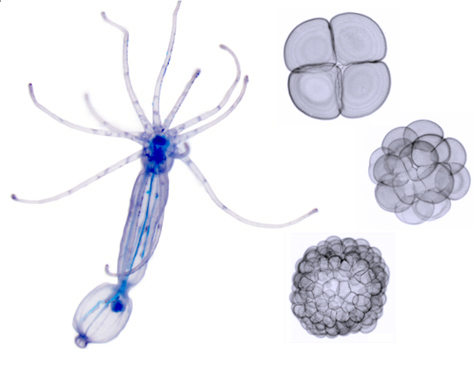
 Invasive cells such as immune and metastatic cancer cells form protrusions known as invadosomes, which mediate adhesion to the underlying substrate and induce extracellular matrix degradation – thus promoting invasiveness. On p.
 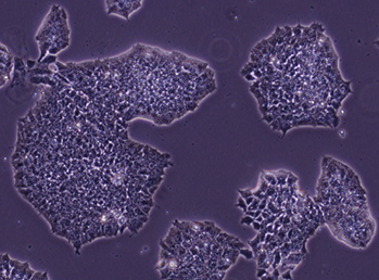 For many years it has been clear that embryonic cells from different mouse strains differ in their properties for generating embryonic stem cells (ESCs). Specifically, ESCs can be generated and maintained from some (‘permissive’) strains in the presence of serum and LIF, whereas these conditions are insufficient to support self-renewal of cells derived from other genetic backgrounds (‘non-permissive’ strains). Here (p.
 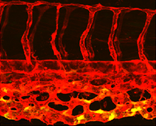 β-catenin is a multifunctional protein that acts both as a downstream mediator of the Wnt signalling pathway and as a core component of adherens junctions. It is also widely expressed during development. Consequently, dissecting out the specific functions of β-catenin in particular contexts can be challenging. Shigetomo Fukuhara, Naoki Mochizuki and colleagues now report a transgenic zebrafish line that allows the visualisation of β-catenin activity in living tissues (p.
 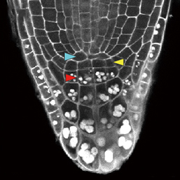 In the plant root meristem, a highly orchestrated pattern of cell divisions controls both root growth and cell fate. A large number of signalling factors and transcriptional regulators have been found to control proliferation and differentiation in the root meristem, including small peptide ligands of the CLV3/CLE family. However, the mechanisms by which these peptides act remain poorly understood. Now (see p.
 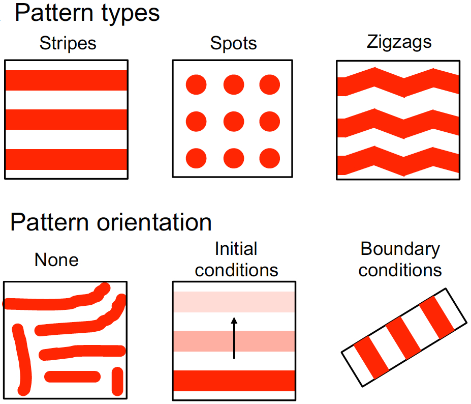 Hiscock and Megason present a mathematical approach to understanding periodic patterning development. They also suggest ways in which different types of model can be tested, illustrating the potential of this methodology using specific biological examples. See the Hypothesis on p.
 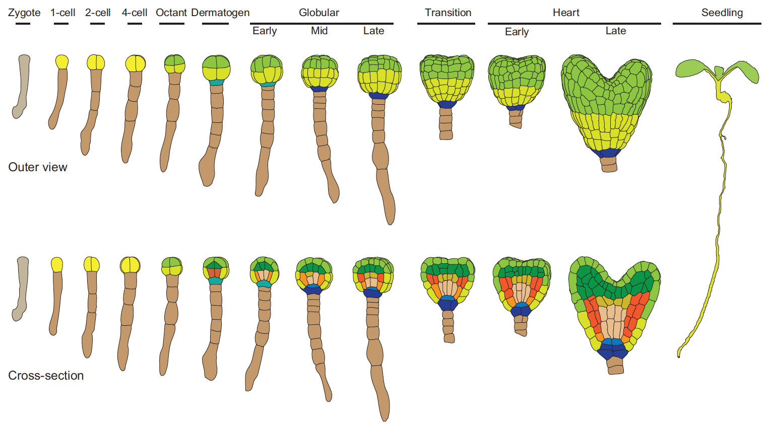 The cell fate decisions and patterning steps that occur during plant embryogenesis are essential reiterated during organogenesis. Hove, Lu and Weijers summarise our current understanding of the early stages of plant embryogenesis, with a focus on how the major lineages are specified. See the Review on p.