Last month, the Node ran a cover competition with images taken by advanced graduate students and postdoctoral fellows from across the Americas who gathered in Chile to participate in the International Course on Developmental Biology, an EMBO Practical Course held at the Marine Biology Station of Quintay (CIMARQ).
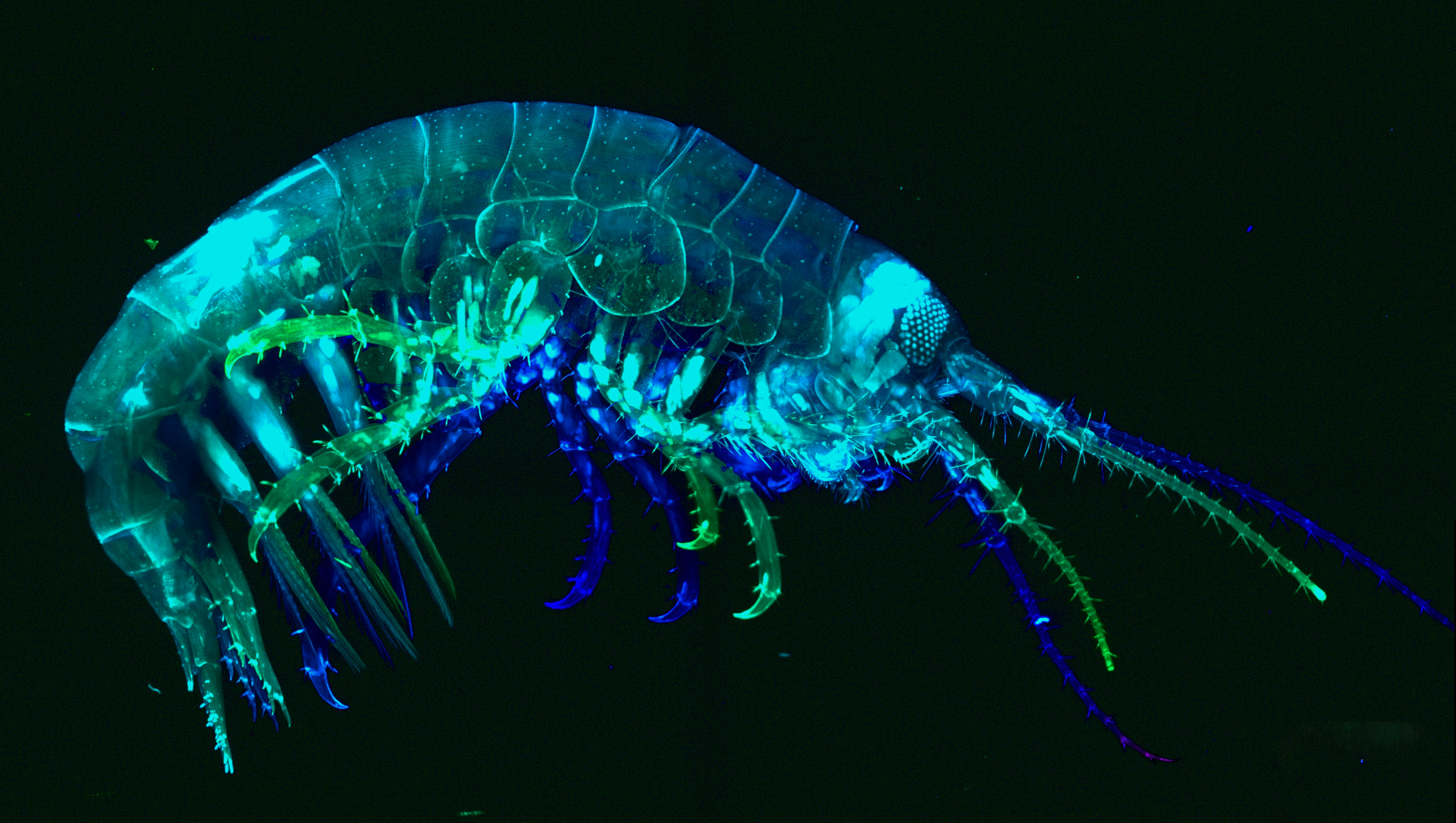
We held a vote over a two-week period. Nine impressive submissions were received from the 2025 student cohort, with images ranging from amphipods to Xenopus laevis and the winners of the popular vote were chosen. Among the great selection of images, the image taken by Natália Heloísa de Oliveira, Fernanda Salce, and Federica Salatiello, Amphipod, received the most votes. Congratulations! Their image will be published on the front cover of Development later in the year.
First, we hear from one of the winners, Fernanda Salce.
Amphipod. Amphipod collected in Quintay Bay by students of the 2025 course, and stained with phalloidin and DAPI.
Fernanda, can you describe your research career so far?
Currently, I am a PhD student in Brazil at the Federal University of Rio de Janeiro, Macaé, RJ. Specifically, I work at the Institute of Biodiversity and Sustainability, affiliated with the Morphological Sciences PhD program.
Can you tell us about your current research?
I am focusing on Developmental Biology as I study the regenerative process of locomotor limbs in the beetle Tribolium castaneum. I am trying to analyse how transcriptional perturbations retain enough plasticity to ensure a given biological process is completed. Thus, I have been studying the limits of leg regeneration in Tribolium larvae under different zld dsRNA concentrations. In previous studies, our group showed that zld is a pioneer transcriptional factor whose activity is pivotal to the success of leg regeneration in the Tribolium larvae model, with zld dsRNA treatment fully inhibiting this process. Here, I am investigating at which point this system might recapitulate function even in the partial absence of zld mRNA. So, more specifically, I am trying to observe whether the regenerative process can be characterized as a bimodal switch-like system and, furthermore, which genes are integral and which are dispensable for leg regeneration in a zld-dependent manner. In parallel, I am also observing changes in morphospace exploration during leg regeneration under these different zld dsRNA concentrations.
What is your favourite imaging technique/microscope?
I frequently use the stereomicroscope for imaging. However, after the course in Chile, I have to say that Light-Sheet microscopy has become my new favorite technique. Not for its accessibility, mind you! But for the amount of morphological data you can extract from your samples. It is simply astonishing!
What is the most impactful thing you learned in the course?
That people are more accessible and approachable than they might seem at first. I would never have thought in my life that Roberto Mayor and I would be sharing drinks and grilling steaks together, but there we were, haha! But on a serious note, the course truly highlights how important social interactions and interpersonal skills are for a scientist.
If possible, describe a bit more about the image that made the cover, including what seems to be capturing wild-caught animals!
This is a crustacean in the genus Parhyale. We caught this bad girl with Nipam Patel’s help. He told us to put a few carrot pieces inside a plastic water bottle with some stones to attract a few of these animals overnight. Keep in mind, this was done inside an aquarium-like system with thousands of red sea urchins. The next day, we actually caught some and thus began our fishing minigame, using Pasteur pipettes and clove oil as anaesthetic. This specimen was particularly interesting because it was a female carrying eggs, so we separated the mother for the Light-Sheet microscope and the eggs for downstream analysis on the confocal microscope. The latter was also brought to Chile with Nipam’s aid.
As a side note, the confocal microscope had an extraordinary logistical undertaking behind it, from what we were told. Apparently, each piece of equipment had to be labelled and registered upon entry into the country, so this particular confocal microscope was designed to be “easily” assembled and disassembled. Truly mesmerizing stuff.
Lastly, for staining, we left our specimen in a phalloidin solution for a week, then stained it with DAPI before our first use of the Light-Sheet microscope during the course. Retroactively, I don’t think that we stained for enough time because you’ll notice that there are myofibers clearly visible in the extremities, but on the animal’s central axis, there was barely any staining – the few observations we caught were restricted to the head and portions of the tail. So, for future participants, my advice would be to begin staining as early as possible and to visualize your possible findings during the last week of the course for better results 😊.
The other two winners, Federica Salatiello, who was funded by the BEOM Department, Stazione Zoologica Anton Dohrn, Italy, and Natália Heloísa de Oliveira, a PhD student at the Federal University of São Paulo, Brazil, under the supervision of Prof. Dr. Marimelia Porcionatto, will be interviewed and featured on the Node later in the year.
Thank you to all the students who participated in the course: Victoria Fernandez, Ana Maria Soria, Candelaria Diaz, David Arancibia, Emilia Failache, Erika Venancio, Federica Salatiello, Fernanda Salce, Fernanda Dalami, Gonzalo Spelzini, Ignacio Casanova, Ingrid Pinto, Jaime Aguayo, Juan Pablo Venegas, Luis Eduardo Sánchez , Marco Mundaca, Mariana Tovar, Natália Heloísa de Oliveira, Nicolás Zúñiga and Pablo Fernández.
Did these images inspire you to take the course? The next cohort of the International Course on Developmental Biology is currently accepting applications until 30 July 2026.
A scientists we tend to move around a lot during our training period. Adjusting to new scientific and social environments can be challenging and intimidating but also exciting. Andrew Ramirez writes about navigating this process with the right mindset and some useful strategies.
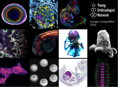
We are delighted to announce the winners of the Young Embryologist Network Image Competition held as part of the YEN 2026 Conference, which took place on the 15th June at the Francis Crick Institute. A huge thank you to all 30 participants who submitted their beautiful images.
The winning image, Lamination in the Zebrafish Retina by Jack Nicholls, will be featured in the YEN 2027 promotional materials. Congratulations to all of our shortlisted entrants!
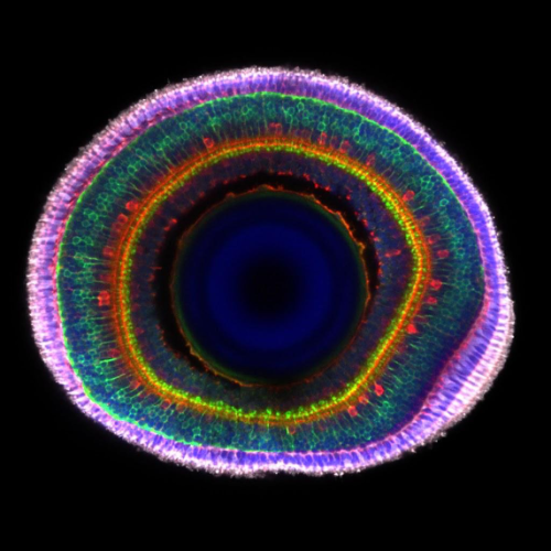
Winning image: Jack Nicholls – Lamination in the Zebrafish Retina
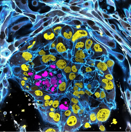
Runner-up: Ipek Gassaloglu Guler – The First Contact: Interaction Between an Embryo Model and Maternal Tissue
Runner-up: David Arancibia-Altamirano – Aristotle’s Egg Through a New Lens
1. Lamination In The Zebrafish Retina
Jack Nicholls, City St George’s, University of London
Immunohistochemistry of an 8-day post-fertilisation larval zebrafish retina stained with anti-ChAT in red (star burst amacrine cells), anti-PKCα in green (ON bipolar cells) and grey (photoreceptors), with a DAPI counterstain. The anti-PKCα signal was manually separated into the bipolar and photoreceptor cell regions and coloured in green and grey to highlight the boundary between the two layers. Imaged on a LSM800 confocal microscope.
2. The First Contact: Interaction Between an Embryo Model and Maternal Tissue
Ipek Gassaloglu Guler, Yale University
This image portrays a stem cell-derived human embryo model (blastoid) cultured on endometrial epithelial cells. This experiment aimed to visualize the initial contact between the blastoid and endometrial epithelium, to capture one of the earliest stages of embryo-maternal interaction.
Within the blastoid, the epiblast is shown in magenta (SOX2) and the trophectoderm in yellow (GATA3). To highlight the interaction between the blastoid and maternal cells, integrin B1 (ITGB1) is shown in white. F-ACTIN, which outlines cellular architecture is shown in blue. The image was acquired using a Leica STELLARIS 5 confocal microscope in tile-scan mode with a 63x objective.
3. Aristotle’s Egg Through a New Lens
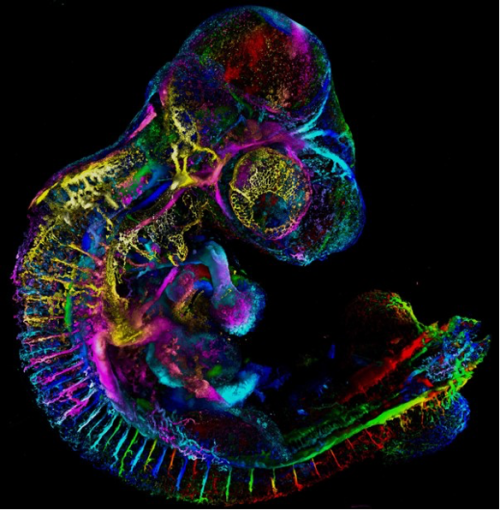
David Arancibia-Altamirano, University College London, Universidad Mayor
Since Aristotle’s studies in embryology, the chicken embryo has captivated the imagination of scientists and spurred exploration of epigenesis and preformation. Echoing classic experiments with India ink, fluorescent ink injection combined with tissue clearing and light-sheet microscopy now allows us to study the beauty and complexity of vascular networks in 3D and with high resolution. Sample displayed by depth colour-coding across 3.1mm; prepared by me and my friend, Jesus Juarez.
4. Dancing Ghosts
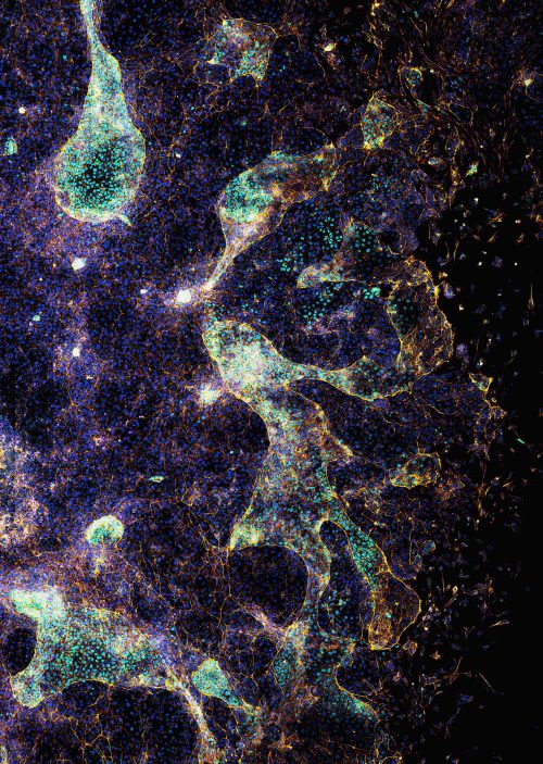
Ornella Clara, Aix-Marseille University
At first glance, the image resembles a ghostly choreography suspended in a cosmic landscape. In reality, it captures the collective cell migration during the early formation of a gut-like tissue structure. These gastruloids spread on a laminin-coated substrate, self-organizing into patterns reminiscent of embryonic development. The transcription factor CDX2 (light blue) marks intestinal identity, while phalloidin staining (yellow-orange) highlights the actin cytoskeleton that drives cell movement, and nuclei are shown in blue-violet.
This image was acquired using confocal microscopy. It illustrates how coordinated cellular behaviour gives rise to complex tissue architecture.
Experimental work by Dalia El Arawi; staining and confocal imaging by Ornella Clara.
5. Growing Into Form
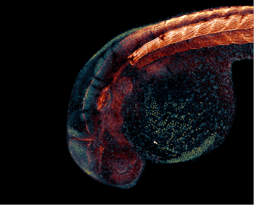
Matyas Bubna-Litic, University College London
A one-day old zebrafish embryo already has recognisable features such as the early forms of the eye and ear as well as the segmented backbone, which will go on to form vertebrae and muscle. Cell nuclei and filamentous actin are visualised in this stained fixed sample. Imaged using a Zeiss LSM980 with an Airyscan2 detector in multiplex mode.
6. Down to the Bone
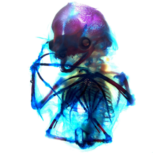
Alexandra Lion, Brigham and Women’s Hospital and Harvard Medical School
A beautiful network of bones and cartilage, which in life would provide support, protection and facilitate movement of the body. The image shows a short-tailed fruit bat (Carollia perspicillata) embryo at embryonic stage 22 which has been cleared and stained with alcian blue for cartilage and alizarin red for bone. This staining allows for visualization of the still-ossifying bones of the bat autopod, and most strikingly of the skull, which appears to smile with clearly visible canine teeth. Imaged during the 2024 Embryology Course at the Marine Biological Laboratory using transmitted light on a Zeiss Axio Zoom.V16 microscope, then further processed using FIJI.
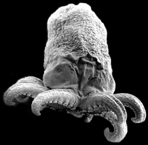
7. Octopus Embryo
Ailen Cervino Len, Baylor College of Medicine
Octopus embryo taken with Scanning Electron Microscopy (SEM).
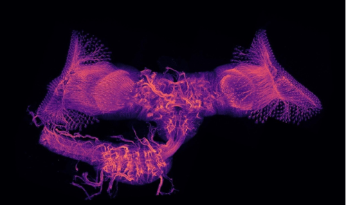
8. Wiring Diagram Ryan Cheng, Centre for Developmental Neurobiology, King’s College London
Maximum intensity projection image of the central nervous system of Drosophila melanogaster during metamorphosis. This sample was collected at around 24 hours after puparium formation and stained with an anti-Neuroglian antibody to visualize the neurite tracks. Multiple z-stacks were imaged on a Zeiss LSM800 and reconstructed in FIJI.
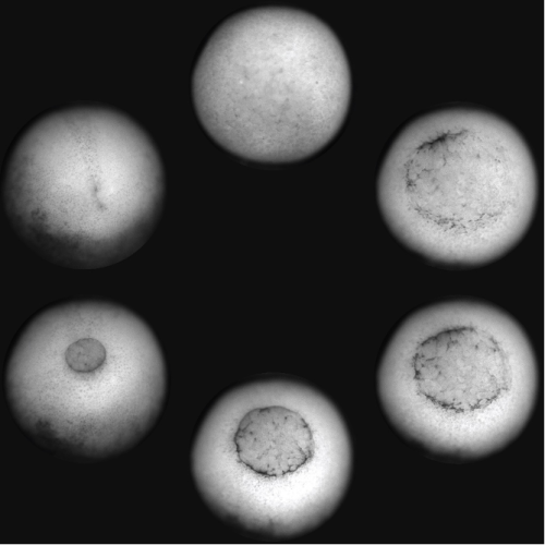
9. The Phases of Gastrulation Hoang Anh Le, University College London
A Xenopus laevis embryo was imaged from the ventral side showing the different phases of gastrulation, from the formation of the blastopore lip to its closure and the beginning of neurulation. The embryo was imaged with an upright brightfield microscope.
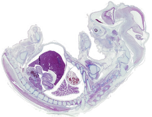
10. beCOWming
Noemi Monferini, Developmental Biology Institute of Marseille
A bovine foetus in histological section stained with Azan trichrome, revealing the delicate architecture of foetal tissues.
We would also like to highlight the winner of the public vote, which has received 128 out of the 773 votes cast:
Weaving a Nervous System
Lamiya Dohadwala, Tata Institute of Fundamental Research
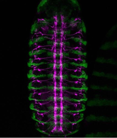
A confocal view of the developing central nervous system in a Drosophila embryo. Green marks engrailed-expressing segmental compartments, while magenta highlights Fasciclin II-positive nerve fibres, tracing the intricate network of axon pathways that form the embryonic nerve cord.
A Perturb-seq screen guided by species divergence uncovers pathways for collateral artery formation Xiaochen Fan, Ronghao Zhou, Brian C. Raftrey, Pamela E. Rios Coronado, Emily Trimm, Erin Clancy, Xinhong Chen, Jamie Bozeman, Maggie S. Chen, Shoxruxxon Alimukhamedov, Juan Alcocer, Idalina Bonham, Stuti Agarwal, Alina Isakova, Vinicio A. de Jesus Perez, Chong Y. Park, Timothy F. Shay, Viviana Gradinaru, Thomas Quertermous, Jesse M. Engreitz, Kristy Red-Horse
Developmental genetic response of the zooplanktonic tunicate Oikopleura dioica to marine noise pollution Eva R. Quintana, Nuria P. Torres-Águila, Ignasi Nou-Plana, Sissel Norland, Valentina Caorsi, Giorgio Blumer, Matteo Bozzo, Elettra Panarari, Giacomo Sabaddin, Simona Candiani, Irene Guarneri, Lucia Manni, Roberta Pennati, Filomena Ristoratore, Giovanni Zambon, Marios Chatzigeorgiou, Rosa Maria Alsina-Pagès, Cristian Cañestro
Modeling human B cell development with pluripotent stem cells Xiaoning Sun, Jamie J. Kwan, Krishna Kothari, Alexandra F. Nazzari, Astrid Kosters, Colin A. Fields, Bao Q. Thai, Deepta Bhattacharya, Michael Atkins, Kelvin Chan Tung, Xinyuan Zhao, Vladimir T. Manchev, Marion Kennedy, Eliver Ghosn, Gordon Keller
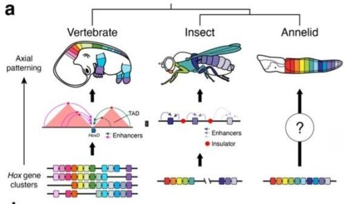
Convergent evolution of cluster-wide Hox gene regulation in Bilateria Billie E. Davies, Francisco M. Martín-Zamora, Tom Frankish, Elise Parey, Nancy Ellis, Noura Maziak, Kero Guynes, Grygoriy Zolotarov, Yi-Jyun Luo, Ferdinand Marletaz, Juan M. Vaquerizas, Arnau Sebé-Pedrós, Nicolae Radu Zabet, Paul J. Hurd, José M. Martín-Durán
The SPARK complex forms the molecular basis of vertebrate fertilization Victoria E. Deneke, Johannes P. Suwita, Haoting Wang, Shingo Tonai, Yonggang Lu, Karin Panser, Alexander Schleiffer, Jeremy A. Hollis, Maria Novatchkova, Gerhard Dürnberger, Karel Stejskal, Gabriela Krssakova, Andreas Blaha, Aleq Adrianne R. Andresan, Muriel Mirus, Hana Marvanova, Hsin-Yi Chang, Taichi Noda, Alejandro Burga, Elisabeth Roitinger, Masahito Ikawa, An-drea Pauli
Nucleolar Dynamics During Oogenesis Ruoyu Li, Grace McKown, Dai Tsuchiya, Mark Mattingly, Anna Galligos, Michay Diez, Jui Feng Lu, Mary C McKinney, Sean McKinney, Boris Rubinstein, Timothy J Corbin, Melainia McClain, Carrie Carmichael, Victoria A Hassebroek, Stephanie H Nowotarski, Jennifer L Gerton, Kamena K Kostova
In vitro sexual dimorphism establishment in schistosomes Rémi Pichon, Magda E Lotkowska, Jude L. D. Bulathsinghalage, Madeleine McMath, Mary Evans, Benjamin J. Hulme, Kirsty Ambridge, Geetha Sankaranarayanan, Simon Kershenbaum, Sarah D. Davey, Josephine E. Forde-Thomas, Karl F. Hoffmann, Matthew Berriman, Gabriel Rinaldi
Comprehensive Lineage Tracing Maps the Landscape of Cell Fate Decisions in Mouse Embryogenesis William N. Colgan, Luke W. Koblan, JoAnne Villagrana, Tien-Chi Jason Hou, Minming Wang, Gokul Gowri, Whitney Chandler, Leonardo A. Sepúlveda, Didar Ciftci, Karina Smolyar, Alicia Young, Lars Wittler, Styliani Markoulaki, Kyle Loh, Xiaowei Zhuang, Nir Yosef, Zachary D. Smith, Jonathan S. Weissman
Written by Ella Knüpling, Juliette Gracia and Jiaming Bi
On the 23rd of June 2026, The Crick and Partner Universities PhD Student Summer Symposium took place at The Francis Crick Institute, UK. More than 165 PhD students from Imperial College London, University College London, King’s College London and The Crick came together to share their research, build new connections and listen to an inspiring keynote lecture and thought-provoking panel discussion.
The student-led symposium showcased an impressive breadth of research. Sixteen PhD students gave oral presentations, while more than 65 students presented posters. Covering topics from neuroscience and developmental biology to cellular and computational biology, attendees gained insight into the outstanding research being conducted at The Crick and its partner universities.
Themed talk sessions
The day started off with four themed talk sessions, each featuring presentations from four students working in related research areas.
The Neuroscience and Behavioural Biology session explored the effects of anxiety disorder in learning patterns, the mapping of myelination dynamics using nanoneedle-based multi-omics, the epigenetic landscapes of different cell types in the Alzheimer’s disease brain, and how layer 5 of the visual cortex receives and interprets inputs.
The Cancer, Immunology and Disease Biology session covered how different diets cause compartment-specific modulation of the gut, a novel intronic enhancer that drives a MYC-dependent feedback loop in leukaemia, nanoneedle medical devices to deliver miRNA, as well as functionally isolating Myc from the broader Myc network.
In the Computational and Quantitative Biologysession, attendees learnt about lymph node ECM changes via vertex modelling, different spatial distributions of homologous recombination deficiency in breast cancer, machine learning transformer models to infer differentiation trajectory, and an integrative multi-omics study that linked hepatic steatosis to carotid atherosclerosis.
Finally, the Developmental, Cell and Molecular Biologysession included presentations about the development of the inner ear, double-strand break repair mechanisms, tools to study extracellular vesicles and the early protein aggregates involved in Alzheimer’s disease.
At the end of each session, the audience voted for their favourite talks. Congratulations to the talk prize winners for their outstanding work:
Cancer, Immunology & Disease Biology: Kaoutar Abaakil (Imperial College London) – Dietary pattern shapes gut physiology along a proximal-to-distal axis: a multi-omics study in mice.
Neuroscience & Behavioural Biology: Tinya Chang (University College London) – The inputs to layer 5 of the visual cortex.
Computational & Quantitative Biology: Emma Champneys (University College London) – Spatially Resolved and Evolutionarily Dynamic Homologous Recombination Deficiency in Breast Cancer.
Developmental, Cell & Molecular Biology: Bowen Chen (King’s College London) – Shaping the Ear: Exploring the Physical and Mechanical Cues.
Poster sessions and short talks
Following the lunch break, students at all stages of the PhD presented their research projects and discussed their work with fellow attendees across two separate poster sessions. Eight poster presenters furthermore had the chance to showcase exciting 3-minute snapshots of their research during the short talk session, demonstrating their ability to communicate complex science in a concise and accessible way.
Poster prizes were awarded to one student from each year group:
1st year: Justine Sansom (King’s College London)
2nd year: Wentao Wang (King’s College London)
3rd year: Leah Zerlin (The Francis Crick Institute, University College London)
4th year: Andria So (University College London)
Keynote speaker and panel discussion
A highlight of the day was the keynote lecture delivered by Dr James Lee, Clinician Scientist Group Leader at The Francis Crick Institute. Stepping in at short notice following a cancellation, James gave a fantastic talk entitled What I Wish I Knew as a PhD Student. Highlighting the lessons he has learned during his scientific career, he offered valuable advice that resonated strongly with the student audience. What stood out was James’ emphasis on the importance of addressing research questions that are motivated by genuine curiosity and personal interest, which is required to persevere through challenging times when experiments may not be working. We are incredibly grateful to James for his time and insights!
The symposium concluded with a panel discussion on Communicating Science in an Era of Public Mistrust. The diverse and distinguished panel featured Dr Wendy Barclay, Professor of Infectious Disease at Imperial College; Dr Leslie McIntosh, VP of Research Integrity and Security at Digital Science; Catriona Clarke, Engagement Editor at Nature; and Vicky Maskell, Strategic Communications and Engagement Consultant. Chaired by Dr Kate Bishop, Senior Group Leader at The Crick, the panel explored how scientists and institutions can build trust and communicate effectively with the public at times in which misinformation, conspiracy theories, extremism and digital media are decreasing public trust in science. The lively audience participation made for a particularly engaging conversation, providing plenty of food for thought. Our thanks go to all the panellists and Kate!
Student Social
The day was rounded off with a social, allowing attendees to enjoy pizza and drinks while making new connections. Juliette and Irmak furthermore led a badge-making activity, repurposing scientific magazines that had been accumulating at the Crick. The activity was well-received, quickly gathering a crowd of students browsing through pages of journal magazines to find the perfect image for a badge.
Acknowledgements
We would like to acknowledge the symposium organising committee: Emily Harders, Ella Knüpling, Jiaming Bi, Irmak Toksoz and Juliette Gracia. This event was funded by The Crick and University Partnership Networking Fund. We would also like to highlight the key administrative support of Anna Lakey and the rest of the Academic Training Team at The Francis Crick Institute for making this event possible.
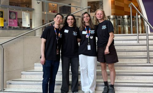
Organising committee present on the symposium day. From left to right: Jiaming Bi, Juliette Gracia, Irmak Toksoz and Ella Knüpling.
Finally, we would like to thank all the students whose scientific contributions, participation and enthusiasm made the symposium such a success. We hope to welcome you back next year!
An important insight from Chen Wang, Ph.D. about finding fulfillment from scientific projects that broadly benefit the research community. Thanks also to the Hobert lab for supporting this kind of study.
Development is plastic, meaning it has the capacity to mold, to allow for change in behavior and physical traits, usually until its form reaches a certain point. The field of developmental biology is fascinating, such that a single cell knows how to make a whole species with a distinct body pattern and shape. Watching the development of a human child in real time is even more astounding. A delicate, not able to do anything form, then watches and learns as it grows, and develops into a capable being. Growing and evolving through school and work, to then become fully equipped with the capacity to be chasing whatever fascinates and supports its being. There is also beauty in watching people grow through school and the workplace. It is reflected in how communities come together to support its young, how individuals share their experiences so others can learn through stories, and especially in how mentors invest their time and guidance in others, often getting little in return.
In recounting how one is constantly learning as a human, I feel happy that at North Carolina Central University (NCCU), I get to make small contributions towards both sides of the development coin. On one side, I study how processes of embryonic development get disrupted with environmental toxins, and on the other side I also get to share some fun science activities with young minds as they are developing. While I have recently celebrated seeing our manuscripts on BioRxiv while they are in review, here I want to share the other side of this development coin. The humbling experience of working with high school students, and the rewarding experience of their recent field trip to our institute.
An hour is not a long drive, but when we consider the exposure and experiences available to high school students, that one-hour distance can sometimes become astronomical. It can mean that while one school has access to resources and opportunities to explore science, another misses out without even realizing the extent of what they are missing. With support from Dr. Kumar and community partners, including Ms. Ruby, we have started a science lab activity initiative that aims to reduce the distance created by that one-hour drive. In alignment with the STEM curriculum, we have designed hands-on activities that high school students can participate in during their classes, learning everything from handling basic laboratory equipment and conducting fun experiments to listening to guest lectures from scientists sharing their current research and practicing the skill of presenting what they have learned.
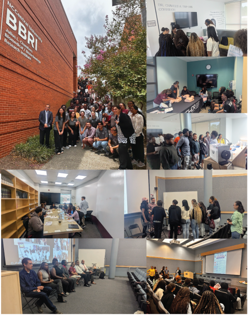
Last week, we welcomed a group of high school students from Enfield, NC, to NCCU for a field trip. My hope for this visit was simple: to create an opportunity for students to connect with people at different stages of STEM careers, see firsthand the work being done at the Julius L. Chambers Biomedical and Biotechnology Research Institute, and perhaps begin to envision themselves pursuing careers in science. We started the day with a panel discussion featuring an undergraduate student, an experienced master’s student, a newer PhD student, a public health educator, and three professors working in different research areas. All of the panelists shared their career trajectories, their motivations for pursuing their specific career paths, what their average day looks like, and what they wished they could change or adjust about it. One professor shared how someone they met during high school remains one of their strongest advocates and a mentor they still keep in touch with today, emphasizing that one should never underestimate the power of connecting with people at any stage of life. A PhD student shared how, as a first-generation student, she constantly has to navigate the hidden curriculum and figure things out along the way. She emphasized that it is okay not to know something, that it is okay to ask questions, and that it is definitely okay to reach out for help, even when it feels like you should already know the answer. A community engagement and public health expert shared how it is important to meet communities where they are, rather than trying to make them fit one’s schedule. As a result, many events intended to serve communities often take place after regular office hours or during weekends. An undergraduate student shared their aspiration to pursue an MD/PhD, describing their personal motivations for becoming a physician while also discovering a passion for research and learning. They explained that they did not realize it was possible to pursue both paths, but that doing so has now become their career goal.
A major theme that emerged from the panel discussion was that many individuals discovered career paths they did not know were available to them. Along the way, they were guided by mentors and learned to overcome the fear of asking questions and reaching out to others. Essentially, as I see it, they are now paying forward the help and guidance they received by sharing their own stories with these young, developing minds.
Following the panel, students rotated through a series of hands-on STEM activities. Each student received a passport that was stamped at every station as they explored different areas of research represented at our institute. They matched zebrafish embryos to different stages of development to learn how rapidly these organisms grow and why they are such a neat research organism. They mixed glue and water to create molds, introducing the concept of 3D scaffolds that support cells and tissues. They made brain hats while learning about the functions of different brain regions, practiced chest compressions on CPR manikins to see its value in saving lives, and explored how capillary action is used in simple blood-screening tools used in community health settings. Personally, watching the students engage with everyone, bravely ask questions, laugh about their guesses during activities, and enthusiastically mix their colorful molds was both a precious and humbling experience. It was inspiring to see our small community of researchers at the institute come together for the high school students and share what we do. It made me hopeful that, through this experience, the students were able to envision what their future aspirations could be. I am truly thankful to everyone who was a part of this event and grateful to the community that helped pay forward the mentorship and guidance that we have all received at various points along the way.
Of all the articles on helpimascientist.com, this one garnered the most views—several thousand within a few days after posting about it on Twitter. And I was reminded of the subject after several conversations this week with several new trainees, who wanted to discuss the topic. As always, I’m somewhat reticent to give very specific advice to others, but not because I don’t have definite opinions. I certainly know what being “successful” in science has required of me. What I don’t generally know is how to answer that question for others. Still, I made a stab at it a few years ago, and I stand by what I wrote. If nothing else, it’s a good starting point for open honest discussions.
I would now add that despite working hard at my job, I essentially never missed my son’s or daughter’s sporting events, concerts, etc. I even helped coach baseball and skiing, took up many new hobbies, and spent plenty of quality time with my wife and family. Achieving a balance is never easy and is more of a “lifetime-average” goal than day-to-day objective. But it’s possible if you work at it.
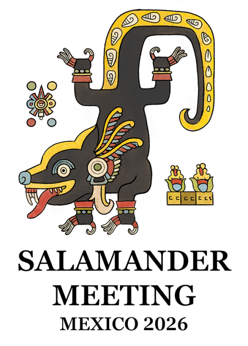
We are pleased to invite you to participate in the Salamander Meeting 2026, which will be held at the Universidad Nacional Autónoma de México (UNAM) in Mexico City from October 6 to 8, 2026.
We invite you to register and submit your abstract on the website.
This meeting will bring together researchers, postdoctoral fellows, and students working on salamander biology, with a particular focus on regeneration, development, and related areas, especially in Ambystoma mexicanum and other salamander models.
The scientific program will include 3 keynote lectures, 17 short talks, and a poster session, providing opportunities for discussion and exchange of ideas across the field.
As this edition will be held in Mexico, the meeting will also include a special cultural component, including a visit to Xochimilco, the axolotl’s native habitat, and a lecture on the broader historical and cultural context of the axolotl in Mexico.
During the meeting, the organizing committee will recognize Dr. Malcolm Maden for his significant scientific contributions to limb regeneration and for clarifying the role of retinoic acid in this process.
IMPORTANT POINTS TO CONSIDER
*The registration fee includes: a visit to Xochimilco, conference materials, coffee breaks, and 2 meals, as well as breakfasts for those staying at the host hotel.
*Accepted abstracts may be included in an e-book to be published in Frontiers in Cell and Developmental Biology (for authors who choose this option) and will be indexed with Crossref and CLOCKSS.
*The conference will include a special issue in Frontiers in Cell and Developmental Biology titled “Salamander Regeneration and Development: Cellular, Molecular, and Evolutionary Insights”; please also consider submitting your work.
We would be delighted to welcome you to Mexico City for this meeting.
As new journal impact factors are released this week, it’s worth considering what an impact factor really is and whether the academic community should put so much emphasis on a single metric.
In the latest issue of Development, Editor-in-Chief James Briscoe and I openly discuss our views on the impact factor and how the field of developmental biology is perceived through its lens. As part of a larger series drawing on real conversations we’ve had with our community, we highlight that the impact factor is not an accurate measure of Development’s real contribution to research and researchers. We detail how authors and readers can take practical steps towards ethical publishing practices that support our communities, as well as the field-specific, society or not-for-profit journals that serve them.
We hope this Editorial ignites discussion and empowers researchers to make change.
“We are all, in a sense, co-authors of the IF. It is not a number that descends from on high; it emerges from millions of individual citation decisions made by researchers like you. If we collectively cite the work that matters to us, in the journals that serve our field, the metrics will follow. We also have a collective responsibility to challenge how IF is used: in grant reviews, in hiring decisions, in promotion committees. If we believe that research should be judged on its own merit, we need to act on that belief, not just as editors, but as reviewers, as panel members, and as colleagues”