Most folks think that our brains don’t produce any more neurons after we’re born, but thankfully they do! A small subset of stem cells within the hippocampus gives rise to neurons in a region of the brain believed to be associated with learning and memory, and a recent paper describes the self-renewal properties and potential of these stem cells, and identifies the use of Sox1 as a marker for them.
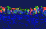
The production of neurons continues throughout adulthood from two sources in the brain—from radial astrocytes in the subgranular zone (SGZ) in the hippocampus and along the lateral walls of the lateral ventricles in the ventricular-subventricular zone (V-SVZ). Neural stem cells (NSCs) within the SGZ divide to produce mature neurons in the granular layer of the hippocampus, which has been suggested to play a role in learning and memory. A recent paper in Development describes the potential of this NSC population, and identifies markers that can be used to identify these stem cells. The transcription factor Sox1 is found throughout early central nervous system development, and according to Venere and colleagues, is found in the NSC population of radial astrocytes. By lineage tracing these Sox1-positive radial astrocytes, Venere and colleagues found that these NSCs give rise to most, if not all, granular neurons and a small number of hilar astrocytes. The number of Sox1-positive radial astrocytes declines with age, consistent with a decrease in neurogenesis. Despite the decline in number, these cells remain transcriptionally active. In the images above, Sox1 (green) is found in a subset of radial astrocytes (blue). Sox1, Sox2 (red), and the radial astrocyte marker GFAP (blue) are all found in one subset of cells (arrowheads), while another subset of non-radial progenitor cells has both Sox1 and Sox2 markers (arrows). Sox2 is known to have overlapping, but distinct, expression with Sox1 during development.
For a more general description of this image, see my imaging blog within EuroStemCell, the European stem cell portal.
Venere, M., Han, Y., Bell, R., Song, J., Alvarez-Buylla, A., & Blelloch, R. (2012). Sox1 marks an activated neural stem/progenitor cell in the hippocampus Development, 139 (21), 3938-3949 DOI: 10.1242/dev.081133
Identifying gene expression signatures that underlay diversification of muscle cells using cell type specific genomic approaches in Drosophila
Muscle network in Drosophila embryos represents an attractive system for studying cell diversification. Each muscle displays a specific size, location, attachment points and innervation. Several studies including those of our group (Junion et al., 2007, Bataillé et al., 2010; reviewed in de Joussineau et al., 2012) have led to identify key factors that control specification of different types of muscles and acting downstream realisator genes. At the same time these studies revealed a need for designing cell type specific genome-wide approaches allowing a more global and unbiased view of cell diversification processes.
The recruited post-doctoral fellow will integrate an ambitious and innovative project designed to address this issue. The main objective of this study is to apply newly developed cell-specific approaches to perform transcriptional and ChIPseq analyses in subsets of embryonic Drosophila muscles. As an outcome, we hope to uncover realisator gene signatures that specify different subsets of muscles and in particular required for setting fusion programs, attachment and innervation of individual muscles.
From a more general point of view this study should provide insights into the logic of the genetic control of cell diversification as such.
We are searching for a highly motivated researcher who will join our team and interact with bioinformatician already working on this project. Experience in transcriptional profiling and ChIPseq approaches as well as in Drosophila developmental genetics will be considered as an advantage but is not a requirement.
Please consider that GReD Institute provides dynamic scientific environment and access to a large spectrum of facilities including up-to-date confocal imaging and Drosophila transgenesis platforms.
Junion, G., Bataillé, L., Jagla, T., DaPonte, J.P., Tapin, R. and Jagla, K. : Genome-wide view of cell fate specification: ladybird acts at multiple levels during diversification of muscle and heartprecursors. Genes & Dev 2007, 21, 3163-80.
Bataillé L, Delon I, Da Ponte JP, Brown NH, Jagla K. Downstream of identity genes: Muscle-type specific regulation of the fusion process. Dev Cell, 2010, 19:317-328.
de Joussineau C, Bataillé L, Jagla T, Jagla K. Diversification of muscle types in Drosophila:upstream and downstream of identity genes. Curr Top Dev Biol. 2012, 98:277-301.
What was your favourite paper in the fields of developmental or stem cell biology this past year?
We’re preparing a little feature on the Node for the month of December: an advent calendar with a new paper recommendation each day from December 1 to 24.
For this, we need your help! What was your favourite paper of the past twelve months?
To keep the calendar selections a surprise, we ask you to email your choice to thenode [at] biologists.com (or use the contact form). It would be great if you could also tell us, in one sentence, why you selected that paper. The deadline is November 22nd.
You can submit multiple papers, but NOT one from yourself or your lab. (Similarly, Development staff are not selecting Development papers as their own choice – but anyone else can!)
Papers you select should have been published after December 1, 2011. It doesn’t matter which journal it’s in, as long as the article covers a topic in developmental biology, stem cells, or a related area (evo-devo, developmental genetics, etc.) Don’t forget the first half of the year – it seems long ago, but I’m sure there were some great papers those months.
If we end up with more than 24 papers, we’ll make a final selection based on the diversity of suggestions we’ve received at that point.
We’re looking forward to showing you the full selection of Node reader-picked papers next month!
Within the Development team, we are seeking to recruit a new Associate Reviews Editor with special responsibility for the stem cell field – reflecting the journal’s increasing visibility in that area. Further details on this exciting opportunity are available on the jobs page.
DMM also have a position for a Scientific Editor, and you can find more information on this opening here. Applicants with previous editorial experience are particularly encouraged.
Both jobs will be based at the Company of Biologists’ office in Cambridge, UK, and both provide a fantastic opportunity to work with a small not-for-profit publisher at the heart of the scientific community.
Scientific Editor Disease Models & Mechanisms
Based in Cambridge, UK
Joining a young journal in an expanding and important area of research, this is an exciting opportunity for a promising scientific editor to make a significant contribution to a growing open access journal serving a major new biomedical community.
The journal is entering the next phase of its development with the appointment of a team of academic editors, having been managed previously by a professional editor. As part of this evolution of the journal model, we will require the new scientific editor to be flexible in their areas of responsibility. Disease Models & Mechanisms publishes both primary research articles and commissioned front section material and we expect the scientific editor to take a role in both of these areas.
Since the journal serves both basic biomedical researchers and clinicians, applicants will have a PhD or MD with some relevant research experience, and a broad knowledge of model organisms and disease issues, and will ideally already hold an editorial role (we also provide on-the-job training).
Core responsibilities are expected to include:
• Support for the academic editors in their assessment and handling of primary research articles
• Commissioning, handling peer review and developmental editing of material for the front section of the journal
• Writing pieces for the journal, conducting interviews and preparing press releases
• Representing the journal at international conferences and within the wider scientific community
• Creative involvement in the journal’s development and marketing
The Scientific Editor will work alongside an experienced in-house team, and additional responsibilities may be provided for the right candidate.
Essential requirements for the job are enthusiasm, commitment, judgement, integrity and a mature attitude. Candidates should have excellent interpersonal skills, excellent oral and written communication skills, and a broad interest in research and the research community.
The position represents a unique opportunity to gain experience on an exciting new biomedical research journal and offers an attractive salary and benefits.
The Company of Biologists (www.biologists.com) is a not-for-profit organisation, publishing five journals in the biological sciences: Disease Models & Mechanisms, Development, Journal of Cell Science, The Journal of Experimental Biology and Biology Open. The organisation has an active programme of charitable giving for the further advancement of biological research, including travelling fellowships for junior scientists, contributions to academic societies and conferences, and a series of scientific workshops.
Applicants should send a CV along with a covering letter that states their salary expectations and summarises their relevant experience and why they are enthusiastic about this opportunity.
Applicants must be able to provide confirmation of entitlement to work in the UK.
Applications should be sent by email no later than 18th November 2012 to miriam@thecob.co.uk
Informal queries to Miriam Ganczkowski on +44 (0)1223 426 164.
Associate Reviews Editor (Stem Cells) Development
Based in Cambridge, UK
Applications are sought for a new role of Associate Reviews Editor for the stem cell field at Development.
Joining an experienced, expanding and successful team, this is an exciting opportunity for a promising scientific editor to make a significant contribution to one of the major journals in the field of developmental biology. Development publishes primary research articles, reviews and other front section content. The journal is expanding its focus in the stem cell field, and is seeking to appoint a specialist editor to cover this growing area.
Applicants will hold a PhD, ideally in stem cell biology, although outstanding candidates with experience in other areas of developmental biology and with a strong interest in the stem cell field will be considered. Post-doctoral and/or previous editorial experience are highly desirable, although we will provide on-the-job training. The successful candidate will have a broad interest in science, the scientific community and publishing. Excellent interpersonal and literary skills, enthusiasm and commitment are also essential requirements for the position.
Core responsibilities:
• Commissioning, handling peer review and developmental editing of material for the front section of the journal
• Writing press releases, article highlights and material for Development’s community website ‘the Node’
• Representation of the journal at international conferences and within the wider scientific community
• Creative involvement in the journal’s development
The new Associate Reviews Editor will work alongside an experienced in-house team, including the Executive Editor and current Reviews Editor, as well as with our international team of academic editors. Additional responsibilities may be provided for the right candidate.
The position represents a unique opportunity to gain experience on a highly successful life-science journal and offers an attractive salary and benefits. The position will be based in our office in Cambridge, and the contract will be for a three-year period.
The Company of Biologists (www.biologists.com) is a not-for-profit organisation, publishing five journals in the biological sciences: the three established journals Development, Journal of Cell Science and The Journal of Experimental Biology, as well as two newer Open Access journals, Disease Models & Mechanisms and Biology Open. The organisation has an active programme of charitable giving for the further advancement of biological research, including travelling fellowships for junior scientists and contributions to academic societies and conferences.
Applicants should send a CV along with a covering letter that summarises their relevant experience and why they are enthusiastic about this opportunity, and states their salary expectations.
Applications should be sent by email no later than November 26th to miriam@thecob.co.uk
Informal queries to Miriam Ganczkowski on +44 (0)1223 426 164
Here are the highlights from the current issue of Development:
Dishevelled: the Notch-Wnt go-between
The Notch and Wnt signalling pathways are used during animal development to generate a diverse array of cell types. The two pathways often have opposing effects on cell-fate decisions but some cells receive inputs from both pathways simultaneously. In these circumstances, it is common for the receiving cell to exhibit a Wnt-ON/Notch-OFF response but how is this response generated? Now, on p. 4405, Keith Brennan and co-workers report that Wnt acts via Dishevelled, a key mediator of Wnt/β-catenin signalling, to inhibit the Notch pathway and that this crosstalk controls cell-fate specification during Xenopus epidermal development in vivo. Dishevelled, they report, binds and directly inhibits the CSL (CBF1, Suppressor of Hairless, Lag-1; RBPJκ in mice) transcription factors that mediate Notch signalling. Moreover, this crosstalk mechanism is conserved between vertebrates and invertebrates. Thus, by acting as both an activator of Wnt signalling and an inhibitor of Notch signalling, Dishevelled sharpens the distinction between opposing Wnt and Notch responses, thereby ensuring that robust cell-fate decisions are taken during development.
MicroRNA regulation of angiogenesis
Secreted signalling molecules regulate cellular communication across tissues during development. For example, during angiogenesis in zebrafish embryos, vascular endothelial growth factor (VEGF) secreted by muscle controls the expansion and remodelling of the vascular network. The precise regulation of VEGF expression in muscle cells is therefore essential for angiogenesis but how is this regulation achieved? On p 4356, Yuichiro Mishima, Antonio Giraldez and colleagues report that miR-1 and miR-206 (miR-1/206; two conserved microRNAs with similar sequences) negatively regulate angiogenesis during zebrafish development. The negative effect of miR-1/206 on angiogenesis, the researchers report, is mediated in part by direct regulation of VegfAa in muscle. Thus, masking the target sites for miR-1/206 in the 3′ UTR of vegfaa has a pro-angiogenic effect similar to that of miR-1/206 knockdown. By contrast, reducing the levels of VegfAa rescues the increase in angiogenesis produced by miR-1/206 knockdown. Together, these findings uncover a novel function for miR-1/206 in the control of developmental angiogenesis and highlight a key role for microRNAs as regulators of cross-tissue signalling.
HIF1α controls collagen secretion under hypoxia
The production of collagen – a major component of the extracellular matrix – depends on hydroxylation of proline residues, a reaction that uses molecular oxygen as substrate. Cells that develop in hypoxic settings can nevertheless produce collagen during embryogenesis. Elazar Zelzer and colleagues begin to resolve this paradox on p. 4473 by identifying the transcription factor hypoxia-inducible factor 1 α (HIF1α) as a central regulator of collagen hydroxylation and secretion by chondrocytes in the hypoxic growth plate of developing mouse bones. The researchers show that Hif1a loss of function in growth plate chondrocytes arrests the secretion of collagen. Hif1α, they report, drives the transcription of collagen prolyl 4-hydroxylase, which mediates collagen hydroxylation and its subsequent folding and secretion. Concurrently, Hif1α also maintains cellular oxygen levels, probably by controlling the expression of pyruvate dehydrogenase kinase 1, an inhibitor of the tricarboxylic acid cycle. This two-pronged mechanism, the researchers suggest, allows chondrocytes to secrete large amounts of collagen in the hypoxic environment of the growth plate.
Cycling towards sepal cell specification
During development, cell division and cell specification must be coordinated to ensure that the tissues and organs of the adult organism are the correct size and contain the right proportions of various cell types. Here (p. 4416), Adrienne Roeder and colleagues investigate how developmental regulators interact with the cell cycle in Arabidopsis sepals to create a characteristic pattern of outer epidermal cells in which elongated giant cells, which are produced by endoreduplication (DNA replication without cell division), are interspersed between small cells, which divide mitotically. They show that distinct enhancers are expressed in giant cells and small cells, which suggests that these cells have different identities as well as different sizes and ploidies. Several members of the epidermal specification pathway control the identity of giant cells, they report, which is established upstream of cell-cycle regulation. By contrast, endoreduplication represses small cell identity. Thus, suggest the researchers, cell type affects cell-cycle regulation but, in addition, cell-cycle regulation can control cell identity.
Epigenetic reprogramming egged on
The generation of cloned embryos by somatic cell nuclear transfer (SCNT) into enucleated mature oocytes is inefficient because epigenetic reprogramming is limited in these embryos. However, treatment of somatic nuclei with a cytoplasmic extract from germinal vesicle (GV) stage oocytes before SCNT is known to improve the efficiency of cloned mouse production. Now (p. 4330), Hong-Thuy Bui, Jin-Hoi Kim and co-workers use an extract from GV stage pig oocytes (GVcyto-extract) to investigate epigenetic reprogramming events in pig fibroblasts. The researchers report that fibroblasts treated with GVcyto-extract express the stem cell-associated proteins Oct4 and Nanog and re-differentiate into three primary germ cell layers in vitro and in vivo. Moreover, the use of donor nuclei treated with GVcyto-extract, they report, increases the number of high quality SCNT-generated blastocysts that exhibit levels of histone H3-K9 methylation and acetylation and Oct4 and Nanog expression similar to those in embryos fertilised in vitro. These results suggest that a combination of epigenetic reprogramming techniques might improve the efficiency of development in SCNT-generated embryos.
Notch pick and mix regulates lung development
In developing mammalian lungs, the terminal buds of elongating airways contain multipotent epithelial progenitors that give rise to the three major lung epithelial cell types: Clara, ciliated and neuroendocrine (NE) cells. Now, Mitsuru Morimoto and colleagues provide new insights into how Notch signalling regulates the distribution of these epithelial cell types in the airway (see p. 4365). Using stepwise removal of Notch1, Notch2 and Notch3 from developing mouse lung epithelium, the researchers show that Notch2 alone mediates the Clara/ciliated cell fate decision whereas all three receptors regulate NE fate selection in an additive manner. All three Notch receptors also additively regulate the size of the presumptive pulmonary neuroepithelial body (pNEB; NEBs are clusters of NE cells) through mutual interactions between NE cells and a population of SSEA-1 (stage-specific antigen 1)-positive epithelial cells that surrounds the pNEB. These and other results indicate that two different assemblies of Notch receptors control the number and distribution of lung epithelial cell types during lung organogenesis.
PLUS…
Drosophila neuroblasts: a model for stem cell biology
As part of the ‘Development: the big picture’ series, Homem and Knoblich explain why Drosophila neuroblasts, the stem cells of the developing fly brain, have emerged as a key model system for neural stem cell biology. See the Primer on p. 4297
Endocytic receptor-mediated control of morphogen signaling
Thomas Willnow and colleaugues describe how low-density lipoprotein receptor-related proteins (LRPs) constitute central pathways that modulate morphogen presentation to target tissues and cellular signal reception, and how LRP dysfunction leads to developmental disturbances in many species. See the Primer on p. 4311
Piecing together the vertebrate skull
As part of the ‘Development Classics’ series, Nicole M. Le Douarin discusses her 1993 Development paper, in which she and her colleagues used the quail-chick chimera system to decipher the embryonic origin of the bones of the head skeleton of the avian embryo. See the Spotlight article on p. 4293
Last week, the Royal Society hosted a meeting entitled “Regulation from a distance: Long-range control of gene expression in development and disease”. The impressive London offices of the Society (complete with double helix-inspired door handles) added a sense of occasion to what was bound to be a fascinating meeting, based on the list of excellent speakers from Europe and the US. It brought together a diverse range of scientists from different backgrounds, united by their common interest in the vast stretches of non-coding DNA in the genome, and the role that it plays in gene regulation. I’ll try to give a general overview of the talks here, but with greater focus on the developmental biology topics.
The meeting was opened by Robert Hill (University of Edinburgh) who described an extreme example of a long-range enhancer: the ZRS enhancer that drives Shh expression in the developing limb bud from a distance of 1 Mb. His group have studied this enhancer’s role in preaxial polydactyly (extra digits). The talks that followed showcased a range of useful methods for identifying such distant enhancers.
Francois Spitz (EMBL) described the use of the GROMIT method (Genome Regulatory Organisation Mapping with Integrated Transposons) to mobilise a transposon containing a “regulatory sensor” around the genome, giving reporter gene expression as a read-out of the tissue-specific enhancer signals at different genomic locations. Ivan Ovcharenko (NIH) presented a computational approach for identifying such tissue-specific enhancers, showing that the lessons learned from analysing the tissue-specific transcription factors bound at promoters are also useful for locating enhancers of those tissue-specific genes. Joanna Wysocka (Stanford) presented a combination of different approaches for enhancer identification including conservation analysis, identification of DNaseI hypersensitive sites and ChIP-seq for histone modifications and p300. Her group has used these methods to identify neural crest-specific enhancers that drive novel regulators of craniofacial development.
Several speakers discussed the chromosome conformation capture or ‘C’ techniques, which are used to visualise physical interactions between distant regions of the genome, such as those between gene promoters and enhancers. Denis Duboule (University of Geneva/EPFL) described the use of 4C to investigate the regulation of the Hoxd gene cluster during limb development, and Wouter de Laat (Hubrecht Institute) spoke about recent advances that have increased the resolution of 4C to allow identification of interactions as close as 10 kb. His group have used this technology to identify a novel enhancer of Oct4 in embryonic stem cells. Wendy Bickmore (University of Edinburgh) emphasised the importance of corroborating findings from the ‘C’ techniques using other methods, such as FISH (fluorescent in situ hybridisation).
Although many methods can be used to identify potential enhancers, it is also important to validate their activity. Len Pennacchio (LBNL) described recent efforts in compiling the VISTA enhancer browser: a publicly available database of enhancers that drive tissue-specific gene expression in vivo in transgenic mouse embryos.
In addition to the developmental biology talks, several speakers focused on the role of long-range gene regulation in human disease, which is an important consideration given that many mutations associated with disease are in non-coding regions of the genome. We heard from Doug Higgs (University of Oxford) on the alpha-globin genes and thallasaemias, Matthew Freedman (Harvard) on the Cancer Genome Atlas, and Gioacchino Natoli (European Institute of Oncology) on enhancers that are activated during the inflammatory response.
Enhancer mutations are also an important driving force in evolution, as was explained by David Kingsley (HHMI/Stanford) who studies the rapid natural evolution of three-spined stickleback populations as they colonise new lakes. Major phenotypic changes in these populations can be explained by mutations in a few key developmental genes, but while mutations that inactivate these genes are lethal, mutations in enhancers can alter their expression patterns in a way that produces an evolutionary advantage. Also on the population genetics theme, Manolis Dermitzakis (University of Geneva) presented human data that he has used to investigate the genetic and epigenetic contributions to gene expression.
The majority of speakers focused on long-range gene regulation by enhancers, but it is also important to consider the role of insulators. This was addressed by Bing Ren (UCSD) in the final talk of the meeting. His group have used the HiC technique to create a genome-wide map of chromosomal interactions, revealing that the three-dimensional structure of the genome is divided in to topological domains. Many long-distance interactions occur within these domains, but very few occur between different domains, suggesting that the domain boundaries represent insulators. However, the role of CTCF as an insulator factor was debated at length in the subsequent discussion session.
The Royal Society meetings have an unusual format, with sets of two half-hour talks followed by half an hour of discussion. These long discussion sessions allowed for much deeper probing of the subject, and encouraged debate that became quite lively at times, showing just how exciting this area of research is! Some topics were revisited throughout the meeting, establishing links between different areas, which is important in driving the field forward. The variety of experimental approaches presented showed how insights can be gained by investigating regulation of gene expression at multiple levels. In his talk, John Stamatoyannopoulos (University of Washington) mentioned that the genome contains over 4 million distinct elements that are associated with gene regulation, so there is clearly a lot of work to be done in this field in the future!
The Royal Society have made the audio of all talks from this meeting available online, so check their website for more details on any of the presentations.
The big news this past month was of course the announcement of the Nobel Prize for Physiology and Medicine, awarded to John Gurdon and Shinya Yamanaka for their work on cellular reprogramming. Katherine Brown wrote a brief post about John Gurdon’s connection to the Company of Biologists – and to the Node!
Hydra Summer School
A few weeks before the Nobel committee acknowledged the importance of the field of stem cells, the Hydra Summer School in Stem Cells and Regenerative Medicine trained a group of young researchers in the field. Two participants wrote about their experience and the highlights of the course. Sarah-Jane writes:
“Personally, I particularly enjoyed the recurring discussion on the definition of a stem cell, and how one must be aware of the language subtleties that exist between different researchers and sub-disciplines. Is a fertilised egg a stem cell? Perhaps, if you’re a developmental biologist.”
Kif added:“There is a huge prospect for the field of stem cell biology in the future, from understanding to alleviating diseases. Lectures and discussions at the summer school gave me confidence that this is a realistic goal.”
Development: Past, Present and Future
Stem cells were also one of the topics discussed at Development’s 25th anniversary symposium. That’s not surprising, considering the theme was “Past, Present and Future”! Several of Development’s current and former editors took the stage to talk about their work, and the day ended with a panel discussion.
Nucleitracker
But let’s not forget the many other advances in the field of developmental biology, that help us study multicellular tissues and organisms. Improvements in imaging and automation techniques are another important development, and we also covered that on the Node this past month: Andrew Chisholm’s group created a method to track nuclei in the developing C. elegans embryo, including the later stages.
“Claudiu Giurumescu, a postdoc in my lab, took a different approach to the problem of tracking nuclei in the crowded environment of the later embryo. Importantly, he decided to use a combination of automatic tracking and manual curation. The tracking relies on the predictable behavior of nuclei in worm embryos: most nuclei do not move around much on the time scale used in 4D movies. Claudiu devised algorithms that took advantage of this predictability to search locally for each nucleus at a given time point, based on the information on where the nucleus is at the previous time point.”
Last month was an exciting one for stem cell research. I’m sure you all saw how stem cells hit international headlines with the announcement of a Nobel Prize for John B. Gurdon and Shinya Yamanaka. I thought you might be interested to read reactions from several leading scientists and check out a film clip about the prize-winning work in our blog on eurostemcell.org
We’ve got a new short film all about reprogramming and iPS cells coming very soon too. Watch out for it going live on our website www.eurostemcell.org at the end of November or early December, in time for the formal Nobel Prize awards ceremony of 10th December.
We were also celebrating in October because it was Stem Cell Awareness day on the 3rd of the month. Researcher Christèle Gonneau helped us celebrate the day by giving us a great window into life as a stem cell biologist on twitter. You can check out a summary of her day’s tweets and pictures in our blog too.
You can keep up to speed on stem cell news and our activities by following @eurostemcell on Twitter or, if Twitter’s not your thing, say hello to us on Facebook or our website.


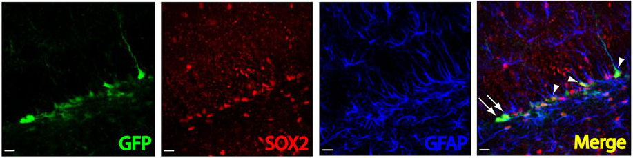
 Venere, M., Han, Y., Bell, R., Song, J., Alvarez-Buylla, A., & Blelloch, R. (2012). Sox1 marks an activated neural stem/progenitor cell in the hippocampus Development, 139 (21), 3938-3949 DOI: 10.1242/dev.081133
 What was your favourite paper in the fields of developmental or stem cell biology this past year?
 The Notch and Wnt signalling pathways are used during animal development to generate a diverse array of cell types. The two pathways often have opposing effects on cell-fate decisions but some cells receive inputs from both pathways simultaneously. In these circumstances, it is common for the receiving cell to exhibit a Wnt-ON/Notch-OFF response but how is this response generated? Now, on p.
 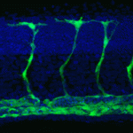 Secreted signalling molecules regulate cellular communication across tissues during development. For example, during angiogenesis in zebrafish embryos, vascular endothelial growth factor (VEGF) secreted by muscle controls the expansion and remodelling of the vascular network. The precise regulation of VEGF expression in muscle cells is therefore essential for angiogenesis but how is this regulation achieved? On p
 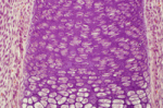 The production of collagen – a major component of the extracellular matrix – depends on hydroxylation of proline residues, a reaction that uses molecular oxygen as substrate. Cells that develop in hypoxic settings can nevertheless produce collagen during embryogenesis. Elazar Zelzer and colleagues begin to resolve this paradox on p.
 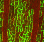 During development, cell division and cell specification must be coordinated to ensure that the tissues and organs of the adult organism are the correct size and contain the right proportions of various cell types. Here (p.
 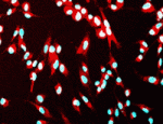 The generation of cloned embryos by somatic cell nuclear transfer (SCNT) into enucleated mature oocytes is inefficient because epigenetic reprogramming is limited in these embryos. However, treatment of somatic nuclei with a cytoplasmic extract from germinal vesicle (GV) stage oocytes before SCNT is known to improve the efficiency of cloned mouse production. Now (p.
  In developing mammalian lungs, the terminal buds of elongating airways contain multipotent epithelial progenitors that give rise to the three major lung epithelial cell types: Clara, ciliated and neuroendocrine (NE) cells. Now, Mitsuru Morimoto and colleagues provide new insights into how Notch signalling regulates the distribution of these epithelial cell types in the airway (see p.
 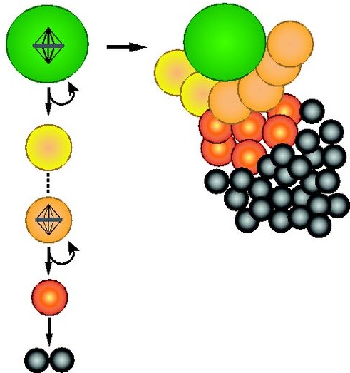 As part of the ‘
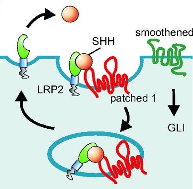 Thomas Willnow and colleaugues describe how low-density lipoprotein receptor-related proteins (LRPs) constitute central pathways that modulate morphogen presentation to target tissues and cellular signal reception, and how LRP dysfunction leads to developmental disturbances in many species. See the Primer on p.
 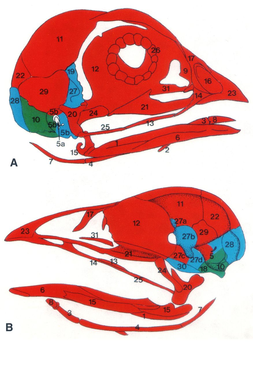 As part of the ‘Development Classics’ series, Nicole M. Le Douarin discusses her 1993 Development paper, in which she and her colleagues used the quail-chick chimera system to decipher the embryonic origin of the bones of the head skeleton of the avian embryo. See the Spotlight article on p.