The community site for and by
developmental and stem cell biologists

Node meet-up at the BSDB meeting

Posted by , on 13 April 2011

NOTE: LOCATION CHANGE!
We’ll meet at the Gulbenkian Cinema Cafe/Bar at 8PM on Thursday, as Mungos is closed
.
This is the cafe that you walk past along the footpath from Elliot College to Woolf College, so when you go from dinner to posters you can drop in along the way.

——

If you’d like to meet some other Node readers (and writers), why not join us for a drink during the BSDB meeting. Bring your lab mates and friends – we like meeting new people!

We’ll get together at 20:00 on Thursday April 28, at Mungos Bar (in Eliot College) Gulbenkian Cinema Cafe Bar.

We’ll be there for about an hour or so, and then we’ll head off to the poster session in the Woolf Building, so don’t worry about missing dinner or posters.

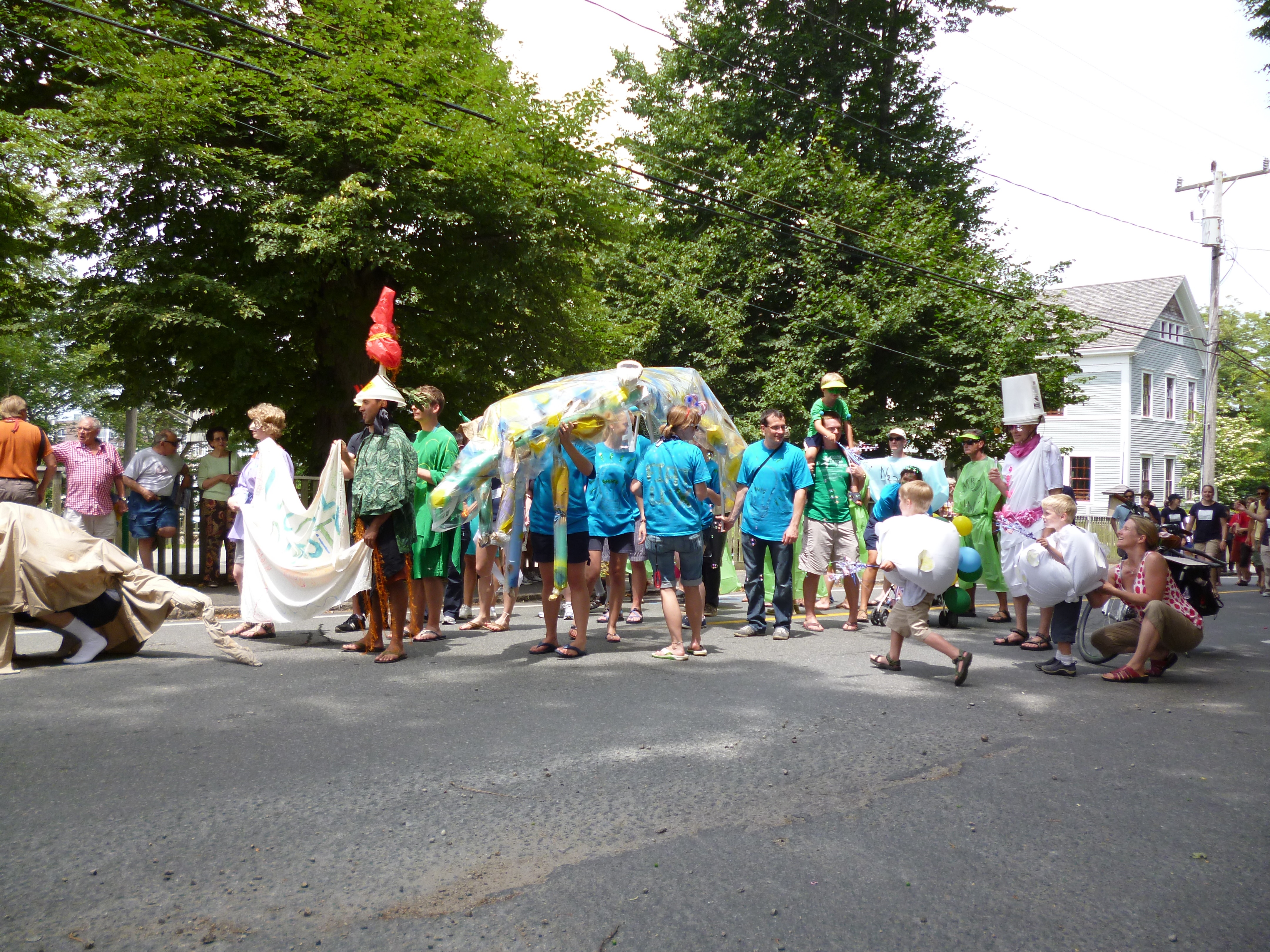
There will be a sign at The Company of Biologists’ stand in the exhibition space about this, and if anything changes (location, times) you can find the information there as well. If it’s not too cumbersome, I’ll try to bring the banner (pictured) to the bar for easy identification, otherwise there’ll be other ways to identify us.

Hope to see you on the 28th at the BSDB meeting!

Thumbs up (2 votes)
Loading...

Categories: Events

Navigate the archive

Use our Advanced Search tool to search and filter posts by date, category, tags and authors.

Establishing a niche

Posted by , on 12 April 2011

If there is hope to fully understand stem cells, then the environment surrounding those stem cells must be understood too.  A recent Development paper describes important results on niche establishment in Drosophila.

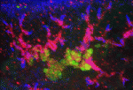
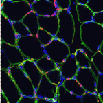
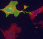
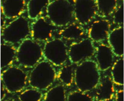
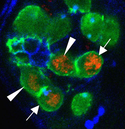
Stem cell niches play an important role in regulating stem cell self-renewal and differentiation.  The Drosophila testis has two populations of stem cells that cluster around a niche called the “hub.”  It was previously known where hub cells originated, but recent work describes how hub cells become specified.  Notch and its two ligands, Delta and Serrate, are required for specifying hub cell fate during gonad development.  Interestingly, endoderm tissue (posterior midgut cells, if you’re keeping score) provides the Delta signal, further emphasizing a well-known relationship between gut and germ cells.  As seen in the above images of larval gonads, flies with mutations in Delta (left, bottom) or  (right, bottom) had fewer hub cells (green) than control gonads (top).

For a more general description of this image, see my post at EuroStemCell, the European stem cell portal.

ResearchBlogging.orgOkegbe, T., & DiNardo, S. (2011). The endoderm specifies the mesodermal niche for the germline in Drosophila via Delta-Notch signaling Development, 138 (7), 1259-1267 DOI: 10.1242/dev.056994

Thumbs up (No Ratings Yet)
Loading...

Tags: , ,
Categories: Images, Research

In Development this week (Vol. 138, Issue 9)

Posted by , on 12 April 2011

Here are the research highlights from the current issue of Development:

Cranial neural crest development: p53 faces up

The tumour suppressor p53 plays multiple roles in the prevention of cancer but its developmental functions are less clear. Here (see p. 1827), Eldad Tzahor and colleagues elucidate the key role that p53 plays in craniofacial development. During embryogenesis, cranial neural crest (CNC) cells give rise to the facial bones, cartilage and connective tissues. Neural crest development involves an epithelial-mesenchymal transition (EMT) that converts epithelial cells into migratory mesenchymal cells, which delaminate from the neural tube. Notably, EMT is an early step in tumour progression. The researchers report that craniofacial development is disrupted in p53 knockout mouse embryos. Then, they show that p53 is expressed in CNC progenitors in chick embryos but that its expression decreases as these cells delaminate from the neural tube. Moreover, p53 gain-of-function results in fewer migrating CNC cells, whereas p53 loss-of-function increases the EMT/delamination of CNC cells. These and other findings suggest that p53 coordinates CNC growth and EMT/delamination processes during craniofacial development.

Oiling the wheels of Hippo signalling

The Hippo signalling pathway, a conserved tumour suppressor pathway, is involved in other developmental processes in addition to proliferation control. For example, Hippo signalling in the posterior follicle cells (PFCs) of Drosophila ovaries is required for oocyte polarisation. Now, Trudi Schüpbach and colleagues report that a phospatidylinositol 4-kinase (PI4KIIIalpha), which catalyses the production of membrane phospholipids, is required in PFCs for oocyte polarisation and Hippo signalling (see p. 1697). The researchers isolated mutations in CG10260, which encodes PI4KIIIalpha, while screening for Drosophila genes required in follicle cells for oocyte polarisation. They show that PI4KIIIalpha loss in PFCs leads to oocyte polarisation defects similar to those caused by mutations in the Hippo signalling pathway, and that PI4KIIIalpha mutations cause misexpression of Hippo targets. Notably, the apical membrane localisation of Merlin, which is required for Hippo signalling, is lost in PI4KIIIalpha mutant PFCs, presumably because of changes in the cell membrane’s lipid composition. Together, these data reveal a new link in the Hippo signalling pathway.

Nu-age transposon silencing

The nuage, a perinuclear structure of unknown function, is present in the germline cells of many organisms. Now, on p. 1863, Haifan Lin and colleagues reveal a function for the Drosophila germline nuage. This structure contains Aubergine and Argonaute 3 (AGO3), two of the three PIWI proteins that are essential for germline development. PIWI proteins bind to PIWI-interacting RNAs (piRNAs) and function in transposon silencing. The researchers report that Partner of PIWIs (PAPI), a novel nuage component, is a TUDOR-domain protein that interacts with all three PIWI proteins through symmetrically dimethylated arginines in their N-terminal domains. This interaction is essential for AGO3 recruitment to the nuage and for transposon silencing. Importantly, the AGO3-PAPI complex associates with the P-body component TRAL/ME31B complex in the nuage and transposon activation occurs in tral mutant ovaries. Thus, the interaction in the nuage between the piRNA pathway and mRNA-degrading P-body components is involved in transposon silencing. The researchers suggest, therefore, that the nuage safeguards the germline genome against deleterious retrotransposition.

Invading heart morphogenesis with NFATC1

During cardiac morphogenesis, proepicardium cells envelop the myocardium to form the epicardium. Some epicardial cells subsequently undergo epithelial-to-mesenchymal transformation and invade the myocardium as epicardium-derived cells (EPDCs). This invasion step underlies the formation of the coronary vessels and fibrous matrix of the mature heart but how is it regulated? Michelle Combs, Katherine Yutzey and co-workers now reveal that NFATC1 promotes EPDC invasion into myocardium (see p. 1747). NFATC1, they report, is expressed in EPDCs in mouse and chick embryos and loss of its expression in EPDCs in mice decreases coronary vessel and fibrous matrix invasion into the myocardium. Other experiments in mouse embryos, chicken embryo hearts and isolated proepicardium cells indicate that NFATC1 activation by RANKL in EPDCs promotes expression of the extracellular matrix-degrading enzyme cathepsin K, promoting EPDC invasion into the myocardium. These new insights into heart morphogenesis, the authors suggest, could aid the development of EPDC-based therapies for cardiac diseases.

Timely neural identity decision making

The timing of cell identity decisions must be closely regulated during brain development. In Drosophila neuroblasts, the sequential expression of several transcription factors, including Hunchback (Hb), controls the temporal generation of diverse neural progeny. Because Hb is necessary and sufficient to specify early-born neurons, its expression has to be downregulated to allow specification of late-born progeny. Now, on p. 1727, Chris Doe and colleagues report that two pipsqueak-domain proteins – Distal antenna (Dan) and Distal antenna-related (Danr) – restrict Hb expression in neuroblasts and limit the numbers of early-born neurons. They show that Dan and Danr function independently of Seven-up (Svp), an orphan nuclear receptor that also regulates Hb expression in neuroblasts. Importantly, Hb misexpression can induce Dan and Svp expression in neuroblasts, which suggests that Hb can limit its own expression through a negative-feedback loop. The researchers conclude that Dan/Danr and Svp act in parallel pathways to limit Hb expression and allow neuroblasts to switch from making early-born to making late-born neurons at the proper time.

Longitudinal axon connections notched up

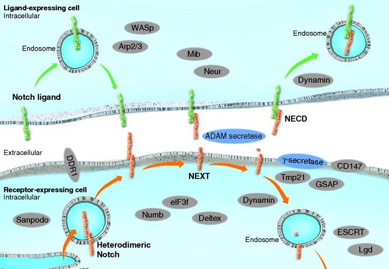
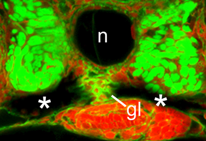
Development of the segmented central nerve cords of vertebrates and invertebrates involves the formation of longitudinal axon connections between successive segments. To establish these connections, a pathway must be marked for pioneer axons to follow and then the pioneers’ motility along that pathway must be promoted. But what are the molecular mechanisms that control these processes? On p. 1839, Edward Giniger and co-workers show how Notch signalling directs both processes in the developing Drosophila CNS. They show that canonical Notch signalling in specialised glial cells causes the extrusion of a mesh of fine filopodia by nearby differentiating neurons and shapes this mesh into a carpet that links adjacent segments. Simultaneously, non-canonical Notch signalling in the pioneer growth cones suppresses Abl tyrosine kinase signalling, which stimulates filopodial development and presumably also reduces substratum adhesion, thereby promoting the ability of pioneer axons to follow the carpet across segment borders. Thus, two parallel but separate Notch functions establish the first longitudinal connections in the fly CNS.

And…

Review: Small RNAs in early mammalian development: from gametes to gastrulation

Small non-coding RNAs, such as microRNAs, endo-siRNAs and piRNAs, are expressed throughout mammalian development and, here, Nayoung Suh and Robert Blelloch review emerging roles for these RNAs in the early stages of mammalian development, from gamete maturation through to gastrulation.

See the review article on p.1653

Thumbs up (No Ratings Yet)
Loading...

Tags: , , , , , , , , , , , , , ,
Categories: Research

Australia Rallies Against Cutbacks To Medical Research Funding

Posted by , on 12 April 2011

Last week, news that the Australian government was planning to slash the budget for medical research by more than half over the next three years leaked out and rocked the scientific community. Only one out of seven grants submitted to the National Health and Medical Research Council (NHMRC), Australia’s major funding body for medical research, is approved with at least another three deemed good enough for funding but ultimately rejected anyway due to lack of funds. With the potential upcoming cuts to medical funding, the NHMRC will only be able to fund one out of ten grants it reviews.

Rally in Melbourne

The gravity of this news cannot be understated. It is a sad disaster. Medical research is of prime importance especially when we take into account terrible disorders such as cancer, HIV/AIDS, cardiovascular diseases, diabetes and infectious diseases which our generation has to face. Cutting funding means saying no to progress against fighting those diseases and the many others. It means leaving people who can be saved to die. Yes, it is a murderous decision.

Developmental biology will not be spared from those cuts. Important and possibly life-changing research related to stem cells, for instance, will be halted, discontinued or rejected, plain and simple. Who will be affected? Well, it could be me, you, your family or friends… it could be anyone.

If the government (elected by the people, for the people) does not realize that it is going to spill its own people’s blood by cutting medical research funding, then it is up to the people to send them a message. And so it did. Rallies were organized today in several major Australian cities including Melbourne, Sydney, Canberra and Adelaide with more to follow in Perth, Darwin and Hobart later this week. A Twitter rally–that successfully trended #protectresearch, the Twitter hashtag adopted by the movement–was also carried out online.

I was at the Melbourne rally where an estimated 4000 demonstrators assembled in front of the State Library. Banners were everywhere, raised high up by outstretched arms, or rulers, meter rules or pipettes. Yes, scientists are an innovative bunch. “Medical research keeps my blood pumping,” “SOS: Save Our Science,” “Cut the nonsense, not the funding!” or “Gillard [the Australian prime minister], we’ll cure your dementia (no seriously… we will)!” were flung around.

Amidst the chorus of “Cures not cuts” and “research saves lives,” were a number of guest speakers. One of them was Nerissa Mapes, a 34-year-old woman who has been living with Parkinson’s disease for the past six years. She addressed the crowd with what was a poignant and deeply powerful speech. Her speech stressed on the important role scientists play in society. It was a wonderful way to remind us all that medical research is for the people. She concluded with this:

“There is no cure for Parkinson’s disease. But there is hope. And hope gives us courage. Don’t let them take this hope away from us.”

Medical research saves life. Cutting its funding is like handling death sentences at will.

For more information visit the Discoveries Need Dollars’ website, twitter or facebook page.

Image credit: strangehours (from flickr).

Thumbs up (5 votes)
Loading...

Tags:
Categories: Funding

2011 Gairdner Awards Recipients Announced

Posted by , on 8 April 2011

The recipients of Canada’s most prestigious science awards, the Gairdner Awards, was recently announced.  The Awards recognize researchers for their contributions to the field of medical research.  The 2011 Gairdner Awards Recipients are:

2011 Canada Gairdner International Awards:

Adrian Peter Bird Ph.D., Howard Cedar M.D., Ph.D., and Aharon Razin Ph.D. for their discoveries on DNA methylation and its role in gene expression.

Shizou Akira M.D., Ph.D. and Jules A. Hoffman Ph.D. for their discoveries and definition of the family of Toll like receptors and the array of microbial compounds that they recognize to provide innate resistance to infection.

2011 Canada Gairdner Global Health Award:

Robert Black M.D., MPH for his contributions to improving child survival and for critical clinical and epidemiological studies to reduce childhood diarrheal deaths.

2011 Canada Gairdner Wightman Award:

Michael Hayden CM, OBC, M.B., Ch.B., Ph.D., FRCP (C), FRSC for his national and international leadership for medical genetics, entreprenuership and humanitarianism.

Awards winners will present lectures in October 2011 as part of the nation-wide celebration of the Gairdner Awards.  A review of the 2010 Gairdner Award recipients and awards lectures can be found in a previous post.  For more informatio about the Gairdners, visit the website.

Thumbs up (2 votes)
Loading...

Tags:
Categories: News, Research

A new view on eye development

Posted by , on 7 April 2011

ResearchBlogging.orgYou’ve seen the news: ES cells generate a 3D retinal structure. But what does this tell us about eye development?

In the developing embryo, the first step toward a functional eye is the formation of the optic vesicle from the neural tube. This optic vesicle then invaginates to form an optic cup, which in turn develops into the outer pigmented layer of the retina and the inner neurosensory layer.

Normally, this all takes place in the context of the developing organism, next to neighbouring tissues. But, in a paper published in Nature this week, the Sasai lab at the RIKEN institute in Japan describes how they generated an optic cup in culture, from mouse embryonic stem cells.

The lab had previously generated retinal precursors from mouse ES cells in culture, but those did not form three-dimensional structures. In this new study, they changed the cultured medium by adding Matrigel (containing basement-membrane components). This initiated the formation of small, polarized, spheres after six days in culture. These spheres then invaginated to form the optic cup structure, as shown in this video from the study:

The immediate relevance of this paper is the increased understanding it offers in the mechanisms behind eye development. The study suggests that formation of the retina occurs to a large extent via an intrinsic order that does not entirely depend on external forces. That does not mean that neighbouring tissues have no influence at all, but this influence appears smaller than previously believed.

While this does not mean that we can make custom eyes on demand just yet, the study does have some other clinical implications: If we can generate functional retina from induced pluripotent stem cells taken from patients’ tissues, these could be used in drug testing or disease modeling, and help increase our understanding of diseases that cause blindness.

Read more:
(more…)

Thumbs up (6 votes)
Loading...

Tags: , ,
Categories: Research, Video

Meet other Node readers at BSDB meeting

Posted by , on 5 April 2011

We’re trying to gauge interest for an informal gathering of Node readers (in the form of drinks after dinner) at the upcoming BSDB meeting. We have a lot of readers among BSDB members, but don’t know if you’re all attending the meeting this year, and whether you’re interesting in meeting other readers and contributors. You can bring your non-Node-reading lab mates along, of course, and just take this as another opportunity to meet some people from other labs.

The meeting program is pretty full, but it looks like there’s a possibility to meet on Thursday April 28 between dinner and poster viewing, or that same night at the end of the poster viewing session. (The Thursday poster session for odd-numbered posters appears to be an hour longer than the time allotted for the even-numbered posters the next day, so this seemed the best moment.)

What do you think? Would you be interested in meeting other Node readers/writers? It will be very informal, and you don’t have to talk about the Node (but you can if you want to, and I’ll be happy to answer questions about the site).

Let us know via the poll if you’d be up for grabbing a drink on Thursday night. If we decide to go ahead with this, we’ll post a notice here, as well as at The Company of Biologists’ stand at the BSDB meeting with time and location details.

Thumbs up (No Ratings Yet)
Loading...

Tags: ,
Categories: Events

Intersection Image Contest Winner

Posted by , on 4 April 2011

Congratulations to Stéphane Vincent of the IGBMC in Illkirch, France, who won the Node’s intersection image competition:

I

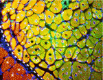
His image showing staining of a gut section of a E17.5 mouse embryo impressed the judges as well as the Node’s readers, receiving more than half of the votes.

Stéphane says: “I took this picture by chance: I was looking at the expression of Sox6 and slow Myosin Heavy Chain in the deep back muscles of a mutant mouse embryo and I saw this very nice “I” popping out in the gut tube… “

With this serendipitous image he has won a TipArt commission, and we hope we’ll get to see the final artwork he receives. In addition, Stéphane’s image will be used in a little project we’re working on to mark the Node’s upcoming first birthday in June.

Thumbs up (5 votes)
Loading...

Tags: , ,
Categories: Images

Marion Silies wins GfE thesis award

Posted by , on 31 March 2011

Every two years, the German society for developmental biology (Gesellschaft für Entwicklungsbiologie – GfE) hands out an award for the best PhD thesis of the previous two years. At their society meeting last week, this award went to Marion Silies, for her PhD thesis on glial cell migration.

I met up with Marion after her talk and asked a few questions about her PhD work in Christian Klämbt’s lab, and whether she had any tips for graduate students.

Congratulations on your award. What was your thesis about?
I worked on glial cell migration in the fly peripheral nervous system. I looked at how neurons and glial cells co-regulate their development. In a screen we found a cell cycle regulator with strong phenotypes in the migration of glial cells, but I showed that it has a post-mitotic function, so a function outside of its function in cell cycle: it controls glial cell migration from the neuron, by regulating distribution of a cell adhesion molecule.

What are you doing now? Are you still working on the nervous system?
For my PhD I studied developmental processes, but for my postdoc I moved on to understand how the nervous system functions. I’m in the lab of Tom Clandinin now, at Stanford University.

Did you have to come back to Germany just to pick up your award?
I would have loved to come just for this meeting, but I was actually in Germany anyway for another meeting, so this just fit very well.

Do you have any tips for current or new PhD students?
My tip for students about to start their PhD would be to pick something that they’re really excited about. I think this is the most important thing: A PhD takes a long time, and you put in a lot of work, so try to find something that you really like. A lot of people think that they have to be at a very prestigious university, or at a very well-known institute. I would say: go wherever you want – just find something that you really like to do, and find a nice boss.
(more…)

Thumbs up (6 votes)
Loading...

Tags: ,
Categories: Careers, Education, Interview

Seeing Further

Posted by , on 29 March 2011

The Royal Society has collected a series of images that illustrate the moment important scientific discoveries were made. This “Moments of Seeing Further” collection includes a notebook sketch from 1980, contributed by Sir John E. Sulston and depicting cell division in C. elegans – work that contributed to the discovery of the fate map of the worm.

Another, much older, image is a 17th century sketch of the process of bean sprouting, by Marcello Malpighi. He didn’t look at plants alone: His microscopy studies in many different organisms has contributed to the early study of development, and his name lives on in several microscopic structures, including the Malpighian corpuscles in the kidneys.

Have a look at the rest of the gallery as well. It even includes a photo of a letter to the Royal Society from Isaac Newton.

(Image © the Royal Society; used with permission)

Thumbs up (No Ratings Yet)
Loading...

Tags: , ,
Categories: Images, Research