“I am a developmental biologist”
Posted by Alex Eve, on 22 August 2023
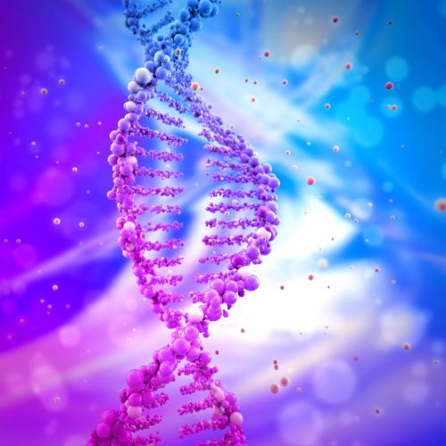
I didn’t become aware of developmental biology (DevBio) until a lecture during my first year of undergraduate studies. From that moment on, however, I was hooked and even changed my degree from forensic biology to biochemistry to have more DevBio modules. DevBio has been the basis of my career ever since. As a first-generation university student with no scientists in my family, I simply didn’t know that DevBio existed nor did I appreciate that there was a whole community of scientists experiencing the joy of working with embryos and other model systems.
I wondered whether my experience was representative of others in the field so, a couple of years ago, I took to Twitter to find out more. Obviously, there are a number of drawbacks to the methodology of this short survey; the nuances of every participant’s background cannot be appreciated and limited poll options restrict detailed (and possibly accurate) feedback. Still, since the DevBio community is (or at least, was) quite active on Twitter, I think the results could be interesting as an initial sample.
First, I asked at what stage of their career members of the DevBio community became aware of the field. At least two flaws to bear in mind here: I was unable to specify whether ‘university’ was considered by participants to be at the undergraduate or postgraduate level and there wasn’t a distinction between learning about DevBio as part of their university studies (e.g. from being a biology student) or just the stage of life (e.g. learning about it from peers or the media while being a student).
I’m curious about some things. So, #devbio Twitter community, when did you first become aware of Developmental Biology as a field?
— Alex Eve (@amjeve) July 21, 2020
Like me, the vast majority of participants (78% of 312) discovered DevBio at university, which is important because most members of the community were in a privileged position of being able to attend university in the first place, highlighting that most people probably aren’t exposed to DevBio in everyday life. It also makes me curious to think how many young students might have chosen to study biology – or go to university at all – if they had known about DevBio beforehand. If, like me, you agree that science is strengthened by diversity every effort needs to be made to provide opportunities for people from all backgrounds. The SDB Choose Development! and the University of Michigan’s Developing Future Biologists programs are excellent examples of promoting DevBio for undergraduates. However, I’d argue that the main message from this poll is that people aren’t aware of DevBio during the formative stages of thinking about their future careers. One of the flaws from the question above is somewhat addressed by the next question: who introduced you to DevBio?
Who introduced you to Developmental Biology?
— Alex Eve (@amjeve) July 21, 2020
Again, the vast majority (80.9% of 94) said that a university lecturer introduced them to the field, indicating that most participants learned of the field through their studies (presumably as an undergraduate in a related subject). Looking back, at least two important options are missing: the media, and extracurricular sources (such as workshops, summer schools, open days and similar outreach and engagement events). Indeed, there are a number of great initiatives to bring DevBio (specifically) to younger students, particularly those from underrepresented backgrounds both inside and outside the classroom. Some examples include Student Scientists, NERD SQUAD Inc., BioEYES, and droso4schools – if you know of others please do mention them in the comments below. I was slightly surprised that very few (1.1%) were introduced to DevBio by family, suggesting that DevBio is not necessarily an inherited vocation!
So, how do we expose the hidden gem of DevBio to more people? Realistically, it won’t be possible for all researchers to create their own outreach and engagement program, although if you’re interested see how some of the ones mentioned above came about by reading some articles with the organisers: Michael Barresi (Student Scientists), Cagney Coomer (NERD SQUAD Inc.), Engaging new audiences with imaging and microscopy (featuring Jamie Shuda from BioEYES) and Developing Future Biologists: developmental biology for undergraduates from underserved communities. Similarly, it might not be feasible to lobby for a change in the biology curriculum in schools around the world, although I hope that, one day, we will see developmental or stem cell biology as a standard topic in the classroom and I think this might positively impact public engagement more generally (for more on this see Exploring the challenges and opportunities of public engagement with fundamental biology). Finally, although we can try to promote DevBio through press releases, it’s not necessarily under our control which stories the media will pick up and how accurately they will report on it. But do we talk about the field at all when outside the company of other biologists? My next question: how would you describe your field when talking to someone outside of academia/industry?
When talking to someone outside of academia/industry, how would you describe your field?
— Alex Eve (@amjeve) July 21, 2020
In this case, there was a roughly 50:50 split in responses. Just under half (49.5% of 93) said that they describe their field as “developmental biology”, while 47.3% preferred a broader term, such as “biology” or “science”. I propose that one simple way to expose more people to DevBio is to tell people that it exists. When someone asks, don’t shy away; choose to say “I am a developmental biologist“, rather than “I am a scientist”, “biologist” or “researcher”. Use the opportunity to explain what DevBio is and what it means to you. Your conversation partner might tell their own children or friends about it and spread awareness of the field to a whole new audience. Word of mouth may be simple, but it’s often an effective way to inspire the next generation of DevBio researchers. Having then potentially recruited a number of new developmental biologists, how easy is it to integrate into the field?
In your experience, is the #devbio community
— Alex Eve (@amjeve) July 22, 2020
Thankfully, none of the 67 respondents considers the community to be “antisocial” and just over half (52.2%) agree that the community is “welcoming”. However, there is still work to be done to make sure that the DevBio community is inclusive because the remaining respondents felt that the community is “exclusive” (10.4%) or “cliquey” (37.3%). There isn’t a magic bullet to solve such issues, but considering and welcoming diversity and inclusion of scientists all backgrounds should be in all our minds and executed to the best of our ability.
Perhaps, then, a more appropriate response to “what do you do?” would be, “I am a developmental biologist and you can be one too“. Indeed, in the words of John Wallingford, “We Are All Developmental Biologists“.
Alex Eve trained as a developmental biologist and is now a Reviews Editor at Development
Reactions from the community
 (No Ratings Yet)
 (1 votes)