The Young Embryologist Network (YEN) Conference was held in May at the Francis Crick Institute. This year, apart from the scientific talks, the programme included a panel discussion on ‘Equality, diversity and inclusion in science’. We caught up with the two panelists, Alison Forbes (Head of EDI, the Francis Crick Institute), and Rafael Galupa (Scientist & social entrepreneur), to talk more in-depth about this important topic.
How did you find the YEN2023 meeting?
Alison: I loved it, great fun to speak to a large audience of early career scientists. I enjoy speaking to scientists as an ‘outsider’ (I studied humanities, I’m in HR, I am not a researcher). It means I can bring a fresh eye to science as an industry, and I think the audience enjoyed that.
Rafael: As a scientist, I really enjoyed myself and was very impressed and excited by all the science presented! This is a great meeting for me to attend regularly in the future and where to send my future lab members. As a scientist concerned with EDI issues, I thought it was great that the organisers included three different moments in the scientific programme to discuss such topics.
Could you tell us more about your background? What made you decide to be involved with EDI initiatives?
Alison: I grew up in Hackney, London and went to a north London grammar school, then a Russell Group university.
I am from a middle-class, academic family. But I always found snobbery, elitism and other ‘-isms’ irritating, which is why I am drawn to this work. I remember my grammar school teachers repeatedly telling us that we were ‘the best of the best’. That attitude never sat right with me.
I worked at UCL for 12 years in widening participation, working on social class and diversity in undergraduate admissions. I ran long term programmes for highly able teenagers from London state schools. They were mostly from low-income backgrounds, lots of refugee or immigrant families, usually parents had not been to university. These students were intelligent, sharp and witty, asked challenging questions, and were tough and resilient.
They were highly ambitious, as were their parents. I learned a lot from these young people, and got to see ‘my kids’ progress to UCL or other excellent universities. I also worked with UCL undergraduates, and saw how working class or minoritised students struggled with imposter syndrome. I remember young people telling me they had nowhere quiet to study, that they had had 4 teachers for Maths A level in one school year, or how their parents pressured them to apply for medicine and would not let them consider any other subjects.
It gave me a better understanding of how class, social mobility and privilege affect careers, and how oblivious we can be of own privilege (‘Well, I worked hard to get here!’).
I moved into EDI through a secondment at UCL. It is a fascinating and evolving field. There is much to be done, and you can have a real impact. I’ve been at the Crick since November 2021, and I love it.
Rafael: I grew up in the suburbs of Lisbon, surrounded by a big extended family. Because my dad went to university and became a power engineer, my household lived comfortably, but this wasn’t the case for the others in the family, or for the households of my school friends. I grew up side by side with these inequalities, and perhaps that’s why I’ve always felt a responsibility to address them.
I started volunteering when I was sixteen, in an orphanage – those kids had a completely different social structure around them (compared to mine), but not necessarily less supportive; this made me understand the importance of a support network, whichever nature it has, and how it is possible to build one if we don’t have the privilege to have one more “naturally”.
Later, in the University of Lisbon, while studying Molecular Biology and Genetics, I was part of a support group for students with disabilities. I had a weekly studying session with a younger undergraduate who was losing his vision. This was a major challenge for him, aggravated by how the classes, the studying materials, basically everything!, were very poorly accessible.
During my PhD, I came across Native Scientists, a nonprofit organisation that connects students with underserved children via interactive workshops, and to this day I have been involved in such activities. During my postdoc I participated in the project Letters to a Prescientist, which promotes letter exchanges between scientists worldwide and students at US-based schools where at least 60% of students qualify for “Free or Reduced Price Lunch”.
This experience inspired me and a friend to start a similar organisation, Cartas com Ciência, targeting students from low-income communities in Portuguese-speaking countries. Many studies show how students from low-income communities are less likely to pursue university studies and choose STEM careers, which promotes the perpetuation of the low-income cycle. The reasons for such stats are many, but include the lack of science role models around those students and often not knowing what a scientific career is. This is something that we as scientists can help eradicate! And there are many other levels at which we can act. So how not to get involved with initiatives that promote such fundamental values as equality, diversity and inclusion?
What do you think is the current biggest challenge in achieving an equal, diverse, and inclusive research culture?
Alison: There isn’t one single challenge, and there isn’t one single solution. It is complex.
Until we have urgency on these issues within government, and within science leadership, progress may well remain slow.
When something is urgent, we make it happen. For example, the sudden pivot by many employers to remote working during COVID. Home working was something disabled employees had asked for before COVID to help manage their conditions, but because allowing remote working was not the norm and was not urgent, it wasn’t seen as possible.
Many organisations are risk averse. They don’t want to be ‘the first’ to do something– they want to their peers to go first, so they can get assurance that an intervention works, and will not cause reputational damage. For example, after some Russell Group universities piloted contextual undergraduate admissions, it became common practise. Similar for interventions like reverse mentoring or various positive action schemes. So, the organisations that do ‘go first’ and take that risk should be credited.
Rafael: As Alison stated, there isn’t one biggest challenge. We all need to change our mindset, but especially people at the higher management of our research institutions. We need a more inclusive, healthier, people-centric work culture. I am sure that things will change – how fast, it remains to be seen. Planck’s principle basically says that change does not occur because individuals change their minds but rather because successive generations have different views (or in more crude terms, “one funeral at a time”). This will certainly be the case, but I also believe in the power of education, and so training in inclusive leadership, for instance, might accelerate things, as a good friend working in EDI, Roshni Mooneeram, taught me. We also need funding agencies and governments to take the lead and set the “tone” – change at those levels is the most likely to have a domino effect. Also, something that is at the root of many of the problems in research institutes (and in society in general) is the frenetic pace at which we live and work – complex problems, such as EDI issues but also most of the scientific questions we address, need us to pause more frequently, to think and reflect.
Why is it important to raise awareness on EDI in scientific meetings?
Alison: Scientists need to understand equity, diversity and inclusion for several reasons:
Equity principles should inform research design. Scientists should be considering sex as a biological variable, the ethnic diversity of patients in clinical trials, and how to design equitable collaborations with scientist colleagues in lower income countries.
Scientists should be recruited by an evidence-based, equitable, transparent process. When a PI recruits a post doc, do they use a process designed to minimise bias and to attract diverse candidates? For example, do they name blind applications, use standard interview questions that are scored, and do they have gender and if possible ethnicity mix on interview panels? There is a lot of evidence that diverse teams are more high performing, but often common recruitment practises in science penalise minoritized applicants.
Most scientists agree public communication is important. But if scientists do not have a nuanced understanding of ‘how they come across’ when speaking to diverse audiences, it can harm public health messages. In the pandemic we saw high vaccine hesitancy in communities of colour in the UK and USA, and among some patient communities with chronic conditions. This was partly because of low trust due to prior experiences of racism, or histories of medical trauma. So, scientists have to understand those contexts when they speak to different audiences.
Rafael: Scientific meetings provide a great platform to discuss EDI because on the one hand, we can reach more people at once; on the other hand, everyone paused their everyday work and is in a new environment, which is more conducive for reflexion and thinking about issues that most of us do not normally think about. Moreover, such complex issues need collective thinking and collective actions, and scientific meetings definitely help with those.
What can early-career researchers do, both individually and as a community?
Alison: As an individual, start with self-education. Read some books and listen to podcasts; I’ve recommended some below.
Then interrogate your small daily actions and decisions. Do you ask questions about diversity in committee? For example, do you ask to see gender or ethnicity pay gap data, or recruitment data, or ask about what inclusion training your institute provides? Who do you defend, advocate for, or invite to join you at conferences? And who do you interrupt or ignore? Who do you seek to build relationships with?
As a wider community, you have power in numbers. Networks like the Node, the EDIS Group or grassroots organisations like Black in Cancer or BBSTEM have a big collective impact and should be nurtured.
Rafael: As Alison stated, join ongoing initiatives – they assume many different forms and flavours, so I’m sure that everyone can find a way to contribute that is aligned with their personality and availability. It’s also important to remember that we all have a certain degree of control over what happens around us (and to us as well) and certainly over how we do things. As we often say at Cartas com Ciência, EDI is about what we do but also how we do it and with whom we do it. Alison’s questions illustrate how we can incorporate such thinking in whichever actions or initiatives we think of doing, from setting a collective journal club, to organising a meeting or a public engagement activity.
Another book I recommend often is The Class Ceiling: why it pays to be privileged by LSE academics Sam Friedman and Daniel Lauriston. They studied labour market data and found clear evidence of a pay gap between middle- and working-class employees in similar roles. They conducted anonymous interviews about experiences of class at work, with staff in a large TV company, an architecture firm and a financial firm. They don’t look specifically at science as an industry, but these themes are common across all sectors. A really insightful and entertaining read.
Rafael: The major scientific journals in life sciences have been regularly publishing special issues on EDI topics, for instance, Cell Press has collections on “Building inclusivity in science”, “Black in science”, “Women in science”, “LGBTQ+ in science” (here). Or the “Diversity, equity and inclusion in science” collection from Nature Human Behaviour (here). Hopefully all those articles are open access! Science published a short and comprehensive article on “How to begin building a culture of diversity, equity, and inclusion in your research group” (open access).
Two amazing books on how science has actually contributed to sexism and racism, I recommend Angela Saini’s books Inferior and Superior (respectively). They present and digest lots of interesting facts and studies, and Angela does a great job in always trying to compare and contrast different perspectives. Plus, it’s fun to read. On improving scientific culture in general, I recommend Uri Alon’s initiatives and available materials (here).
Want to find out what else happened at the YEN Conference? Read the detailed meeting report written by Ioakeim (Makis) Ampartzidis, Courtney Lancaster, Danielle Liptrot, and Rosie Marshall.
To learn more about YEN, follow them on twitter @YEN_community.
In the final episode of the Human Developmental Biology Initiative’s podcast, hip-hop artist Aubz meets Oxford University scientist Shankar Srinivas and they discuss questions such as what is human developmental biology and why is it important?
At the end of the episode, the pair will write and record an original piece of music inspired by their meeting, exploring science in a brand new way.
“We speak of science as one thing, and it’s not. It’s many different things.”
– Shankar Srinivas
About the participants
Shankar is Professor of Developmental Biology at the University of Oxford, at the Department of Physiology Anatomy and Genetics, in the Institute for Developmental and Regenerative Medicine. His group uses mouse and human embryos to study how the body is shaped, and how the heart forms and starts to beat. Shankar is also passionate about science outreach and public engagement. His group participates regularly in science festivals, and collaborates with dancers, choreographers and Virtual Reality specialists to generate movement based art, to explore different perspectives on how the form of the body is determined.
Aubz is a Manchester-based hiphop artist.
Please subscribe and listen to Made the Same Way on Apple podcasts, Spotify, or wherever you get your podcasts. If you enjoy the podcast, please rate and review us on Apple podcasts to help others find us!
On 31 May 2023, Development hosted a webinar on the topic of in vitro and stem cell-based models of development. Below are the talks and Q&As hosted by our Executive Editor, Katherine Brown.
Anchel de Jaime Soguero (COS, University of Heidelberg)
Talk and Q&A by Anchel de Jaime Soguero
Elena Camacho Aguilar (Rice University)
Talk and Q&A by Elena Camacho Aguilar
Tyler Huycke (UCSF)
Talk and Q&A by Tyler Huycke (No Ratings Yet) Loading...
One of the crowd-favourite giveaways here at the Node is our collection of postcards. With our supplies dwindling, we are planning to reprint some of the postcards, and take this opportunity to add some more #devbio favourites to our collection.
We ran a public vote for the Node postcard competition to select the top four images to be printed on new postcards. The image with the most votes will also be featured on the cover of a ‘Development’ issue in 2023.
We are now delighted to announce the results:
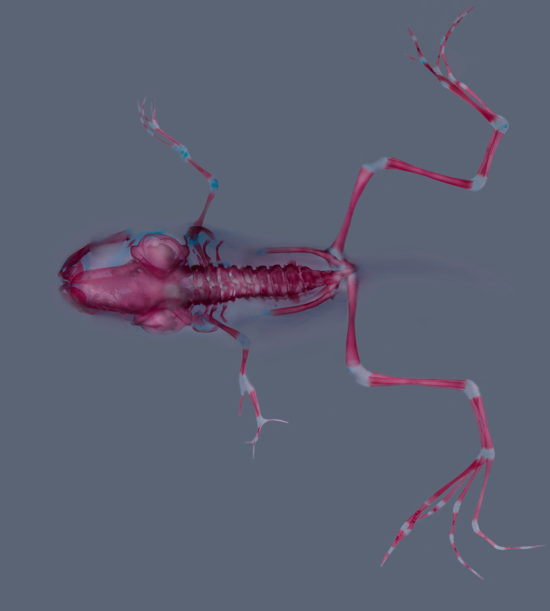
Winner: Xenopus laevis skeleton
Skeletal staining (alizarin red and alcian blue) of a Xenopus laevis at stage 62. Stage 51 larva was treated with a Cyp26a inhibitor during forelimb regeneration. Notice proximo-distal duplication in the left forelimb. Credit: JC Marin-Llera and MV Chimal-Montes de Oca
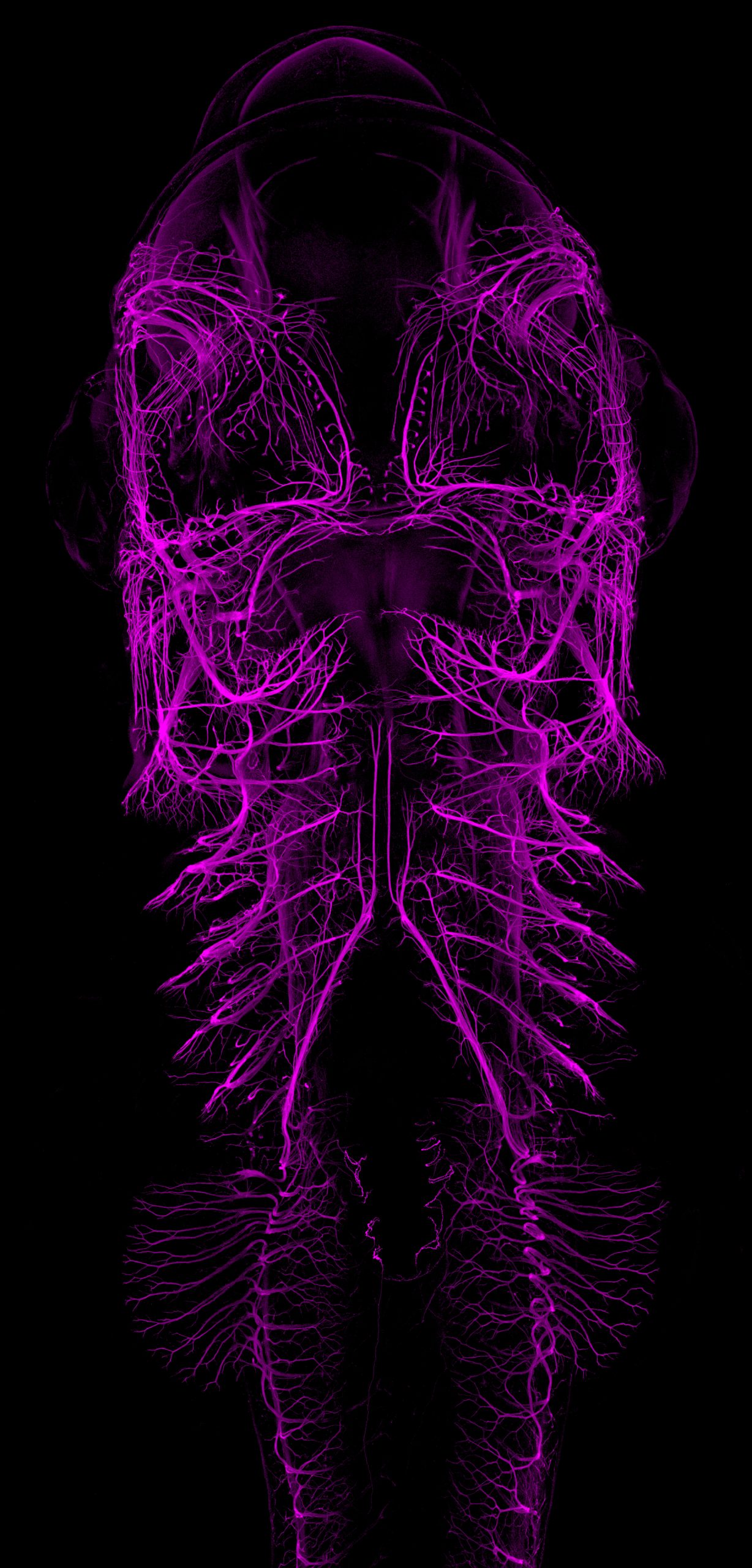
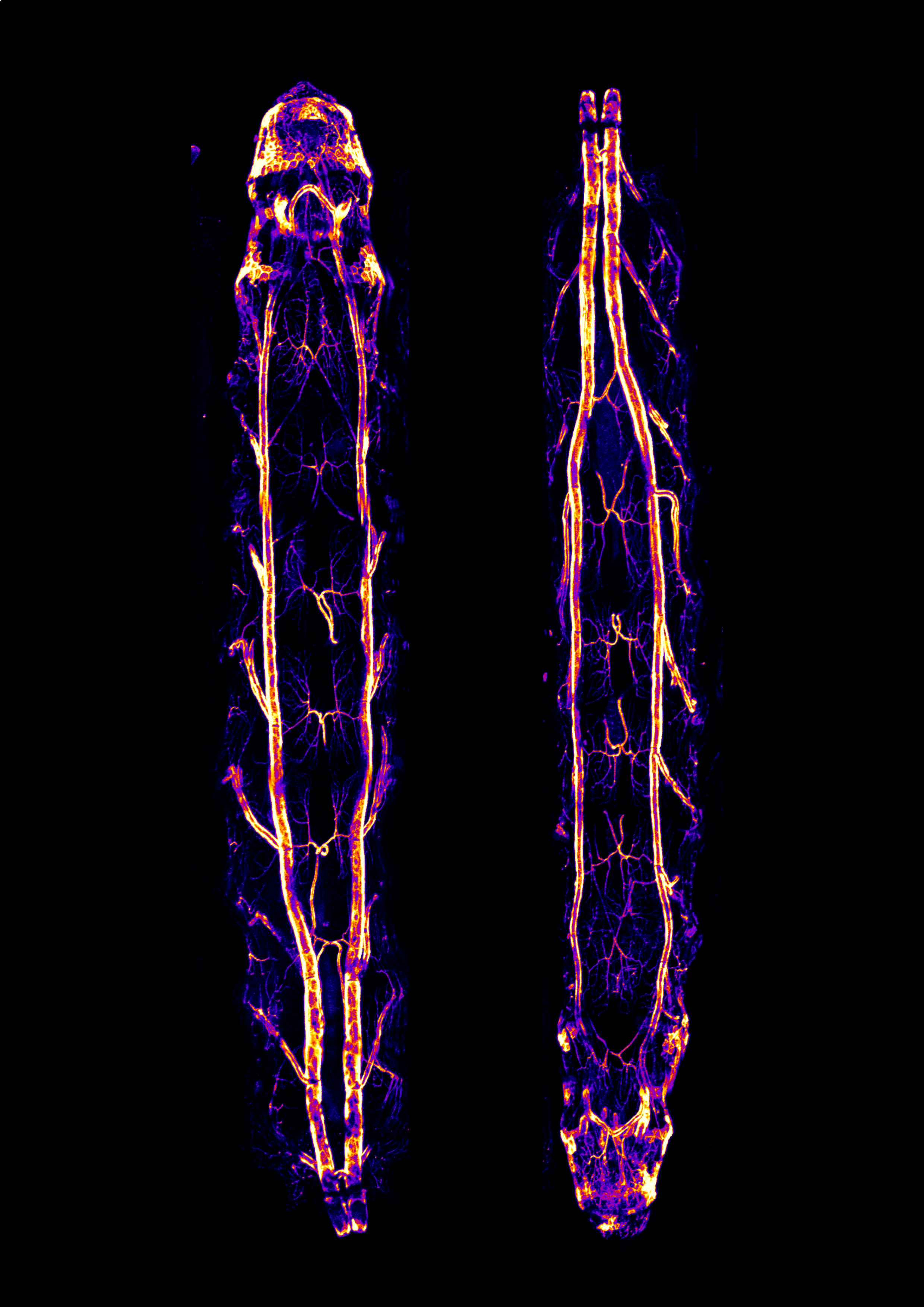
The winning image and the following images will be printed on new postcards:
Catshark embryo Ventral view maximum intensity projection from an immunofluorescence staining labeling the developing nervous system (primarily nerves and ganglia) of a stage 30 small-spotted catshark embryo (Scyliorhinus canicula). The image was acquired using a ZEISS LSM980 with Airyscan2 confocal microscope, stitched and processed using ZEN software from the same microscope. Credit: E. Escamilla-VegaDrosophila larvae This shows heat-fixed Drosophila larvae expressing an infrared fluorescent protein (IFP) in the tracheal system using the Gal4/UAS system. Images were acquired in a confocal microscope (Nikon A1R+) with a 10x objective and using the mosaic modality. Stitching was done using the microscope’s software (Nikon NIS-Elements). The images were Z-projected and pseudocolored in Fiji and further processed using Inkscape. Credit: D. RiosArabidopsis leaf Cells on the epidermis of a 3 day old Arabidopsis leaf. This is an adaptation of a linocut print created based on a microscopy image. Credit: M. Smit
Congratulations to the winners of the competition.
Thank you to everyone who submitted their images to the competition, and to everyone who participated in the voting.
This article summarises a series of recent studies around the mental health crisis in science and the toxic research culture behind it. What has been done to address these issues? What can universities, funders, and other academic institutions do to drive larger, systemic changes in research culture? How can we make better use of platforms like the Node to normalise talking about mental health?
The disturbing data and stories in this report make all the more important that @UKRI_News seizes the opportunity of the FRAP (Future of Research Assessment Programme) to create incentives for positive culture change. https://t.co/EUGqLx1N9s
This editorial written by Science Editor-in-Chief H. Holden Thorp has resonated with many people on Twitter. There is still lots to do in order to build a scientific workforce that reflects the public it serves.
Take a look at what people think.
"Scientists should embrace their humanity rather than pretending they are a bunch of automatons who instantly reach perfectly objective conclusions." Fact is, it matters who does the science – as it determines how and what science gets done. https://t.co/CMptrB7tHL
— Dean Lola Eniola-Adefeso (@Lola_UIC) May 12, 2023
Yes, it matters who does science. And, I am adding, it also matters HOW we do science.
A massive work culture change is required. The cut throat competition needs to stop. The focus on individuals when they could not have achieved alone, needs to stop. https://t.co/5PV2iaBWS6
"the public has been taught scientific insight occurs when old white guys run out of bathtubs shouting 'Eureka!'…That’s not how it works…scientists work in teams [who] share findings with other scientists who make more refinements" https://t.co/eSy0XUm3XZ
I think @hholdenthorp recent piece "It matters who does science." hits the nail on the head, "Scientists should embrace their humanity…" & part of that is working to meet basic needs of well being for the people that do the science. https://t.co/yA2QuZnZ9L
— Siegenthaler_Lab (@SiegenthalerLab) May 17, 2023
A selection of preLights posts relevant to the Node readers. Head over to preLights to see what other recent preprints have been highlighted.
Laboratory evolution of flies to morphogen dosage via rapid maternal changes reveals predictable outcomesby Xueying C. Li et al. To ‘big embryo’ (BE), or not to BE, that’s the question. Selected by Girish Kale. Read the preLight here.
Coordinated growth of linked epithelia is mediated by the Hippo pathwayby Sophia Friesen, Iswar K. Hariharan Roses are red, violets are blue, when the disk proper grows, hippo stretches peripodial epithelium too. Selected by Girish Kale. Read the preLight here.
Conserved Chamber-Specific Polyploidy Maintains Heart Function in Drosophilaby Archan Chakraborty et al. The importance of being in the right place, with the right ploidy, at the right time- and how not to mend broken hearts. Selected by Anastasia Moraiti. Read the preLight here.
Insm1 regulates the development of mTECs and immune toleranceby Wehuai Tao et al. A new player in establishing immune tolerance: How Insm1 regulates mTEC gene expression. Selected by Marina Schernthanner and Jessica Chevallier. Read the preLight here.
Nutrient-regulated dynamics of chondroprogenitors in the postnatal murine growth plateby Takeshi Oichi et al. Fasted bones grow fast later: chondroprogenitors in the growth plate of murine long bones adapt to dietary restriction, leading to catch-up growth during refeeding. Selected by Alberto Rosello-Diez and Boya (Hannah) Zhang and Chee Ho H’ng. Read the preLight here.
Small leucine-rich proteoglycans inhibit CNS regeneration by modifying the structural and mechanical properties of the lesion environmentby Julia Kolb et al. Who is the culprit? Small leucine-rich proteoglycans inhibit axonal regrowth in the lesioned zebrafish spinal cord by changing the structure and mechanics of the extracellular matrix. Selected by Laura Celotto. Read the preLight here.
The phosphodiesterase 2A regulates lymphatic endothelial development via cGMP-mediated control of Notch signalingby Claudia Carlantoni et al. An essential function for the phosphodiesterase 2A during regulation of lymphatic vessel maturation. Selected by Andreas van Impel and Sanjay Sunil Kumar. Read the preLight here.
A transcriptional and regulatory map of mouse somitogenesisby Ximena Ibarra-Soria et al. Somitogenesis: common and divergent maturation programmes along the anteroposterior axis. Selected by Sergio Menchero. Read the preLight here.
Bovine blastocyst like structures derived from stem cell culturesby Carlos A. Pinzón-Arteaga et al. A new livestock embryo model from a self-renewing source. Selected by Carly Guiltinan. Read the preLight here.
Mechanical forces across compartments coordinate cell shape and fate transitions to generate tissue architectureby Clémentine Villeneuve et al. It takes two to tango: coordinated mechanical contributions from epithelium and dermal fibroblasts help break symmetry for downgrowth and fate patterns in mouse hair follicles. Selected by Sudeepa Nandi. Read the preLight here.
A patterned human heart tube organoid model generated by pluripotent stem cell self-assemblyby Brett Volmert et al. A patterned human heart tube organoid model generated by pluripotent stem cell self-assembly. Selected by Silvia Becca. Read the preLight here.
Gene complementation analysis suggests that dodder plants (Cuscuta spp.) do not depend on the host FT protein for floweringby Sina Mäckelmann et al. Host-independent flowering of Cuscuta spp. reignites the search for a ‘Florigen’. Selected by Gwendolyn K. Kirschner and Marc Somssich. Read the preLight here.
Plasmodesmal connectivity in C4 Gynandropsis gynandra is induced by light and dependent on photosynthesisby Tina B. Schreier et al. Light and photosynthesis trigger plasmodesmal formation in C4 dicotyledons. Selected by Yueh Cho. Read the preLight here.
Tristan Frum, Peggy P. Hsu, Renee F.C. Hein, Ansley S. Conchola, Charles J. Zhang, Olivia R. Utter, Abhinav Anand, Yi Zhang, Sydney G. Clark, Ian Glass, Jonathan Z. Sexton, Jason R. Spence
Jeremie Oliver Pina, Daniela M Roth, Resmi Raju, Emma Wentworth Winchester, Parna Chattaraj, Fahad K Kidwai, Fabio R Faucz, James Iben, Cameron Padilla, Justin L Cotney, Rena N D’Souza
Rebecca M. Green, Lucas D. Lo Vercio, Andreas Dauter, Elizabeth C. Barretto, Jay Devine, Marta Vidal-García, Marta Marchini, Samuel Robertson, Xiang Zhao, Anandita Mahika, M. Bilal Shakir, Sienna Guo, Julia C. Boughner, Wendy Dean, Arthur D. Lander, Ralph S. Marcucio, Nils D. Forkert, Benedikt Hallgrímsson
Chiemela Ohanele, Jessica N. Peoples, Anja Karlstaedt, Joshua T. Geiger, Ashley D. Gayle, Nasab Ghazal, Fateemaa Sohani, Milton E. Brown, Michael E. Davis, George A. Porter Jr., Victor Faundez, Jennifer Q. Kwong
Derek C. Liberti, Hongbo Wen, Kwaku K. Quansah, Prashant Chandrasekaran, Josh Pankin, Nigel S. Michki, Annabelle Jin, MinQi Lu, Maureen Peers De Nieuwburgh, Lisa R. Young, Rajan Jain, David B. Frank
Derek C. Liberti, Hongbo Wen, Kwaku K. Quansah, Prashant Chandrasekaran, Josh Pankin, Nigel S. Michki, Annabelle Jin, MinQi Lu, Maureen Peers De Nieuwburgh, Lisa R. Young, Rajan Jain, David B. Frank
Rachel Queen, Moira Crosier, Lorraine Eley, Janet Kerwin, Jasmin E. Turner, Jianshi Yu, Tamil Dhanaseelan, Lynne Overman, Hannah Soetjoadi, Richard Baldock, Jonathon Coxhead, Veronika Boczonadi, Alex Laude, Simon J. Cockell, Maureen A. Kane, Steven Lisgo, Deborah J. Henderson
Jennifer N. Chousal, Srimeenakshi Srinivasan, Katherine Lee, Cuong To, Kyucheol Cho, Wei Zhang, Ana Lisa Yeo, V. Gabriel Garzo, Mana M. Parast, Louise C. Laurent, Heidi Cook-Andersen
Christopher J. Johnson, Akhil Kulkarni, William J. Buxton, Tsz Y. Hui, Anusha Kayastha, Alwin A. Khoja, Joviane Leandre, Vanshika V. Mehta, Logan Ostrowski, Erica G. Pareizs, Rebecca L. Scotto, Vanesa Vargas, Raveena M. Vellingiri, Giulia Verzino, Rhea Vohra, Saurabh C. Wakade, Veronica M. Winkeljohn, Victoria M. Winkeljohn, Travis M. Rotterman, Alberto Stolfi
Carmen L. Diaz Soria, Teresa Attenborough, Zhigang Lu, Jennie Graham, Christopher Hall, Sam Thompson, Toby G. R. Andrews, Kate A. Rawlinson, Matthew Berriman, Gabriel Rinaldi
Abdulvasey Mohammed, Priscila Ferreira Slepicka, Benjamin Solomon, Kelsea M Hubka, Hanh Dan Nguyen, Michael G Chavez, Christine Y Yeh, Virginia D Winn, Casey A Gifford, Purvesh Khatri, Andrew Gentles, Katja Gabrielle Weinacht
Eric Bartell, Kuang Lin, Kristin Tsuo, Wei Gan, Sailaja Vedantam, Joanne B Cole, John M Baronas, Loic Yengo, Eirini Marouli, Tiffany Amariuta, GIANT Consortium, Nora E Renthal, Christina M Jacobsen, Rany Salem, Robin G Walters, Joel N Hirschhorn
Allegra Angeloni, Skye Fissette, Deniz Kaya, Jillian M. Hammond, Hasindu Gamaarachchi, Ira W. Deveson, Robert J. Klose, Weiming Li, Xiaotian Zhang, Ozren Bogdanovic
Darren Blackburn, Korin Sahinyan, Aldo Hernnandez Corchado, Felicia Lazure, Vincent Richard, Laura Raco, Rene Zahedi, Christoph Borchers, Christoph Lepper, Hiroshi Kawabe, Arezu Jahani-asl, Hamed S Najafabadi, Vahab D Soleimani
Duygu Payzin-Dogru, Sarah E. Wilson, Steven J. Blair, Aaron M. Savage, Emil Kriukov, Victor Cat, Louis V. Cammarata, Burcu Erdogan, Shifa Hossain, Noah Lopez, Julia Losner, Juan C. Velazquez Matos, Sangwon Min, Kelly Dooling, Bobby Groves, Alan Y. Wong, Petr Baranov, Hani Singer, Isaac M. Chiu, Brian J. Haas, Jessica L. Whited
Regenerating axolotl limbs from Payzin-Dogru et al.
Natalia A. Veniaminova, Yunlong Jia, Adrien M. Hartigan, Thomas J. Huyge, Shih-Ying Tsai, Marina Grachtchouk, Seitaro Nakagawa, Andrzej A. Dlugosz, Scott X. Atwood, Sunny Y. Wong
Mary Bergwell, Amy Smith, Ellie Smith, Carter Dierlam, Ramon Duran, Erin Haastrup, Rebekah Napier-Jameson, Rory Seidel, William Potter, Adam Norris, Jyoti Iyer
Bess P. Rosen, Qing V. Li, Hyunwoo Cho, Dingyu Liu, Dapeng Yang, Sarah Graff, Jielin Yan, Renhe Luo, Nipun Verma, Jeyaram R. Damodaran, Michael A. Beer, Simone Sidoli, Danwei Huangfu
Neha Zutshi, Bhopal C Mohapatra, Pinaki Mondal, Wei An, Benjamin T Goetz, Shou Wang, Scong Li, Matthew D Storck, David Mercer, Adrian R Black, Sarah P Thayer, Jennifer Black, Chi Lin, Vimla Band, Hamid Band
Neha Zutshi, Bhopal C. Mohapatra, Pinaki Mondal, Wei An, Benjamin T. Goetz, Shuo Wang, Sicong Li, Matthew D. Storck, David F. Mercer, Adrian R. Black, Sarah P. Thayer, Jennifer D. Black, Chi Lin, Vimla Band, Hamid Band
Biao Huang, Zipeng Zeng, Hui Li, Zexu Li, Xi Chen, Jinjin Guo, Chennan C. Zhang, Megan E. Schreiber, Ariel C. Vonk, Tianyuan Xiang, Tadrushi Patel, Yidan Li, Riana K. Parvez, Balint Der, Jyun Hao Chen, Zhenqing Liu, Matthew E. Thornton, Brendan H. Grubbs, Yarui Diao, Yali Dou, Ksenia Gnedeva, Nils O. Lindström, Qilong Ying, Nuria M. Pastor-Soler, Teng Fei, Kenneth R. Hallows, Andrew P. McMahon, Zhongwei Li
Katherine S Stewart, Kevin AU Gonzales, Shaopeng Yuan, Matthew T Tierney, Alain R Bonny, Yihao Yang, Nicole R Infarinato, Christopher J Cowley, John M Levorse, Hilda Amalia Pasolli, Sourav Ghosh, Carla V Rothlin, Elaine Fuchs
Leticia F. Ferigolo, Mateus H. Vicente, Joao P. O. Correa, Carlos H. Barrera-Rojas, Eder M. Silva, Geraldo F.F. Silva, Airton Carvalho Jr, Lazaro E.P. Peres, Guilherme B. Ambrosano, Gabriel R. A. Margarido, Robert Sablowski, Fabio T.S. Nogueira
S Manrique, A Cavalleri, A Guazzotti, GH Villarino, S Simonini, A Bombarely, T Higashiyama, U Grossniklaus, C Mizzotti, AM Pereira, S Coimbra, S Sankaranarayanan, E Onelli, S Masiero, RG Franks, L Colombo
Doosan Shin, Veronica C Perez, Gabriella K Dickinson, Haohao Zhao, Ru Dai, Breanna M Tomiczek, Keun Ho Cho, Ning Zhu, Jin Koh, Alexander Grenning, Jeongim Kim
María Ángeles Rodríguez de Cara, Paul Jay, Quentin Rougemont, Mathieu Chouteau, Annabel Whibley, Barbara Huber, Florence Piron-Prunier, Renato Rogner Ramos, André V. L. Freitas, Camilo Salazar, Karina Lucas Silva-Brandão, Tatiana Teixeira Torres, Mathieu Joron
Samuel E. Ross, Javier Vázquez-Marín, Krista R.B. Gert, Álvaro González-Rajal, Marcel E. Dinger, Andrea Pauli, Juan Ramon Martínez-Morales, Ozren Bogdanovic
Mary Bergwell, Amy Smith, Ellie Smith, Carter Dierlam, Ramon Duran, Erin Haastrup, Rebekah Napier-Jameson, Rory Seidel, William Potter, Adam Norris, Jyoti Iyer
Neha Zutshi, Bhopal C. Mohapatra, Pinaki Mondal, Wei An, Benjamin T. Goetz, Shuo Wang, Sicong Li, Matthew D. Storck, David F. Mercer, Adrian R. Black, Sarah P. Thayer, Jennifer D. Black, Chi Lin, Vimla Band, Hamid Band
Benjamin Liffner, Ana Karla Cepeda Diaz, James Blauwkamp, David Anaguano, Sonja Frölich, Vasant Muralidharan, Danny W. Wilson, Jeffrey Dvorin, Sabrina Absalon
The seventh episode of Made the Same Way, a podcast produced by the Wellcome-funded Human Developmental Biology Initiative, features sociologist and writer Marieke Bigg discussing the ethics of research with early human embryos with Mancunian poet and rapper Meduulla. The pair discuss the legacy of Anne McLaren and muse on future implications of this area of research.
At the end of the episode, the pair collaborate on an original piece of music inspired by their conversation.
“What things can we consider to be right and wrong, and who makes that decision?”
– Meduulla
About the participants
Marieke Bigg writes about bodies and culture. She holds a PhD in Sociology from the University of Cambridge, where she studied the technological transformation of human reproduction, with a focus on Dr Anne McLaren’s role in the human embryo research debates. She now writes both non-fiction and fiction about the cultural dimensions of biology and bodies. In addition to her books, Marieke writes freelance, hosts podcasts and panels, and collaborates with scientists and biologists to discuss and produce art that conjures new social worlds.
Hailing from North Manchester, Meduulla is a 23 year old Zimbabwean-born Rapper, Poet and DJ paving her way through the UK rap scene. Meduulla marries her modern flows and witty lyrics with jazz inspired hip hop instrumentals to create music that reflects the present day whilst carrying a nostalgic air.
Despite having been a writer for 10 years, she only released her first single in 2021 which then led to her appearance on BBC’s The Rap Game UK as a finalist. Her independently released single, Mish Muulla was selected as Track of the Week on BBC 1Xtra Radio,resulting in Meduulla performing at Reading and Leeds Festival in 2022. The wordsmith is a 2023 Sound and Music Seed Award recipient and her poetry won first prize in TogetherintheUK’s migrant writers competition. Her passion for using her lyricism as a force of positive change continues to be recognised by various cultural organisations.
In 2023, Meduulla will release her debut project entitled Oblongata.
Please subscribe and listen to Made the Same Way on Apple podcasts, Spotify, or wherever you get your podcasts. If you enjoy the podcast, please rate and review us on Apple podcasts to help others find us!
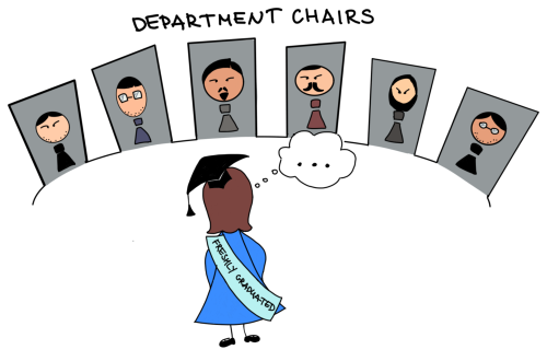
Gender problems in STEM are familiar to women researchers in every corner of the world. Japan is no exception. In a culture that seeks harmony and balance with people around, where conflict is avoided at all costs, it is often difficult to express someone’s needs. Social pressure is working in very subtle ways. You have an event in a room with photos of the previous heads of the department, twenty or thirty of them, which are all men; you go to a conference and see the overwhelming majority of the keynote speakers being male researchers; you notice the appointed leaders of the diversity and inclusion groups are mostly male when the volunteer groups have hardly any. The hours researchers stay in the laboratory make you feel like you are reliving the same day again and again; the boisterous communication style and jokes and comments are too harsh for you. The passion you once had for research is slowly withering away, leaving you with the feeling of unhappiness and unworthiness, and you give up, thinking that you are not made for this work, you are not a good fit, with a question in your mind: “How did I end up here?”.
I was lucky to grow up in a bubble where science was never gendered, there were no sciences that were inherently for women or men, and there were no subjects that some genders were naturally bad at. My parents were working in science-related areas, and they would divide school subjects between them. My mother would help me with mathematics, chemistry, and biology. My father with English, physics, and music. So, it never occurred to me that there was a need for societies supporting women researchers until I moved to Japan. My intrinsic belief that science is for everyone was challenged by a different culture, where many people of both genders believed otherwise for one reason or another. The confusion and frustration led me to research the problem (what else a scientist can do!?). And what I discovered – amazed me.
The universities in Europe and North America have women in STEM organisations active on social media, organising events, inviting young women scientists, actively connecting, and searching for opportunities to widen the network. These societies are vocal and visible, making the world know they exist. They constantly push for change. There are university-based workshops, training programs, and symposiums specifically for women in research. There is this feeling of women researchers trying to unite and support each other, pave the way for future generations, and improve the working environment.
I wished something like this existed in Japan. Most of the groups I found were quite exclusive: only for students, or mainly in Japanese, with no easily accessible information on how to join and what kind of events they are planning. There was little presence on social media, and you would need to make a targeted search to find them. I rarely hear about women researchers-oriented events or workshops, and yet to hear about women researcher-oriented training university programs.
So, I searched for women researchers’ communities outside the university and found ‘’Women in Science Japan’’, the young community founded by Elizabeth Oda, Dr. Sarah K. Abe and Lauren Hartz in 2019. I looked through their website and joined on the spot. I had a chance to talk with one of the co-founders, Elizabeth, and asked her about the motivation behind creating “Women in Science Japan”:
I and my co-founders created this community to address what we have witnessed as gender inequality in Japan, both the statistics you can often hear in the media and micro- and macroaggressions women experience. We also wanted to start with students and give a voice to students in high schools and universities. Because we knew both from the literature and from our own experience that in Japan, there are many negative stereotypes around women pursuing STEM and girls are discouraged from pursuing these fields from the young age.
Elizabeth says that one of the reasons most people join “Women in Science Japan”, and the one she thinks is very important for the future of the community and hopes to improve – is a mentorship program. That was one of my reasons for joining too. Even without talking about being an international researcher in a country that doesn’t speak your native language and has completely different social structures, finding your way in a field that wasn’t designed for you is difficult. I often feel that mentoring comes naturally to male researchers, whereas women researchers need it even more but receive it very rarely and are expected to figure out many things on their own. The community evolved, and apart from students, it started to focus more on early-career women. Elizabeth notes that it was important to create a space where members can be vulnerable, authentic, and empowered, without the fear of retribution, discipline or ostracisation, to feel heard and to have someone else say, “You are experiencing that too? I thought it was just me.”
The reality is that gender inequality is a systemic problem that an individual person can’t solve, so the idea was to create a culture that is more aware of the issues, willing to discuss the issues, face the issues, and hopefully raise these issues outside of the Women in Science Japan community.
Women in Science Japan now unites scientists, educators, women working in start-ups and the corporate world, and students at high schools and universities. Geology, biology, engineering, IT – all fields of science are welcome. And it is beautiful. It is invigorating to have such diversity, to be with people from all walks of life, with different backgrounds and different life stories. It is empowering and inspiring to hear what fellow members have overcome and where they are heading. It is not easy to be vulnerable and share your story, but if the person decides to do it – it is a treasure, a path for growth for both the person sharing and the person listening.
Women in Science Japan offers various activities: career-related events, mentorship mentioned earlier, casual events, and the book club (!). Elizabeth says that the book club is one of the things she is proud of. The book is chosen by members interested in joining the club and is related to gender inequality, science, and Japan. Currently, the book club is reading “How to Be an Inclusive Leader: Your Role in Creating Cultures of Belonging Where Everyone Can Thrive” by Jennifer Brown, which touches on diversity, equity, and inclusion. Being a part of the book club is a fantastic experience for me. The same text often generates different responses in different people, and this experience is the diversity in action. It is eye-opening to hear what people think and how the same words are heard differently because of the different backgrounds. Reading the book together, rather than alone, creates deep conversations, challenges to see the text from different angles and helps to navigate difficult questions.
I think we can only overcome our hardships and glass ceilings by holding each other’s hands and supporting and helping each other. This is one of the things Women in Science Japan is trying to achieve, a support system to help members to navigate complicated work situations or decisions, get feedback, provide clarity about career paths, and create a network that helps to build their businesses or solve work-related problems, or for international members to settle in Japan. And I wanted to use my chances to speak to the world and encourage women researchers to unite, to join communities like “Women in Science Japan”, to create new communities of like-minded people, say for women scientists in developmental biology or tissue engineering, or working on a specific problem. To be visible, vocal, advocate for your needs, become more confident, and create a welcoming future for the new generations of women in science. Or make a safe space for sharing your thoughts, finding your way, and knowing that you are not alone.
You are very welcome to join “Women in Science Japan” if you are currently working in science-related areas in Japan.
But if you are in countries other than Japan, here are some links that can get you started on your journey of finding a safe space. (Thank you to my fellow correspondents, The Node community manager, and my friends for helping me with this list.)
“Cancer rates vary wildly across the world, and we don’t know why. To solve this mystery, scientists are tracking down causes of cancer by the fingerprints they leave in the genome”
Dr Kat Arney
In the latest episode of the Genetics Unzipped podcast, we’re chasing down the perpetrator of a scientific Whodunnit with the DNA detectives – the Mutographs of Cancer team, who are on the hunt for the causes of cancer
In sixth episode of HDBI’s podcast, Made the Same Way, scientist Katie Long explores the topic of human brain development with spoken word artist Harmony.
At the end of the episode, Harmony creates an original spoken word piece based on their conversation.
If we look at every single person’s brain, most of these wrinkles will be in the same place.”
-Katie Long
About the participants
Katie’s labhas been at King’s College London since 2019, and their research focuses on how the human neocortex develops with the correct size, shape and organisation. To address this they use an interdisciplinary approach using human fetal cortex tissue models to look at the cellular and mechanical mechanisms that drive the development of the human neocortex, including the formation of the folds present on the surface of the neocortex, and how dysregulation of these functions can lead to neurodevelopmental disorders. They also use our human fetal tissue culture models to investigate the effect of injury on the developing human brain.
In her spare time, Katie likes to get outdoors and she is a keen cyclist and runner.
Harmony is a spoken word artist who has been interested in the arts since she watched her first movie.
Please subscribe and listen to Made the Same Way on Apple podcasts, Spotify, or wherever you get your podcasts. If you enjoy the podcast, please rate and review us on Apple podcasts to help others find us!
 (4 votes)
 (No Ratings Yet)