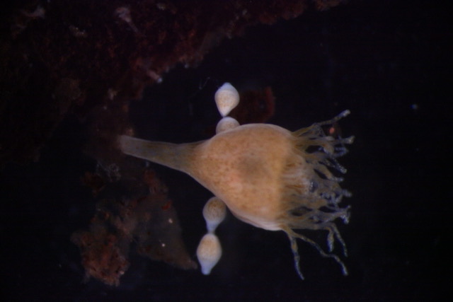
April in preprints
Posted by the Node, on 3 May 2023
Welcome to our monthly trawl for developmental and stem cell biology (and related) preprints.
The preprints this month are hosted on bioRxiv – use these links below to get to the section you want:
- Patterning & signalling
- Morphogenesis & mechanics
- Genes & genomes
- Stem cells, regeneration & disease modelling
- Plant development
- Evo-devo
Developmental biology
| Patterning & signalling
Stromal netrin-1 coordinates renal arteriogenesis and mural cell differentiation
Peter M. Luo, Xiaowu Gu, Christopher Chaney, Thomas Carroll, Ondine Cleaver
Oleic acid decouples fecundity and longevity via DAF-12 steroid hormone signaling in C. elegans
Alexandra M. Nichitean, Frances V. Compere, Sarah E. Hall
EOGT Enables Residual Notch Signaling in Mouse Intestinal Cells Lacking POFUT1
Mohd Nauman, Shweta Varshney, Jiahn Choi, Leonard H. Augenlicht, Pamela Stanley
Sanjay Karki, Mehdi Saadaoui, Valentin Dunsing, Elise Da Silva, Jean-Marc Philippe, Cédric Maurange, Thomas Lecuit
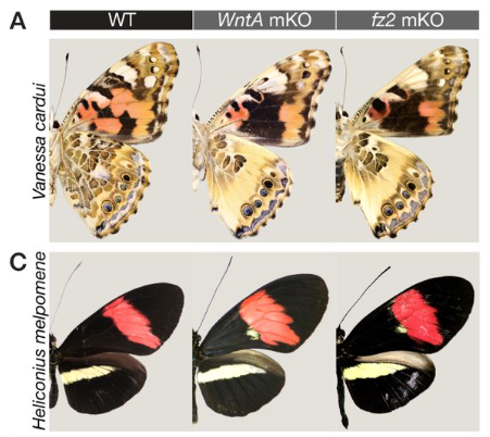
Frizzled2 receives the WntA morphogen during butterfly wing pattern formation
Joseph J Hanly, Ling S Loh, Anyi Mazo-Vargas, Teomie S Rivera-Miranda, Luca Livraghi, Amruta Tendolkar, Christopher R Day, Neringa Liutikaite, Emily A Earls, Olaf BWH Corning, Natalie D’Souza, José J Hermina-Perez, Caroline Mehta, Julia Ainsworth, Matteo Rossi, W. Owen McMillan, Michael W Perry, Arnaud Martin
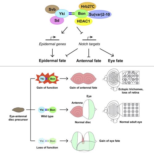
Polycomb safeguards imaginal disc specification through control of the Vestigial-Scalloped complex
Haley E. Brown, Brandon P. Weasner, Bonnie M. Weasner, Justin P. Kumar
Maya N. Evanitsky, Stefano Di Talia
Atoosa Amel, Simoné Rossouw, Mubeen Goolam
Juvenile hormones direct primordial germ cell migration to the embryonic gonad
Barton Lacy J, Sanny Justina, Dawson Emily P, Nouzova Marcela, Noriega Fernando Gabriel, Stadtfeld Matthias, Lehmann Ruth
SPATIO-TEMPORAL DYNAMICS OF EARLY SOMITE SEGMENTATION IN THE CHICKEN EMBRYO
Ana Cristina Maia-Fernandes, Ana Martins-Jesus, Tomás Pais-de-Azevedo, Ramiro Magno, Isabel Duarte, Raquel P. Andrade
Identification of overlapping and distinct mural cell populations during early embryonic development
Sarah Colijn, Miku Nambara, Amber N. Stratman
Tirtha Das Banerjee, Antónia Monteiro
Qiang Lan, Ewelina Trela, Riitta Lindström, Jyoti Satta, Mona M. Christensen, Martin Holzenberger, Jukka Jernvall, Marja L. Mikkola
Alexandra E. Rader, Battuya Bayarmagnai, Maxim V. Frolov
The Role of MAP3K1 in the Development of the Female Reproductive Tract
Eiki Kimura, Maureen Mongan, Bo Xiao, Jingjing Wang, Vinicius S Carreira, Brad Bolon, Xiang Zhang, Katherine A. Burns, Jacek Biesiada, Mario Medvedovic, Alvaro Puga, Ying Xia
Adam Presser, Olivia Freund, Theodora Hassapelis, Ginger L Hunter
Netrin-1 directs vascular patterning and maturity in the developing kidney
Samuel Emery Honeycutt, Pierre-Emmanuel Yoann N’Guetta, Deanna Marie Hardesty, Yubin Xiong, Shamus Luke Cooper, Lori Lynn O’Brien

| Morphogenesis & mechanics
Medioapical contractile pulses coordinated between cells regulate Drosophila eye morphogenesis
Christian Rosa Birriel, Jacob Malin, Victor Hatini
Jacob Malin, Christian Rosa Birriel, Victor Hatini
Tirtha Das Banerjee, Suriya Narayanan Murugesan, Antόnia Monteiro
DRMY1 promotes robust morphogenesis by sustaining translation of a hormone signaling protein
Shuyao Kong, Mingyuan Zhu, M. Regina Scarpin, David Pan, Longfei Jia, Ryan E. Martinez, Simon Alamos, Batthula Vijaya Lakshmi Vadde, Hernan G. Garcia, Shu-Bing Qian, Jacob O. Brunkard, Adrienne H. K. Roeder
Astroglial Hmgb1 regulates postnatal astrocyte morphogenesis and cerebrovascular maturation.
Moises Freitas-Andrade, Cesar H Comin, Peter C Van Dyken, Julie Ouellette, Joanna Raman-Nair, Nicole Blakeley, Quing Yan Liu, Sonia Leclerc, Youlian Pan, Ziying Liu, Micael Carrier, Karan Thakur, Alexandre Savard, Gareth M Rurak, Marie-Eve Tremblay, Natalina Salmaso, Luciano Da F Costa, Gianfilippo Coppola, Baptiste Lacoste
Andrew J Aman, Lauren M Saunders, August A Carr, Sanjay R Srivatsan, Colten Eberhard, Blake Carrington, Dawn E Watkins-Chow, William Pavan, Cole Trapnell, David M. Parichy
Pathways that affect anterior morphogenesis in C. elegans embryos
Balasubramaniam Boopathi, Irini Topalidou, Melissa Kelley, Sarina M. Meadows, Owen Funk, Michael Ailion, David S. Fay
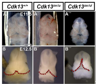
M Hampl, N Jandova, D Luskova, M Novakova, J Prochazka, J Kohoutek, M Buchtova
Xenopus Ssbp2 is required for embryonic pronephros morphogenesis and terminal differentiation
Ailen S. Cervino, Mariano G. Collodel, Ivan A. Lopez, Daniel Hochbaum, Neil A. Hukriede, M. Cecilia Cirio
| Genes & genomes
Single-cell analysis of shared signatures and transcriptional diversity during zebrafish development
Abhinav Sur, Yiqun Wang, Paulina Capar, Gennady Margolin, Jeffrey A. Farrell
Building functional circuits in multispecies brains
Benjamin T. Throesch, Muhammad Khadeesh bin Imtiaz, Rodrigo Muñoz-Castañeda, Masahiro Sakurai, Andrea L. Hartzell, Kiely N. James, Alberto R. Rodriguez, Greg Martin, Giordano Lippi, Sergey Kupriyanov, Zhuhao Wu, Pavel Osten, Juan Carlos Izpisua Belmonte, Jun Wu, Kristin K. Baldwin
Martin Minařík, Melinda S. Modrell, J. Andrew Gillis, Alexander S. Campbell, Isobel Fuller, Rachel Lyne, Gos Micklem, David Gela, Martin Pšenička, Clare V. H. Baker
Roberta Migale, Michelle Neumann, Richard Mitter, Mahmoud-Reza Rafiee, Sophie Wood, Jessica Olsen, Robin Lovell-Badge
Edward J. Grow, Ying Liu, Zhiqiang Fan, Iuri Viotti Perisse, Tayler Patrick, Misha Regouski, Sean Shadle, Irina Polejaeva, Kenneth L. White, Bradley R. Cairns
A single-cell transcriptional timelapse of mouse embryonic development, from gastrula to pup
Chengxiang Qiu, Beth K. Martin, Ian C. Welsh, Riza M. Daza, Truc-Mai Le, Xingfan Huang, Eva K. Nichols, Megan L. Taylor, Olivia Fulton, Diana R. O’Day, Anne Roshella Gomes, Saskia Ilcisin, Sanjay Srivatsan, Xinxian Deng, Christine M. Disteche, William Stafford Noble, Nobuhiko Hamazaki, Cecilia B. Moens, David Kimelman, Junyue Cao, Alexander F. Schier, Malte Spielmann, Stephen A. Murray, Cole Trapnell, Jay Shendure
Ivan Imaz-Rosshandler, Christina Rode, Carolina Guibentif, Mai-Linh N. Ton, Parashar Dhapola, Daniel Keitley, Ricard Argelaguet, Fernando J. Calero-Nieto, Jennifer Nichols, John C. Marioni, Marella F.T.R. de Bruijn, Berthold Göttgens
Abby S Primack, Jack F Cazet, Hannah Morris Little, Susanne Mühlbauer, Ben D Cox, Charles N David, Jeffrey A Farrell, Celina E Juliano
Anoushka Joglekar, Wen Hu, Bei Zhang, Oleksandr Narykov, Mark Diekhans, Jennifer Balacco, Lishomwa C Ndhlovu, Teresa A Milner, Olivier Fedrigo, Erich D Jarvis, Gloria Sheynkman, Dmitry Korkin, M. Elizabeth Ross, Hagen U. Tilgner
Braulio Valdebenito-Maturana
Christina M. Smith, Edward J. Grow, Sean C. Shadle, Bradley R. Cairns
The Drosophila drop-dead gene is required for eggshell integrity
Tayler D. Sheahan, Amanpreet Grewal, Laura E. Korthauer, Edward M. Blumenthal
Characterization of factors that underlie transcriptional silencing in C. elegans oocytes
Mezmur D. Belew, Emilie Chien, W. Matthew Michael
daf-42 is an evolutionarily young gene essential for dauer development in Caenorhabditis elegans
Daisy S. Lim, Jun Kim, Wonjoo Kim, Nari Kim, Sang-Hee Lee, Daehan Lee, Junho Lee
Spatiotemporal transcriptome atlas of human embryos after gastrulation
Jiexue Pan, Yuejiao Li, Zhongliang Lin, Qing Lan, Huixi Chen, Man Zhai, Shengwei Sui, Gaochen Zhang, Yi Cheng, Yunhui Tang, Qingchen Wang, Ying Zhang, Fuhe Ma, Yue Xu, Yiting Mao, Qinfang Chen, Yichun Guan, Nan Meng, Haiqian Lu, Xiangjuan Li, Tingting Zheng, Xiaoying Yao, Qiuyu Qin, Bin Jiang, Yuxing Ren, Meiqi Luo, Ji Nancuo, Xin Jin, Jianzhong Sheng, Congjian Xu, Xinmei Liu, Yanting Wu, Chenming Xu, Lijian Zhao, Hongbo Yang, Ya Gao, Guolian Ding, Xun Xu, Hefeng Huang
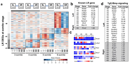
Spatial transcriptome profiling uncovers metabolic regulation of left-right patterning
Hisato Yagi, Cheng Cui, Manush Saydmohammed, George Gabriel, Candice Baker, William Devine, Yijen Wu, Jiuann-huey Lin, Marcus Malek, Abha Bais, Stephen Murray, Bruce Aronow, Michael Tsang, Dennis Kostka, Cecilia W. Lo
David Bolumar, Javier Moncayo-Arlandi, Javier Gonzalez-Fernandez, Ana Ochando, Inmaculada Moreno, Carlos Marin, Antonio Diez, Paula Fabra, Miguel Ángel Checa, Juan José Espinos, David K. Gardner, Carlos Simon, Felipe Vilella
Wnt activity reveals context-specific genetic effects on gene regulation in neural progenitors
Nana Matoba, Brandon D Le, Jordan M Valone, Justin M Wolter, Jessica Mory, Dan Liang, Nil Aygün, K Alaine Broadaway, Marielle L Bond, Karen L Mohlke, Mark J Zylka, Michael I Love, Jason L Stein
A transient dermal niche and dual epidermal programs underlie sweat gland development
Heather L. Dingwall, Reiko R. Tomizawa, Adam Aharoni, Peng Hu, Qi Qiu, Blerina Kokalari, Serenity M. Martinez, Joan C. Donahue, Daniel Aldea, Meryl Mendoza, Ian A. Glass, Birth Defects Research Laboratory (BDRL), Hao Wu, Yana G. Kamberov
| Stem cells, regeneration & disease modelling
Human pluripotent stem cells-derived inner ear organoids recapitulate otic development in vitro
Daniela Doda, Sara Alonso Jimenez, Hubert Rehrauer, Jose F. Carreño, Victoria Valsamides, Stefano Di Santo, Hans Ruedi Widmer, Albert Edge, Heiko Locher, Wouter van der Valk, Jingyuan Zhang, Karl R. Koehler, Marta Roccio
Birth, cell fate and behavior of progenitors at the origin of the cardiac mitral valve
Batoul Farhat, Ignacio Bordeu, Bernd Jagla, Hugo Blanc, Karine Loulier, Benjamin D. Simons, Emmanuel Beaurepaire, Jean Livet, Michel Pucéat
Angela Papalamprou, Victoria Yu, Wensen Jiang, Julia Sheyn, Tina Stefanovic, Angel Chen, Chloe Castaneda, Melissa Chavez, Dmitriy Sheyn
Debabrata Jana, Priya Singh, Purnima Sailasree, Nithyapriya Kumar, Vijay V Vishnu, Hanuman T Kale, Jyothi Lakshmi, Asha Kumari, Divya Tej Sowpati, P Chandra Shekar
Ellen F. Gregory, Shilpi Kalra, Trisha Brock, Gisèle Bonne, G.W. Gant Luxton, Christopher Hopkins, Daniel A. Starr
Overactivated epithelial NF-κB disrupts lung development in human and nitrofen CDH
Florentine Dylong, Jan Riedel, Gaurang M. Amonkar, Nicole Peukert, Paula Lieckfeldt, Katinka Sturm, Benedikt Höxter, Wai Hei Tse, Yuichiro Miyake, Steffi Mayer, Richard Keijzer, Martin Lacher, Xingbin Ai, Jan-Hendrik Gosemann, Richard Wagner
Chaochang Li, Mireia Alemany Ribes, Rosanne Raftery, Uzochi Nwoko, Matthew L. Warman, April M. Craft
PRDM16 functions as a co-repressor in the BMP pathway to suppress neural stem cell proliferation
Li He, Jiayu Wen, Qi Dai
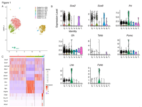
Karine Rizzoti, Probir Chakravarty, Daniel Sheridan, Robin Lovell-Badge
Characterization of regeneration initiating cells during Xenopus laevis tail regeneration
Sindelka Radek, Abaffy Pavel, Zucha Daniel, Naraine Ravindra, Kraus Daniel, Netusil Jiri, Smetana Karel Jr., Lukas Lacina, Endaya Berwini Beduya, Neuzil Jiri, Psenicka Martin, Kubista Mikael
Stephanie N. Oprescu, Nick Baumann, Xiyue Chen, Qiang Sun, Yu Zhao, Feng Yue, Huating Wang, Shihuan Kuang
Dedifferentiating germ cells regain stem-cell specific polarity checkpoint prior to niche reentry
Muhammed Burak Bener, Autumn Twillie, Mayu Inaba
p53 promotes revival stem cells in the regenerating intestine after severe radiation injuryv
Clara Morral, Arshad Ayyaz, Hsuan-Cheng Kuo, Mardi Fink, Ioannis Verginadis, Andrea R. Daniel, Danielle N. Burner, Lucy M. Driver, Sloane Satow, Stephanie Hasapis, Reem Ghinnagow, Lixia Luo, Yan Ma, Laura D. Attardi, Costas Koumenis, Andy J Minn, Jeffrey L. Wrana, Chang-Lung Lee, David G. Kirsch
Derivation of trophoblast stem cells unveils unrestrained potential of mouse ESCs and epiblast
Debabrata Jana, Purnima Sailasree, Priya Singh, Mansi Srivastava, Vijay V Vishnu, Hanuman T Kale, Jyothi Lakshmi, Gunda Srinivas, Divya Tej Sowpati, P Chandra Shekar
Radical fringe facilitates NOTCH1 and JAG1 cis interactions to sustain Hematopoietic stem cell fate
Roshana Thambyrajah, Maria Maqueda, Wen Hao Neo, Kathleen Imbach, Yolanda Guillen, Daniela Grases, Zaki Fadlullah, Stefano Gambera, Francesca Matteini, Xiaonan Wang, Fernando J. Calero-Nieto, Manel Esteller, Maria Carolina Florian, Eduard Porta, Rui Benedito, Berthold Göttgens, Georges Lacaud, Lluis Espinosa, Anna Bigas
Arsheen M. Rajan, Nicole L. Rosin, Elodie Labit, Jeff Biernaskie, Shan Liao, Peng Huang
Shruthi Subramanian, Julie A.I. Thoms, Yizhou Huang, Paola Cornejo, Forrest C. Koch, Sebastien Jacquelin, Sylvie Shen, Emma Song, Swapna Joshi, Chris Brownlee, Petter S. Woll, Diego Chacon Fajardo, Dominik Beck, David J. Curtis, Kenneth Yehson, Vicki Antonenas, Tracey O’ Brien, Annette Trickett, Jason A. Powell, Ian D. Lewis, Stuart M. Pitson, Maher K. Gandhi, Steven W. Lane, Fatemeh Vafaee, Emily S. Wong, Berthold Göttgens, Hamid Alinejad Rokny, Jason W.H Wong, John E. Pimanda
A quantitative characterization of early neuron generation in the developing zebrafish telencephalon
Glòria Casas Gimeno, Ekaterina Dvorianinova, Carla-Sophie Lembke, Emma SC Dijkstra, Hussam Abbas, Yuanyuan Liu, Judith TML Paridaen
Atoosa Amel, Mubeen Goolam
| Plant development
Monika Kubalová, Karel Müller, Petre Ivanov Dobrev, Annalisa Rizza, Alexander M. Jones, Matyáš Fendrych
Arabidopsis NF-YCs interact with CRY2 and PIF4/5 to repress blue light-mediated hypocotyl growth
Wei Wang, Lin Gao, Tianliang Zhao, Jiamei Chen, Ting Chen, Wenxiong Lin
Physcomitrium patens SMXL homologs are PpMAX2-dependent negative regulators of growth
Ambre Guillory, Mauricio Lopez-Obando, Khalissa Bouchenine, Philippe Le Bris, Alain Lécureuil, Jean-Paul Pillot, Vincent Steinmetz, François-Didier Boyer, Catherine Rameau, Alexandre de Saint Germain, Sandrine Bonhomme
Amanda K Broz, Daniel B Sloan, Iain G Johnston
A transcriptome analysis of OsNAC02 Ko-mutant during vegetative endosperm development
Mei Yan, Guiai Jiao, Gaoneng Shao, Ying Chen, Maodi Zhu, Lingwei Yang, Lihong Xie, Peisong Hu, Shaoqing Tang
Ping-Hung Hsieh, Jennifer M. Frost, Yeonhee Choi, Tzung-Fu Hsieh, Daniel Zilberman, Robert L Fischer
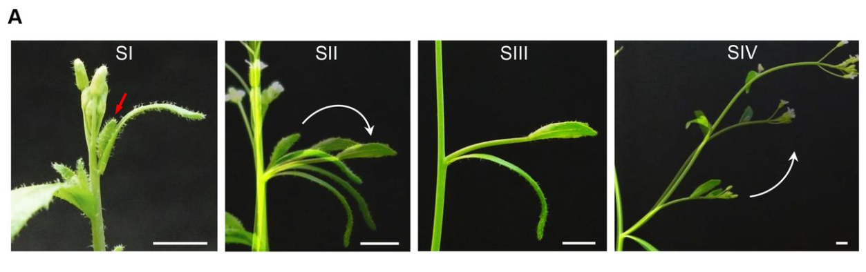
Arabidopsis lateral shoots display two distinct phases of growth angle control
Martina De Angelis, Stefan Kepinski
Autophagy in maternal tissues contributes to Arabidopsis thaliana seed development
Ori Erlichman, Shahar Weiss, Maria Abu-Arkia, Moria Ankary Khaner, Yoram Soroka, Weronika Jasinska, Leah Rosental, Yariv Brotman, Tamar Avin-Wittenberg
Brennan Hyden, Dana L. Carper, Paul E. Abraham, Guoliang Yuan, Tao Yao, Leo Baumgart, Yu Zhang, Cindy Chen, Ronan O’Malley, Jin-Gui Chen, Xiaohan Yang, Robert L. Hettich, Gerald A. Tuskan, Lawrence B. Smart
| Evo-devo
Evolution of chemosensory tissues and cells across ecologically diverse Drosophilids
Gwénaëlle Bontonou, Bastien Saint-Leandre, Tane Kafle, Tess Baticle, Afrah Hassan, Juan Antonio Sánchez-Alcañiz, Roman J. Arguello
Johannah Rickman, Abigail E Burtner, Tate J Linden, Sharlene E Santana, Chris J Law
Cell Biology
Drishya Iyer, Diandra Mastrogiacomo, Kunyu Li, Richa Banerjee, Ying Yang, Joshua P. Scallan
Sexually dimorphic dynamics of the microtubule network in medaka (Oryzias latipes) germ cells
Mariko Kikuchi, Miyo Yoshimoto, Tokiro Ishikawa, Yuto Kanda, Kazutoshi Mori, Toshiya Nishimura, Minoru Tanaka
Yuichiro Hara, Takuma Kumamoto, Naoko Yoshizawa-Sugata, Kumiko Hirai, Song Xianghe, Hideya Kawaji, Chiaki Ohtaka-Maruyama
Cara McCormick, Alicia K. Rogers
Tsubasa Itoh, Mari Uehara, Shinnosuke Yura, Jui Chun Wang, Akiko Nakanishi, Takashi Shimizu, Masahiko Hibi
Suppression of ferroptosis by vitamin A or antioxidants is essential for neuronal development
Juliane Tschuck, Vidya Padmanabhan Nair, Ana Galhoz, Gabriele Ciceri, Ina Rothenaigner, Jason Tchieu, Hin-Man Tai, Brent R. Stockwell, Lorenz Studer, Michael P. Menden, Michelle Vincendeau, Kamyar Hadian
Tristetraprolin promotes survival of mammary progenitor cells by restraining TNFα levels
Stedile Micaela, Lara Montero Angela, García Solá Martín Emilio, Goddio María Victoria, Beckerman Inés, Bogni Emilia, Ayre Marina, Naguila Zaira, Coso Omar, Edith C. Kordon
Kinesin-1 promotes centrosome clustering and nuclear migration in the Drosophila oocyte
Maëlys Loh, Fred Bernard, Antoine Guichet
Corentin Mollier, Joanna Skrzydeł, Dorota Borowska-Wykręt, Mateusz Majda, Vincent Bayle, Virginie Battu, Jean-Chrisologue Totozafy, Mateusz Dulski, Antoine Fruleux, Roman Wrzalik, Grégory Mouille, Richard S. Smith, Françoise Monéger, Dorota Kwiatkowska, Arezki Boudaoud
RhoA GEF Mcf2lb regulates rosette integrity during collective cell migration
Hannah M. Olson, Amanda Maxfield, Nicholas L. Calistri, Laura M. Heiser, Alex V. Nechiporuk
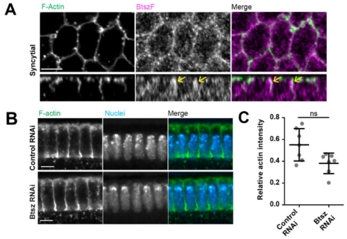
Bitesize bundles F-actin and influences actin remodeling in syncytial Drosophila embryo development
Anna R. Yeh, Gregory J. Hoeprich, Bruce L. Goode, Adam C. Martin
Robert Hardt, Alireza Dehghani, Carmen Schoor, Markus Gödderz, Nur Cengiz Winter, Shiva Ahmadi, Ramesh Sharma, Karin Schork, Martin Eisenacher, Volkmar Gieselmann, Dominic Winter
Modelling
The time integral of BMP signaling determines fate in a stem cell model for early human development
Seth Teague, Gillian Primavera, Bohan Chen, Emily Freeburne, Hina Khan, Kyoung Jo, Craig Johnson, Idse Heemskerk
Stanley E. Strawbridge, Agata Kurowski, Elena Corujo-Simon, Alastair N. Fletcher, Jennifer Nichols, Alexander G. Fletcher
Matthew Stower, Felix Zhou, Holly Hathrell, Jason Yeung, Shifaan Thowfeequ, Jonathan Godwin, Falk Schneider, Christoffer Lagerholm, Marco Fritzsche, Jeyan Thiyagalingam, Xin Lu, Jens Rittscher, Shankar Srinivas
Sandy Lee, Huichun Zhan
Ran Wang, Xianfa Yang, Jiehui Chen, Lin Zhang, Jonathan A. Griffiths, Guizhong Cui, Yingying Chen, Yun Qian, Guangdun Peng, Jinsong Li, Liantang Wang, John C. Marioni, Patrick P.L. Tam, Naihe Jing
Turing pattern prediction in three-dimensional domains: the role of initial conditions and growth
Soha Ben Tahar, Jose J Muñoz, Sandra J Shefelbine, Ester Comellas
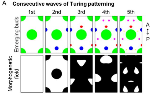
Gap junctions in Turing-type periodic feather pattern formation
Chun-Chih Tseng, Thomas E. Woolley, Ting-Xin Jiang, Ping Wu, Philip K. Maini, Randall B. Widelitz, Cheng-Ming Chuong
Tools & Resources
Optimized husbandry and targeted gene-editing for the cnidarian Nematostella vectensis
João E. Carvalho, Maxence Burtin, Olivier Detournay, Aldine R. Amiel, Eric Röttinger
Tomohiro Tamari, Yoshihisa Ikeda, Kento Morimoto, Keiko Kobayashi, Saori Mizuno-Iijima, Shinya Ayabe, Akihiro Kuno, Seiya Mizuno, Atsushi Yoshiki
Soumyaroop Bhattacharya, Caroline Cherry, Gail Deutsch, Birth Defects Research Laboratory (BDRL), Ian A. Glass, Thomas J. Mariani, Denise Al Alam, Soula Danopoulos
A Suite of Mouse Reagents for Studying Amelogenesis
Tomas Wald, Adya Verma, Victoria Cooley, Pauline Marangoni, Oscar Cazares, Amnon Sharir, Evelyn J. Sandoval, David Sung, Hadis Najibi, Tingsheng Yu Drennon, Jeffrey O. Bush, Derk Joester, Ophir D. Klein
An Image-Guided Microfluidic System for Single-Cell Lineage Tracking
Aslan Kamil Mahmut, Fourneaux Camille, Yilmaz Alperen, Stavros Stavrakis, Parmentier Romuald, Paldi Andras, Gonin-Giraud Sandrine, J Andrew deMello, Gandrillon Olivier
Nichole Link, J Michael Harnish, Brooke Hull, Shelley Gibson, Miranda Dietze, Uchechukwu E Mgbike, Silvia Medina-Balcazar, Priya S Shah, Shinya Yamamoto
An AI-based segmentation and analysis pipeline for high-field MR monitoring of cerebral organoids
Luca Deininger, Sabine Jung-Klawitter, Petra Richter, Manuel Fischer, Kianush Karimian-Jazi, Michael O. Breckwoldt, Martin Bendszus, Sabine Heiland, Jens Kleesiek, Ralf Mikut, Daniel Hübschmann, Daniel Schwarz
Cheng-Yu Li, Helena Boldt, Emily Parent, Jax Ficklin, Althea James, Troy J. Anlage, Lena M. Boyer, Brianna R. Pierce, Kellee Siegfried, Matthew P. Harris, Eric S. Haag
Developmental staging and future research directions of the model marine tubeworm Hydroides elegans
Katherine T. Nesbit, Nicholas J. Shikuma
Research practice & education
Gwendolyn Bogard, Erin Saybolt, Moraima Castro-Faix, Adriana Bankston
Purchases dominate the carbon footprint of research laboratories
Marianne De Paepe, Laurent Jeanneau, Jerôme Mariette, Olivier Aumont, André Estevez-Torres
The landscape of biomedical research
Rita González-Márquez, Luca Schmidt, Benjamin M. Schmidt, Philipp Berens, Dmitry Kobak
Twitter and Mastodon presence of highly-cited scientists
Maximilian Siebert, Leonardo Maria Siena, John P.A. Ioannidis
Self-referencing rates in biological disciplines
Sean M. Cascarina
 (No Ratings Yet)
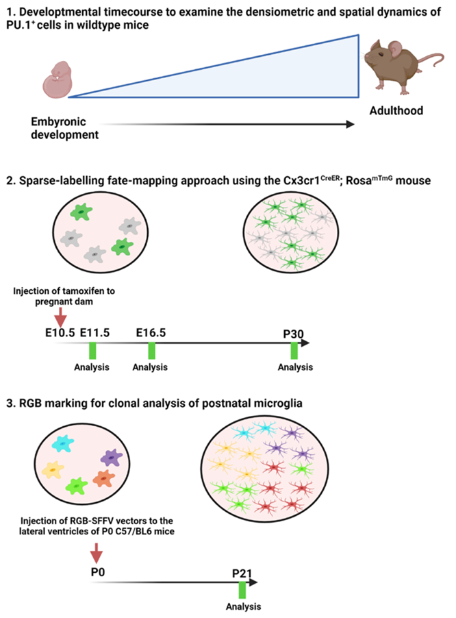

 (3 votes)
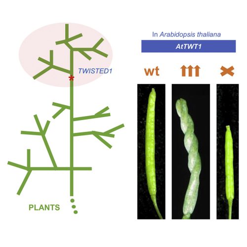
_by_Claes_(Nicolaes)_Jansz_Visscher.jpg)