Meet the Node correspondents: Brent Foster
Posted by the Node, on 3 April 2023
We recently announced that we will be working with three newly appointed the Node correspondents, who will be helping us to develop and write content for the Node in 2023. We caught up with each of them to chat about their research backgrounds and the topics that they’re excited to write about over the course of the coming year.
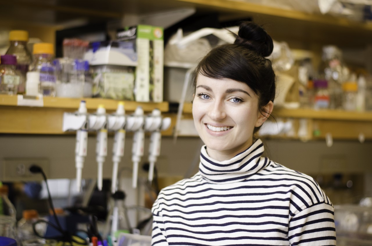
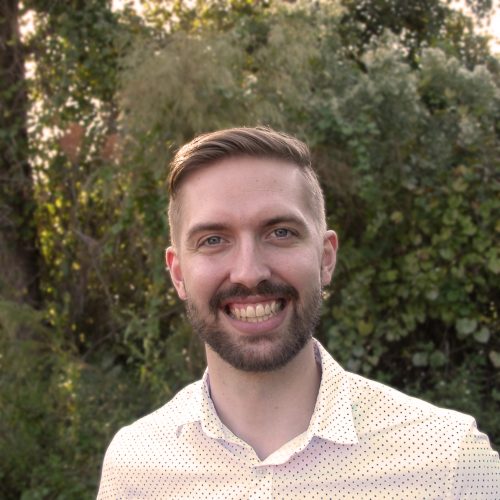
First up is Brent Foster, a technician at the Whitney Laboratory for Marine Bioscience, University of Florida, where he is studying the evolutionary origin of nervous systems using a range of marine invertebrates. As you will see, Brent has a longstanding interest in creative writing alongside his scientific career and began to blend the two by writing for his local newspaper. We talked about how his experience of using unconventional experimental organisms influences his writing, and the importance of being part of a science writing community.
Congratulations on being selected as a the Node correspondent. Why did you decide to apply for the role?
I decided to apply for it mostly because I had such a good experience with The Company of Biologists’ Creative Science Writing Workshop that I went to last year. I respect the mission of The Company of Biologists and their whole philosophy, and I thought this would be a really cool opportunity to expand beyond the readership I’ve had so far with my science writing.
What led you to first become interested in science writing? And what sort of science writing have you been doing so far?
I’ve always liked science. My dad was a high school biology teacher, and we lived in a rural area. I got to know the local wildlife, local trees and all of that fairly well. I really enjoyed the outdoors and I’ve always loved learning how nature works. And then on the flip side, once I started my undergraduate degree, I discovered that I enjoyed writing as well. And I had never really thought of pairing the two – I just considered writing as a hobby and assumed science would be my career, and that was my understanding going forward.
When I started working in the lab at the UF Whitney Laboratory, I met up with a local news editor. We struck up a conversation and I found out he was interested in science, but didn’t have a science background, and I was interested in writing, but hadn’t really done any formal writing, other than a few personal essays in college. And he basically issued a challenge to write something for the Palm Coast Observer, which is the local newspaper here. So, I approached my supervisor, and I asked him if it would be ok to write up some articles about what the lab is doing for the newspaper. From there, I discovered that I really enjoyed writing about the sciences, maybe even a little bit more than I enjoyed doing the experiments in the laboratory, and so I’ve expanded from there. The past two years I’ve presented some work at the Society for Integrative and Comparative Biology (SICB) meeting, and there I met some people who write for the SICB blog, and so I’ve contributed a few posts there. Last year I applied to the Creative Science Writing Workshop, hosted by The Company Biologists, loved it, and I’ve kind of snowballed into larger and larger projects. In October, I went to a science writing conference in Memphis, where I met a few folks and learned a little bit about the life of a science writer.
Thinking about what you were saying there about going to workshops and conferences focused on science writing, have you found that meeting likeminded scientists who also have an interest in creative writing has influenced you?
Yeah, absolutely. That was one of the big things from the Workshop last year: I was kind of shocked at how all of these brilliant scientists really just seemed to want to have some sort of creative outlet. And not always just about the work that they’re doing themselves, which showed me how creative science and scientists can be. So that was really eye opening. And then in January, when I was at SICB, I announced that I was a correspondent for the Node. I had a couple of people come up to me afterwards and we were throwing ideas back and forth about some different writing projects. That was a lot of fun. So, it has opened up some doors as well as friendships that I don’t think I would have found otherwise.
In terms of your own research, what has your career path been so far? And what’s your current research focus?
This seems to be the story for a lot of people now, but my career path is very, very windy. When I got started as an undergraduate student, I wanted to be an epidemiologist. After my first semester, I deferred my education and lived in Brazil for two years. I learned the language and the richness of that culture, and after just two months I noticed that I started dreaming in Portuguese. When I returned to my University, I attended a club meeting for deaf and hearing-impaired students. I only know rudimentary American Sign Language (ASL), so I’m sure my fingers were just fumbling with an accent. To offset the clumsiness of my hands, I would mouth the words I was trying to spell. It was only after the meeting when I realised that the words I mouthed were in Portuguese, not English. Somehow ASL and Portuguese had gotten tangled up in my brain, and both of them seemed sectioned off from English. I thought that was pretty fascinating. So, in the end, my undergraduate degree was in neuroscience, with minors in linguistics and creative writing.
I was especially interested in neurolinguistics, so how language is processed in the brain. That was my attempt to try to marry my interest in writing and science. I did some eye tracking studies, MRI studies, and EEG. And as I was getting all these great research experiences, I was a little disappointed in how little we could actually tell from those particular methods. I mean, you can tell a lot by each of them, but there were no clear causal effects, right? It was all “well, this is happening, and we are interpreting it to mean maybe this.” It wasn’t very definite, and that was a little disappointing for me when trying to understand some basic biology.
Then my wife was accepted to a PhD programme at the University of Florida, and so we moved down here. Originally, we were on the main campus, and I was in an MRI lab where we did a lot of cognitive neuroscience research related to attention and anxiety. They were really, really cool projects, and I’m grateful for the experience that I had there, but again, I had the same feeling of, “man, I’m just not sure how much we can actually tell from what we’re doing.” My wife and I stayed on main campus for the first year of her PhD. And then she moved out here to the Whitney Lab, and I didn’t want to have to commute back and forth. So, I actually reached out to the director of the lab, Mark Martindale (who’s now my supervisor), and I told him about my research experience. I had no wet lab experience – all of the studies that I’d done were human studies and nothing at all relevant to what they do here at the Whitney Laboratory. But I was interested in learning and asked if I could come down even just on the weekends to get some wet lab experience. And he turned around and offered me a job, and that’s how I started here.
So, I’ve switched from neurolinguistics to basic marine biology and developmental biology. And I love it. I feel like I can understand the outcomes of the experiments that I do a little bit clearer than I could within the broader cognitive sciences. I don’t mean to disparage other sciences as I think there are a lot of valuable things that we can learn from them – it just wasn’t for me.
Do you find that the science you’re doing now is more inspiring for your writing than what you were studying before? Because I imagine the sort of organisms you’re working on are a lot more interesting from that creative science writing perspective.
Yeah, that’s a great question. I think people could relate more to what I was doing before, when I was asking questions like “what’s the brain doing?” and “how do people process language?” In the eye-tracking lab, I came to understand a little bit about how people read, which has been helpful in the technical part of crafting my writing in a way that’s easy to read. But thinking back to what I found cool about science when I was a kid, I was always drawn to weird, quirky animals that do weird, quirky things – nature’s a real creative playground. Now I get to tell people that I work with sea anemones, and they’ll say, “I don’t know if I know anyone who works with a sea anemone,” or I mention that I work with comb jellies, and they say, “oh, what’s a comb jelly?” That’s a really fun type of interaction, to hear people saying, “that’s so cool. I never knew that.” And I try to capture that fun as much as I can in my writing.
Thinking about the sort of things that you’re going to be writing for the Node, what topics are you excited to write about? And do they relate to your own work or are you going to be branching out a bit into other areas?
Well, like I said, the quirky biology is what gets me excited, so I’m hoping to focus on non-model organisms, organisms that not everyone knows or hears about. This will probably involve a little bit of the work in our lab, because there are a lot of us here at the Whitney Laboratory who work with non-model organisms that are great for answering very specific questions but often get glossed over just because they’re not so well established.
My secondary focus is related to the idea of working with non-model organisms. Because these organisms are not as well established, I want to highlight labs that have to develop their own technologies or develop their own techniques or adapt existing techniques for their animals. This is hard and I think a lot of times underappreciated, both by the public and even by other scientists. With Drosophila, for example, you can do all sorts of things which you might take for granted. By contrast, I just talked to a scientist a few weeks ago who is trying to adapt transgenic approaches for cuttlefish, and they’ve spent almost a year trying to get something to work. So, I want to show those two aspects of something that I don’t think gets shown a lot, or at least doesn’t get into the major headlines. I want to give non-model organisms their due, I guess!
What are you hoping to gain from the experience of being a the Node correspondent?
Most of my science writing so far has been either hyperlocal or very specific to one group of biologists from a single conference, so one thing I’m hoping to gain as a correspondent for the Node is reaching a broader audience. I know the Node is pretty popular internationally, and I haven’t written for an international audience before. So I hope to stretch myself a bit in that area. I am looking forward to meeting other scientists in my field, hearing their stories and helping to share those stories.
I am also excited to interact with the Node team and other correspondents to learn about the craft of science writing a bit more. I don’t really have any formal training in science communication so I’m looking forward to participating in workshops. And of course, I’ve mentioned this several times now, but just having a community is huge. I didn’t realise how big the science writing community is until last year, and it’s gratifying to feel welcomed to it. Before I discovered it, I almost felt like an outsider. I was always asking myself, “is there a place for these two different aspects of my identity that everyone else seems to think of as a contradiction?” It’s fun to see that there are indeed other people who are combining their interests in science and writing.
Apart from writing, what do you enjoy doing in your spare time?
Lately, I have been doing a lot of writing! But I do enjoy the outdoors, particularly kayaking or canoeing – there are some lovely springs around here. In fact, I have a kayak trip this weekend and I’m hoping to see some manatees. I enjoy playing the piano, too. Primarily classical piano, though I don’t get a lot of opportunities to do that these days. And I enjoy reading. As a student, you kind of feel as if you either don’t have the time to read or that sometimes you’re reading about things that you’re not too excited about. In the gaps between studies, it’s been fun to be able to read some things just for the pleasure of reading.
Do you find that this sampling of other people’s writing has an influence on your own work?
Yeah, absolutely. Because I’ve gotten so interested in science writing, a lot of what I’ve been reading lately is by other science writers. That’s been informative in learning how other people approach science writing, but I also enjoy regular literature. I’ll pick up a piece of fiction or even poetry sometimes. And to be honest the poetry reading that I do, and the creative writing classes I took as an undergraduate that taught me a little bit about poetry, have actually informed my ability to write concisely in a way that I’m not sure I would have picked up from other genres. So, I think that by dabbling a little bit in a novel here, a short story there, some poetry, all of those things, you can take something from each one of those genres that together can make a science writing piece pretty powerful.
 (3 votes)
 (No Ratings Yet)