It’s musical chairs at The Company of Biologists at the moment (though fortunately without any chairs actually being removed!) – after Seema Grewal left Development last month to become the new Executive Editor of Journal of Cell Science, we’re delighted to have appointed Laura Hankins, currently the Company’s Science Communication Officer, as the new Reviews Editor on Development. Laura started working with us a little over a year ago following a PhD with Jordan Raff in Oxford, and I’m really pleased that she’ll be joining the journal in the near future – having worked with her in her current role, I know she’s going to be a great member of the team. And Helen Zenner – who you’ll know as the Node’s Community Manager – is going to be moving over to our sister site, FocalPlane. Helen’s got a very strong microscopy research background, and has some great ideas for developing FocalPlane.
All this means that we now have two openings for scicomm-type roles! Both these positions are suitable either for current researchers looking to move away from the lab, or for people who already have some scicomm experience. They provide a great opportunity to learn about the scientific publishing world, and to contribute to a not-for-profit organisation that really believes in supporting the biological community. To find out more, take a look at the job adverts for the Node Community Manager and the Science Communications Officer positions, and please feel free to reach out if you want to find out more about either role.
The European Developmental Biology Congress: a distributed meeting across Oxford, Barcelona and Paris 25th – 28th September 2023
The quadrennial European Developmental Congress usually positions itself neatly at the mid-way point between meetings of the International Society for Developmental Biology, or at least that was the case, until the pandemic-induced postponement of the latest ISDB meeting to October 2021.
This left less than a year between the ISDB and EDBC meetings. We at the British Society for Developmental Biology, the hosts for #EDBC2023, see this not as a problem but an opportunity – a chance to experiment with some alternatives to the usual international conference format.
For one thing, we have decided to focus EDBC on the ‘rising stars’ of European developmental biology. Most of our invited speakers will be outstanding and engaging speakers who started their own labs within approximately the last five years. We are currently finalising our programme and are always happy to take suggestions of excellent speakers based in European labs from our community, so please do get in touch through our twitter @_BSDB_ or our shiny new mastodon account @BSDB@mstdn.science or by emailing our Meetings Officer sally.lowell@ed.ac.uk. The meeting will also contain all the usual highlights of BSDB meetings, including talks from the winners of the Waddington medal, the Cheryll Tickle medal and the Beddington medal.
Furthermore, in the spirit of experimenting with more accessible conference formats1 we will distribute the meeting across three different locations in Europe. The main hub of the meeting will be in Oxford, and there will be interlinked one-day ‘spoke’ meetings in Paris, led by Sigolène Meilhac, Nicola Festuccia, Tanya Foley, Tom Cumming & Guillaume Frasca and Barcelona, led by Alejo Fraticelli & colleagues tba. Talks will stream back and forth between the Oxford hub and the spoke locations, and each of the three sites will also host its own social and scientific events.
We have exciting plans for the scientific and social programmes at all three locations, including lots of opportunities to present your work through short talks chosen from abstracts, flash talks, and lively poster sessions. All will be revealed early next year when the #ECDB2023 website goes live – watch this space!
We encourage anyone who can’t travel to any of the three locations to consider organising a small scale ‘watch party’, perhaps streaming your favourite sessions to a departmental seminar room and combining this with a local poster session. We particularly hope that PhD students and postdocs might be interested in organising local events of this type – do get in touch (sally.lowell@ed.ac.uk) if you’d like to discuss how BSDB can support you with this.
The making of colorful neuromesodermal progenitors
During the embryonic development, while the gastrulation process is taking place, cells within embryos self-organize by creating groups and layers of cells, where each one will develop into different adult tissues. Human and mouse embryos are called triploblastic organisms as they are characterized by the development of three germ layers: ectoderm, which will form epithelial and neural tissues; mesoderm, which will differentiate into muscle, bone, circulatory system and spleen, among other tissues; and endoderm that will produce organs such as gut, lungs, and pancreas.
During gastrulation, when the germ layers are being organized, progenitor cells commit to the generation of one of these three structures and their specific cell types. For example, an ectodermal progenitor will be committed to the generation of either epithelial or neural cell types. However, some studies have described the presence of dual-fated progenitors that could produce both mesodermal and neuroectodermal lineages (Tzouanacou et al., 2009; Wymeersch et al., 2021). These cells were named neuromesodermal progenitors (NMPs) and they are identified by the co-expression of Sox2 and Brachyury (T) genes.
NMPs have risen a lot of interest due to their exceptional behavior and they are being studied with the aim to increase our knowledge about cell fate and cell differentiation, both in developmental biology and biomedical research (Binagui-Casas et al., 2021). Therefore, there has been a lot of effort on the development of in vitro models of these cells, as an easier tool to study their characteristics.
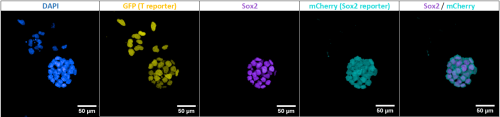
These in vitro NMP-like cells can be risen from Mouse Embryonic Stem Cells (mESC) using protocols such as Gouti et al., 2014, whose outcome has been described to be around an 80% of NMPs, within a mix of other cells in various states of differentiation. In Figure 1 we can see an example of these cultures where in vitro NMP-like cells are identified with Sox2 and T co-expression while there is also the presence of cells committed towards mesodermal lineages, identified with single T expression.
These cultures have been able to then produce both mesodermal and neural lineages at the population level, where mesodermal lineages are identified with single T expression, while neural lineages are characterized by single Sox2 expression. However, there has been a lot of controversy on whether these in vitro progenitors are also dual-fated at a single cell level as described for in vivo NMPs (Tzouanacou et al., 2009). This means that, in a group of in vitro NMP-like cells, some could be more committed to neural lineages while others to mesodermal, and it is not known yet if a single progenitor can generate both lineages.
Figure 1. In vitro NMP-like cells at day three of the differentiation protocol to NMPs from mESCs. DAPI in blue, GFP reporter of T gene-expression in yellow, Sox2 in magenta and mCherry reporter of Sox2 gene-expression in cyan.
To better characterize the potency and other characteristics of these in vitro NMP-like cells, Prof Wilson’s lab developed a new double reporter mESC line for Sox2 and T expression. In this case, mCherry protein will report Sox2 expression while GFP protein will report T expression. The knock-in experiments were performed by using gene targeting, to optimize the fidelity of expression, while leaving the endogenous gene’s expression intact. The use of this cell line allows the possibility to obtain a pure population of in vitro NMP-like cells that are double positive for Sox2 and Bra expression by using Fluorescent-activated Cell Sorting (FACS).
During my summer internship in Prof Wilson’s lab and under the supervision of Dr Anahí Binagui-Casas I have been characterizing this new cell line by describing the differentiation outcome of a pure population of in vitro NMP-like cells when cultured in different conditions and plated at different cell densities.
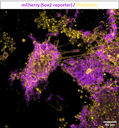
Firstly, in Figure 2 we can see the culture of a pure population of in vitro NMP-like cells in media supplemented with retinoic acid (RA) and smoothened agonist (SAG), as described in Gouti et al. 2014, to induce spinal cord differentiation. We can see the formation of axons and neural rosettes, and they can be identified with Sox2 expression and depleted expression of T, suggesting neural differentiation.
Figure 2. Culture of 7.000 in vitro NMP-like cells, sorted using FACS and maintained for five days in N2B27 supplemented with RA and SAG. mCherry reporter of Sox2 in magenta, and Phalloidin in yellow to visualize the cytoskeleton.
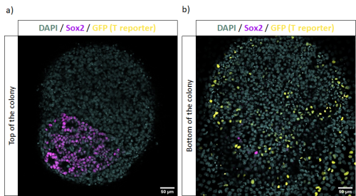
Figure 3 represents another condition where positive cells for Sox2 and Bra were cultured in media supplemented with FGF and CHIR for five days, as described in Tsakiridis & Wilson, 2015 to induce both neural and mesodermal lineages. In this case, there is the presence of cells differentiating towards neural tissues, assembled in rosette-like structures and with single expression of Sox2. Meanwhile, cells differentiating towards mesoderm are identified with single expression of T, or its gene-expression reporter GFP.
Figure 3. Culture of 1.000 in vitro NMP-like cells, sorted using FACS and maintained in N2B27 media supplemented with FGF and CHIR for five days. Images representing different planes of the same colony. Sox2 expression in magenta, GFP reporter of Bra expression in yellow and DAPI in cyan.
These experiments gave us insight about the differentiation outcome of this new cell line towards different lineages by testing multiple protocols. Furthermore, I was able to characterize the culture of in vitro NMP-like cells plated at different cell densities after sorting them using FACS. This knowledge will contribute to future experiments requiring the use of this double reporter mESC line as a tool to characterize in vitro NMP-like cells’ clonal fate and to understand if these cultures are a good model for in vivo NMPs.
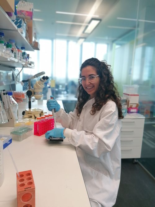
After all this work testing multiple conditions for in vitro NMP-like cells cultures and optimizing their growth, I found out that I really enjoyed working with stem cells and differentiation protocols. Although I spent many hours in the cell culture facility losing track of time, it was very exciting to see how cells grew and changed every day. Specially, I was very excited to see how they were able to organize themselves and create amazing structures, which I then learned how to immunostain and obtain beautiful images under the confocal microscope.
Figure 4. Me working on immunostaining differentiated in vitro NMP-like cells.
To conclude, this studentship and Prof Wilson’s lab have been a great opportunity for me to learn a lot about how biomedical research is driven and to confirm that I would like to continue developing my career in developmental biology. I would like to encourage students to do these internships because it is an enjoyable way of starting to apply the knowledge acquired during your bachelor’s degree and to investigate if you would enjoy developing your career in this field.
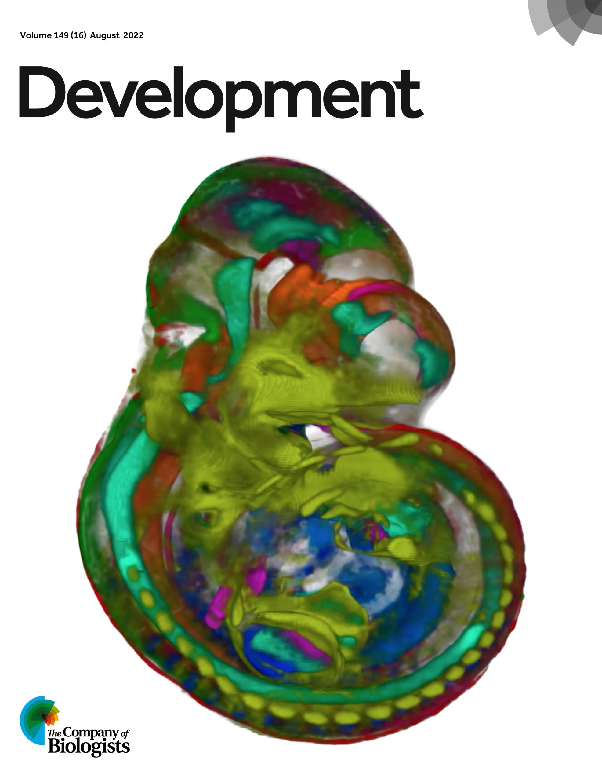
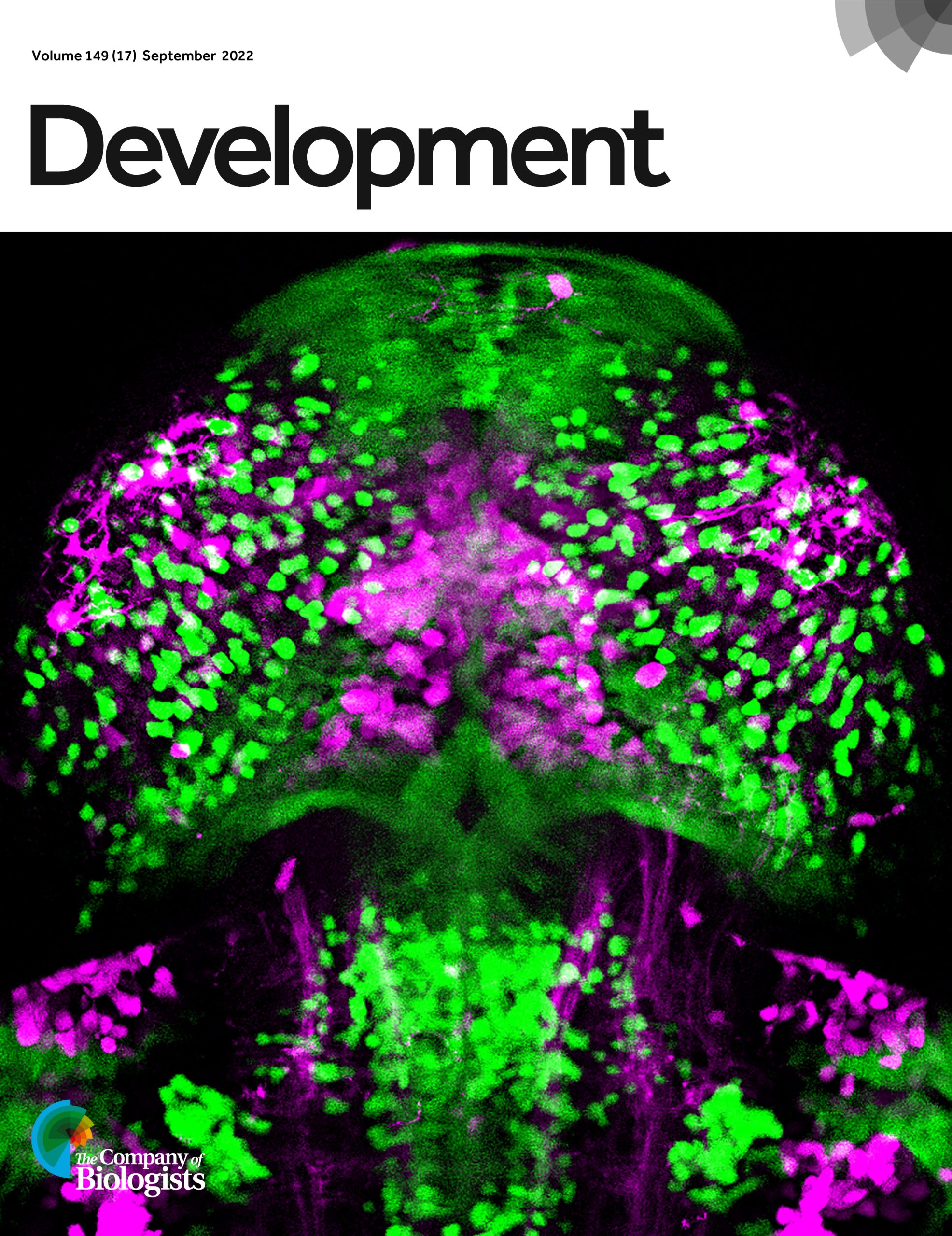
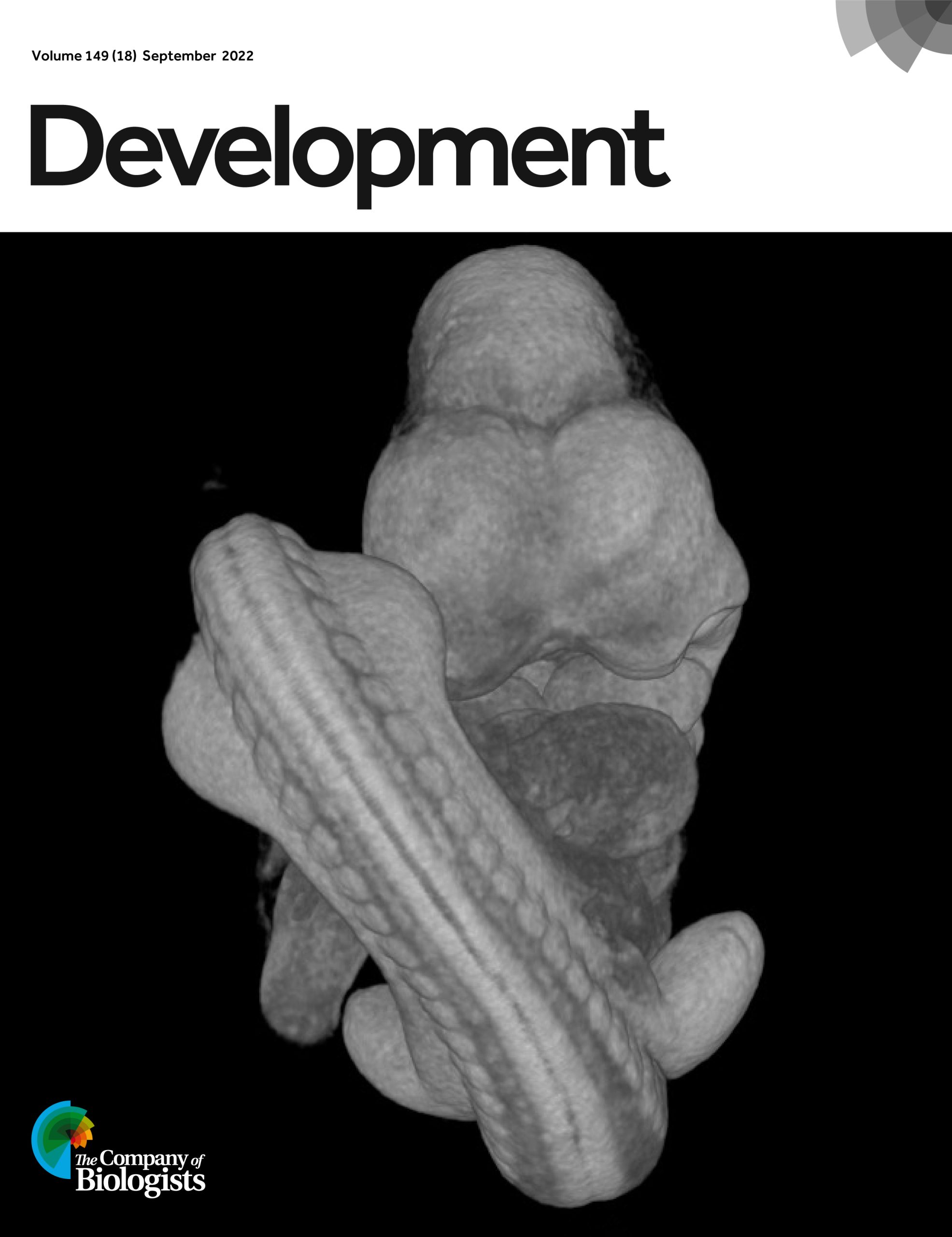
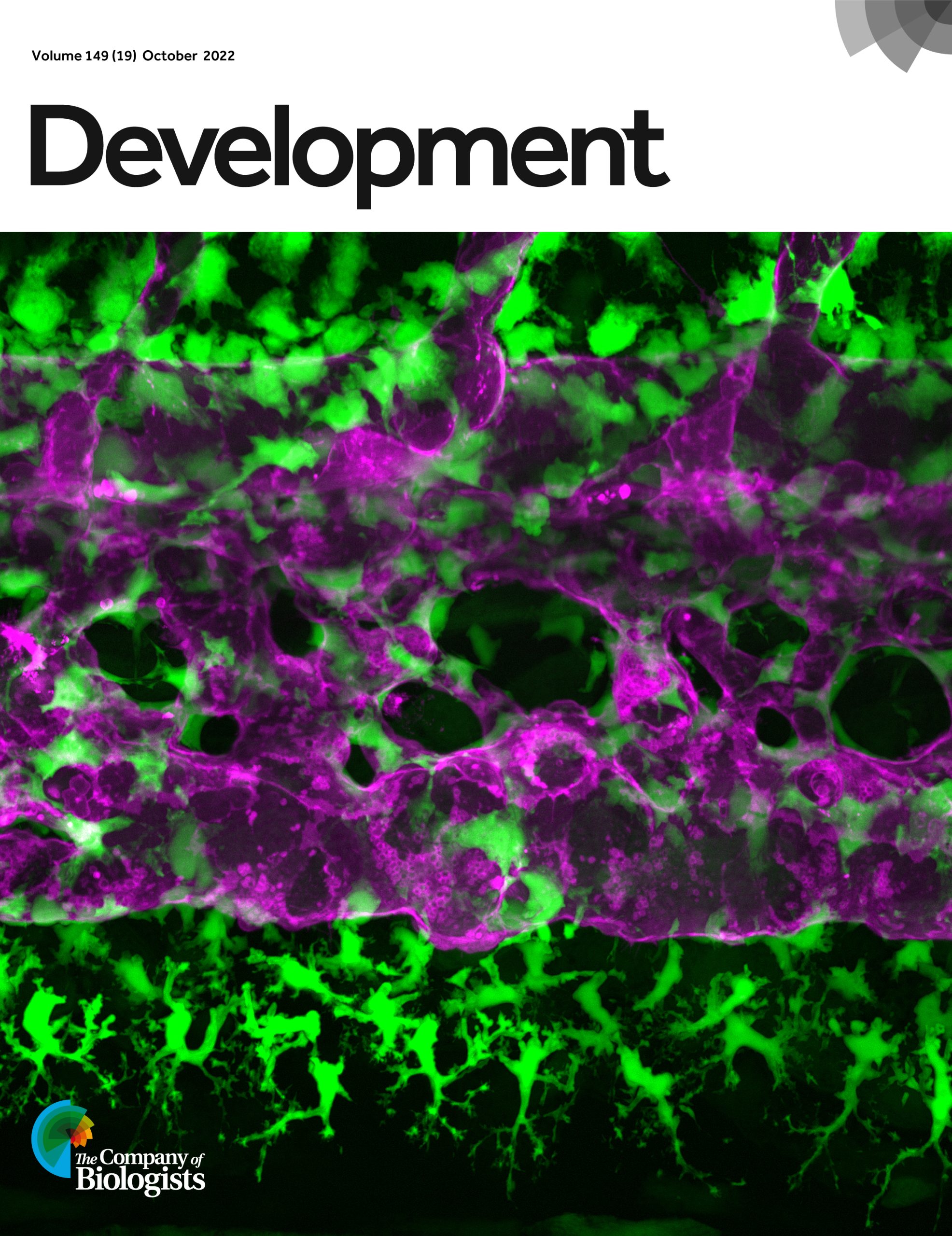
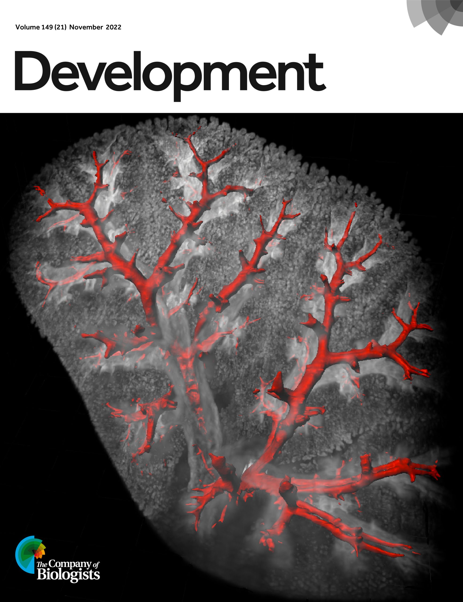
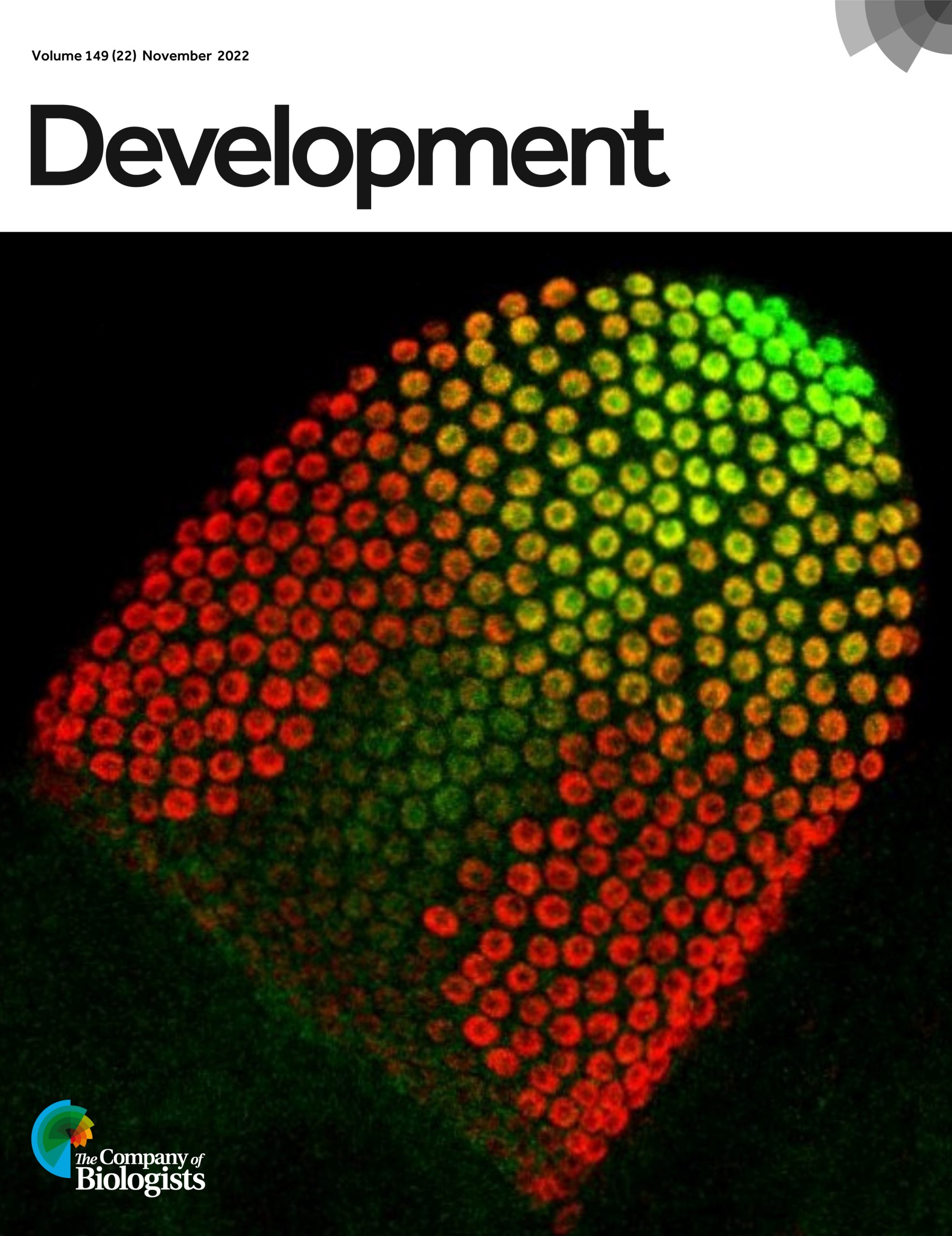
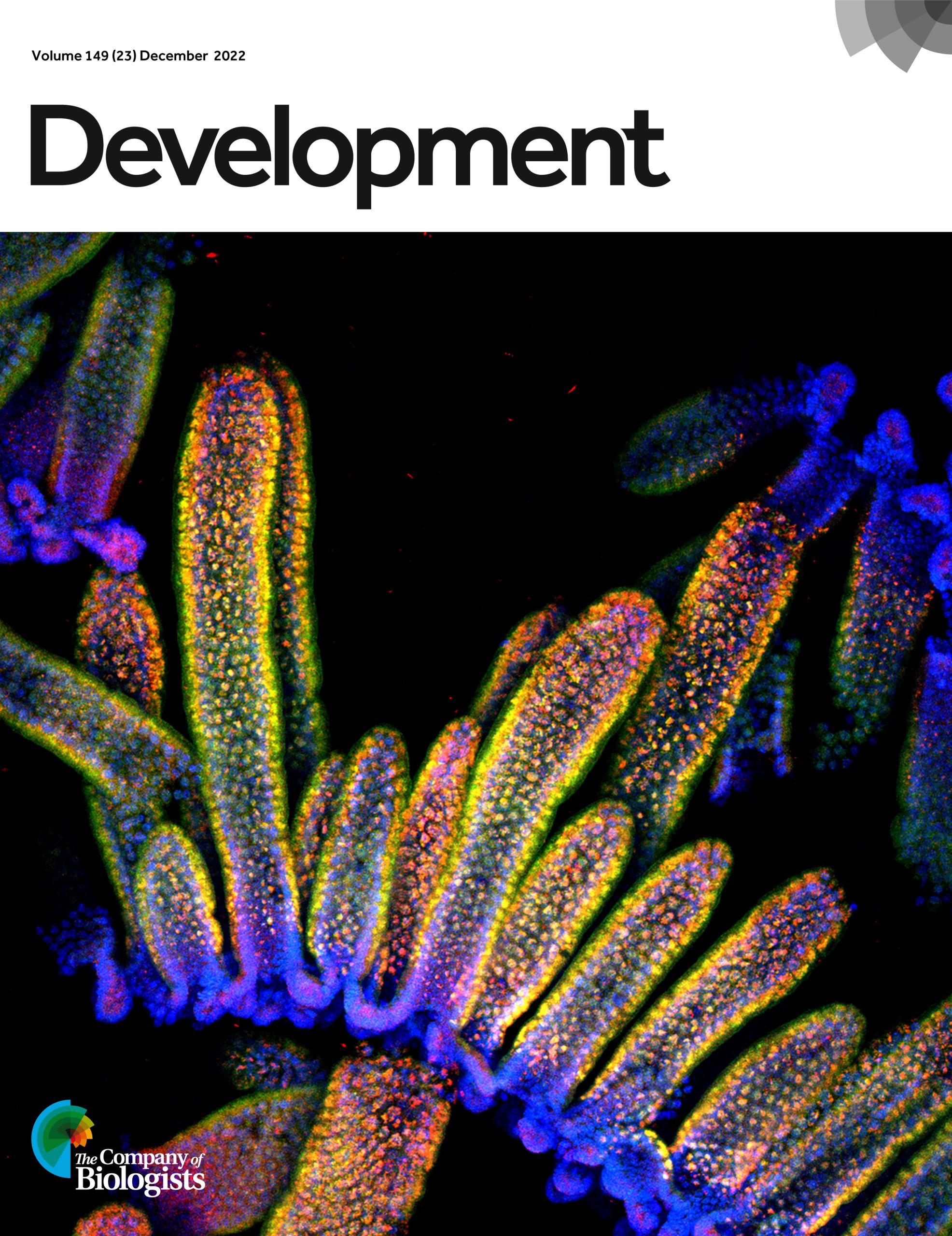
We were lucky enough to have 24 amazing cover images for Development over the past 12 months. Now, it’s your chance to vote for your favourite. To vote, click your choice in the text following the images and then click save. Voting is open until 7 January 2023.
In the battle of the model organisms, mouse comes out on top of the most featured list, followed closely by Drosophila, with honourable mentions for zebrafish, Arabidopsis and Xenopus.
This poll is now closed.
Thanks to everyone that voted and of course, everyone who contributed to such a fantastic year of cover images for Development.
On Wednesday 7 December, Development hosted three talks on metabolic and nutritional control of development and regeneration, the topic of our upcoming Special Issue.
Below you’ll find each of the talks and Q&As hosted by our Associate Editor, Irene Miguel-Aliaga (Imperial College London and MRC-LMS, UK)
Natalia López Anguita (PhD student in the Stem Cell Chromatin Group at the Max Planck Institute for Molecular Genetics) ‘Role of hypoxia in pluripotent cells and during differentiation via gastruloid formation’
Hannah Brunsdon (Postdoctoral Research Fellow in Liz Patton’s group at the IGC, University of Edinburgh) ‘Aldh2 is a metabolic gatekeeper in melanocyte stem cells’
Benjamin Jackson (MD-PhD Candidate in Lydia Finley‘s group at Memorial Sloan Kettering Cancer Center) ‘A non-canonical tricarboxylic acid cycle underlies cellular identity’
Exploring the role of planar cell polarity in the regulation of the patterning of human axial progenitors
I first became interested in stem cell biology and development while studying Medical Sciences at the University of Edinburgh. During the lockdown, I attended a very inspiring seminar in which Dr Guillaume Blin discussed in-vitro models of developmental patterning. Despite being unable to join the lab in my second year, I explored the topic, and we discussed some ideas from the literature. The BSDB summer studentship provided me with a perfect opportunity to explore these ideas in a lab setting. This summer, I got a chance to work alongside an exceptional team in the Blin lab on my own project at the Centre for Regenerative Medicine (CRM).
I have been interested in the role of planar cell polarity (PCP) in health and disease and decided to investigate its role in developmental patterning. PCP dysregulation can play a significant role in cancers and congenital disorders (Wang, de Marco, Capra and Kibar, 2019). In particular, in congenital malformations of spinal structures, such as neural tube defects (NTDs), where aetiology is still to be elucidated (Chen et al., 2018). A proportion of individuals with NTDs (Chen et al., 2018) and idiopathic scoliosis (Wise et al., 2020) have mutations in some of the core components of the PCP pathway, namely Wnt11, Vangl1 and Vangl2, and Celsr. These components localise asymmetrically within the cell to define cell polarity along the epithelial plane (Butler and Wallingford, 2017).
This pathway is involved in early development, notably when spinal progenitors are established in a structure called the anterior primitive streak (APS) (Andre et al., 2015). As these transient and rapid events occur within a complex 3D environment in-utero, much remains to be understood about cell fate decisions in the APS (Wymeersch et al., 2021). One way to study these processes in a human context is to use human embryonic stem cells to model early development (Blin, 2021). During my project, I worked on a novel in-vitro model that mimics the early stages of human axis elongation. I used this system to test the hypothesis that PCP regulates the patterning and balanced proportion of early spinal progenitors in humans.
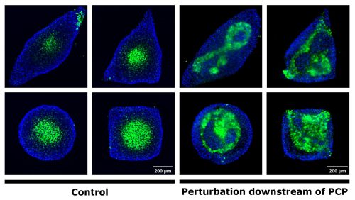
My experimental strategy consisted in confining hESC onto custom-made micropatterns and treating the colonies with a spinal fate-inducing medium. In these conditions, hESC self-organise into spatial domains of cell fates and initiate axial growth. I first tested a set of antibodies directed against PCP components. These antibodies did not provide a specific signal, but I obtained images that looked magical, so I was not too disappointed! Next, I decided to use small molecule inhibitors to perturb the PCP pathway. I inhibited either cytoskeleton remodelling downstream of PCP or the secretion of PCP ligands. When I stained for neurectoderm and endodermal cell fate markers, I observed a very severe patterning defect when cytoskeleton remodelling was inhibited (Figure 1).
I was very excited with these clear preliminary results and I wish I had the time to perform additional experiments that would demonstrate the involvement of PCP more specifically, such as PCP components staining and knockdown experiments. I have also assisted my colleagues in testing new micropatterning methods and creating cell lines using transfection methods.
Figure 1. Perturbation of cytoskeletal remodelling downstream of PCP leads to neurectoderm patterning defects. Immunostaining of micropatterned colonies 48h after the induction of differentiation. The endoderm is shown in blue. Neurectodermal cells (green) cluster at the centre in the control condition (a-d) regardless of colony shape, while patterning is perturbed in the treated condition (e-h).
Thinking back to my first days in the lab I was very excited to work alongside experienced scientists but also felt anxious as I needed to use techniques that I was not yet familiar with. During my first week, I worked out how to employ the techniques routinely used in the lab and presented my initial research plan to discuss my approach. Once I became acquainted with the protocols, they became a natural part of my routine. For example, I became proficient in micropatterning, a microfabrication method that makes it possible to standardise the size and geometry of hESC colonies (Blin, 2021). This project also allowed me to try several imaging techniques and perform image analysis using Nessys (Blin, 2019) and PickCells. I really enjoyed organising, sharing, and optimising the protocols in an online lab book. I am so happy I could contribute and that my designs are now used by the lab. We have also collaborated with a computational lab and worked on shared scripts in python.
During my internship, we also attended the Mammalian Synthetic Biology congress taking place in Edinburgh. It was a great opportunity to hear about novel data and techniques from researchers around the world and discuss how we could apply them in our lab. Networking and making friends in a professional environment like the CRM gave me the opportunity to gain unique perspectives from postgraduate students and researchers from various groups. Presenting and discussing my ideas and data allowed me to gain more insight into the dynamics of working in research and academia. These 8 weeks inspired and prepared me to pursue a career in science.
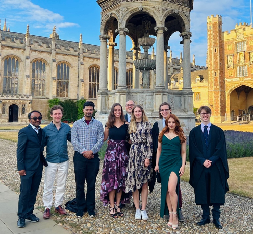
My greatest gratitude goes to my amazing supervisor, Guillaume, for everything that he has done for me. I am so grateful to Miguel for always being there for me, Heather for caring for all of us, and Fatma for the priceless moral support (all below).
Figure 2. The Blin lab (from left): Me, Fatma, Miguel, Guillaume, and Heather.
I want to express my gratitude to the amazing people at the CRM who were always keen to help and explain their experiments with so much passion. Many thanks to the BSDB for giving students like me such a wonderful opportunity to kickstart research careers. I would recommend all students to start looking for a lab they would be interested in doing a BSDB-founded internship in!
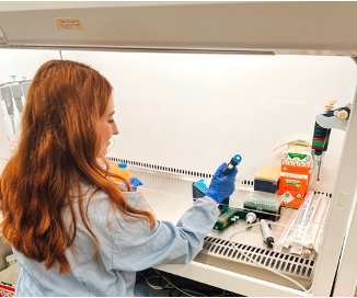
Over summer 2022, I had the opportunity to work with Dr Olena Riabinina in Insect Neuro Lab at Durham University. Her team specialises in neurobiology and neuroecology of insects with established work in mosquito olfaction and bumblebee olfactory neurobiology and ecology. My project was working with PhD student Matthew Quinn, assisting with a chapter of his PhD research project to molecularly characterise the development of the larval visual systems in the Anophelesgambiae mosquito.
Anopheles gambiae are a group of species which include the most significant vectors of deadly disease malaria in sub-Saharan Africa. As malaria poses one of the most significant public health threats worldwide, research surrounding mosquito sensory systems could prove vital in informing vector management techniques. One avenue of Insect Neuro Lab’s previous research has focussed on mosquito olfaction. Significant because research indicates sense of smell is crucial in host seeking behaviour (Wheelwright et al., 2021)(Riabinina et al., 2016). However, as mosquito larvae and pupae are also responsive to visual stimuli, a deeper understanding of their visual development has potential to provide valuable methods of mosquito-driven disease prevention. Visual perception of environmental cues is crucial for insects to, in combination with other senses, avoid predators, source food, mates and ovipositioning.
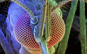
Figure 1- The mosquito compound eye structure. Image by David Scharf/Corbis
Compared to the mammalian ‘single-aperture’ eye, mosquitos have poor image resolution, however, can detect comparatively fast movement in a large viewing angle. Each unit is composed of a cornea, lens, and photoreceptor cells which sense light wavelength (colour) and intensity. Opsins are a highly conserved photoreceptor molecules observed in mosquitos and across the animal kingdom. They are membrane-bound proteins which absorb photons and change their conformation, initiating a signalling cascade known as phototransduction (Shichida & Matsuyama, 2009). Previous studies have shown that An. gambiae have 11 opsin genes, 6 of which detect long wave light. This is more than typical for insects which usually have 4. There has been a suggested association of increased long wave opsins being found to accommodate more complex light conditions within aquatic environments (Giraldo-Calderón et al., 2017).
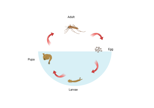
Figure 2- Mosquito life cycle (made on Biorender)
Shown in figure 2, mosquitos have four different stages in their life cycle: Egg, larvae, pupa, and adult. For the larval and pupal stages of development, mosquitos live in an aquatic environment. In contrast, adults are flying insects. It follows that their visual systems will be differently adapted to better assist survival in the contrasting environments. Previous studies involving the larvae of dengue and yellow fever transmitting mosquito Aedes aegypti show that mosquitos develop adult eye cells in late larval and pupal stages. It is predicted that both sets of eyes contribute to visual capabilities but have different photoreceptor cell types (Mysore et al., 2014). This difference in gene expression over time enables genetic targeting and quantification of components of the two visual systems. The aim of this project was to use molecular techniques to characterise expression of opsin genes in larval and pupal An. gambiae and to test the hypothesis that opsin expression profile changes throughout development.
Methods
The project involved synthesizing cDNA from RNA extracted from An. Gambiae at different time stages ranging from egg to adult. CDNA synthesis was necessary because amplification of RNA requires conversion into double stranded form. This was done using a kit containing Moloney murine leukaemia virus reverse transcriptase. The resulting cDNA was used to perform qPCR using ten different opsin and two housekeeping genes as templates. The method utilised real-time fluorescence of a double stranded DNA binding dye to detect amplification at each cycle of PCR. When fluorescent signal is detected above a decided threshold of background fluorescence, a quantification value/ Cq value is determined which calculates relative abundance between samples.
Results
We found that, consistent with previous findings (Jenkins & Muskavitch, 2015), expression of long wavelength-sensing opsin genes was distinct between larval and adult stages. With notably high expression of Opsin 6 in larval stages and high expression of opsins 1,3 and 4 in adult An. gambiae. Opsin 8 which encodes an ultraviolet sensing photoreceptor showed expression in both larval and adult forms which slightly increased in the latter. Lastly, opsin 9 which detects short-wavelength light had very low expression in the early larval stages increasing steadily with time into the adult. This research will be followed up with behavioural assays using knockout mutants to ascertain the roles of the different opsin genes in larvae survival behaviour.
Conclusions and Looking Forward
If I were to have the opportunity to pursue this research further, I would be interested to conduct immunolocalization imaging on larvae and pupae retinas comparative to research seen in figure 3 on adult An. gambiae. To map the different opsin gene expression of the larval and pupal eye structures would shed insight into how the expression levels observed translates structurally.
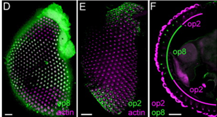
Figure 3- Edited figure showing antibody staining on the An. gambiae retina with Opsin 8 (D) Opsin 2 (E) and sectioned view of both 8 & 2 (Hu et al., 2009).
The project taught me much about experimental design at a PhD level. As I am considering a career in research, it was incredibly useful for me to observe this in action. I understand better how to seek and try to fill a gap in the literature and how to use a range of molecular techniques to test my hypothesis. Furthermore, the project gave me a lot of confidence in the lab and in my own abilities. After studying for the first two years of my bachelors in the climate of covid, our laboratory time had been limited and we spent much of our studies at home watching our lectures on our computers. Hence, experiencing two months of being a part of a friendly and welcoming lab community was so good for me. Under Matthew’s patient tutelage I added some fundamental molecular skills to my repertoire and deepened my understanding of an area of interest. This knowledge will undoubtably be useful for my degree modules this year and certainly for my level 4 lab-based research year. Additionally, because of being allowed to do this project I have chosen to do my level 3 literature review module within the subject areas of insect neurology and development. The summer has sparked an interest in new areas for me and shown me the benefits of insects as model organisms. Many thanks to everyone at Insect Neuro Lab and the BSDB for facilitating this experience.
Giraldo-Calderón, G. I., Zanis, M. J., & Hill, C. A. (2017). Retention of duplicated long-wavelength opsins in mosquito lineages by positive selection and differential expression. BMC Evolutionary Biology, 17(1). https://doi.org/10.1186/s12862-017-0910-6
Hu, X., England, J. H., Lani, A. C., Tung, J. J., Ward, N. J., Adams, S. M., Barber, K. A., Whaley, M. A., & O’Tousa, J. E. (2009). Patterned rhodopsin expression in R7 photoreceptors of mosquito retina: Implications for species-specific behavior. The Journal of Comparative Neurology, 516(4), 334–342. https://doi.org/10.1002/cne.22114
Jenkins, A. M., & Muskavitch, M. A. T. (2015). Crepuscular behavioral variation and profiling of opsin genes in Anopheles gambiae and Anopheles stephensi (diptera: Culicidae). Journal of Medical Entomology, 52(3), 296–307. https://doi.org/10.1093/jme/tjv024
Mysore, K., Flannery, E., Leming, M. T., Tomchaney, M., Shi, L., Sun, L., O’Tousa, J. E., Severson, D. W., & Duman-Scheel, M. (2014). Role of semaphorin-1a in the developing visual system of the disease vector mosquito Aedes aegypti. Developmental Dynamics, 243(11), 1457–1469. https://doi.org/10.1002/dvdy.24168
Riabinina, O., Task, D., Marr, E., Lin, C. C., Alford, R., O’Brochta, D. A., & Potter, C. J. (2016). Organization of olfactory centres in the malaria mosquito Anopheles gambiae. Nature Communications, 7(1), 1–12. https://doi.org/10.1038/ncomms13010
Shichida, Y., & Matsuyama, T. (2009). Evolution of opsins and phototransduction. In Philosophical Transactions of the Royal Society B: Biological Sciences (Vol. 364, Issue 1531, pp. 2881–2895). Royal Society. https://doi.org/10.1098/rstb.2009.0051
Wheelwright, M., Whittle, C. R., & Riabinina, O. (2021). Olfactory systems across mosquito species. In Cell and Tissue Research (Vol. 383, Issue 1, pp. 75–90). Springer Science and Business Media Deutschland GmbH. https://doi.org/10.1007/s00441-020-03407-2
In 2023, FASEB will host 22 Science Research Conferences (SRCs). SRCs are multiday, in-person meetings featuring discussion of scientific advances and sharing of cutting-edge research through lectures, posters, informal discussions, and social events. The complete SRC schedule is available on our website. You can sort the schedule by month or topic area. 2023 topics include cell biology, neuroscience, clinical and translational medicine, immunology, genetics and genomics, and many other focus areas. Click on the title of the conference you are interested in to see a description, the location, information about registration fees and deadlines, abstract submission instructions, and other key details. The individual conference websites will be updated with additional information over the next few months. For more information, visit www.faseb.org/meetings-and-events/src-events
Determining the Effects of FOXG1 Mutations on Early Neurodevelopmental Structures Using iPSCs
I am an undergraduate Neuroscience student at University College London interested in researching neurodevelopmental and neuropsychiatric disorders. I am grateful for the opportunity that the Gurdon grant gave me to undertake a summer studentship in the lab of Dr Srinjan Basu at the Wellcome-MRC Cambridge Stem Cell Institute under the supervision of Dr Deep Adhya. The lab focuses on imaging organoids to study how chromatin regulators influence stem cell differentiation in the neurodevelopmental conditions autism and epilepsy.
iPSCs and brain organoids as a model for neurodevelopmental conditions
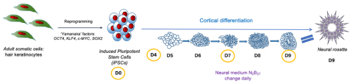
The central nervous system develops from a monolayer of neuroepithelial cells which folds to form the neural tube. Neural stem cells organise themselves within the tube to form neural rosettes, rose-like structures which have been reported both in vivo and in vitro (Hříbková et al. 2018). Recent advances in in vitro neuronal differentiation and organoid technology provide a model system for addressing how normal development of these rosettes is disrupted in conditions such as autism or epilepsy and for dissecting the molecular mechanisms governing these changes. Induced pluripotent stem cells (iPSCs), adult somatic cells reprogrammed back into their pluripotent stem cell stage, can be induced to differentiate into specific cell fates (Fig.1). Using this approach, it has been shown that iPSCs generated from individuals with autism show significant cellular and molecular abnormalities (Adhya et al. 2021). Intriguingly, defects begin much earlier than expected. Autistic iPSCs form abnormal neural rosette structures long before neural stem cells differentiate towards excitatory/inhibitory neurons (Adhya et al. 2021), but how this occurs at the molecular level remains poorly understood.
My project: FOXG1, a transcription factor implicated in epilepsy and autism
The aim of my project was to determine if atypical neural rosette structures form in early cortical organoids from FOXG1-mutant iPSCs. FOXG1 is an essential transcription factor responsible for normal neurodevelopment. Mutations in FOXG1 are significantly associated with syndromic forms of autism and ~90% of FOXG1 syndrome patients show epileptic seizures (Seltzer et al. 2014). Therefore, autism and epilepsy are considered comorbid.
Methods and techniques learned
During my project, I learned how to perform tissue culture (TC): I grew iPSCs and differentiated them into neurons. Working in the sterile TC hood made me a better scientist as I became more conscious about ways to prevent contamination, which is especially important when handling live cell cultures.
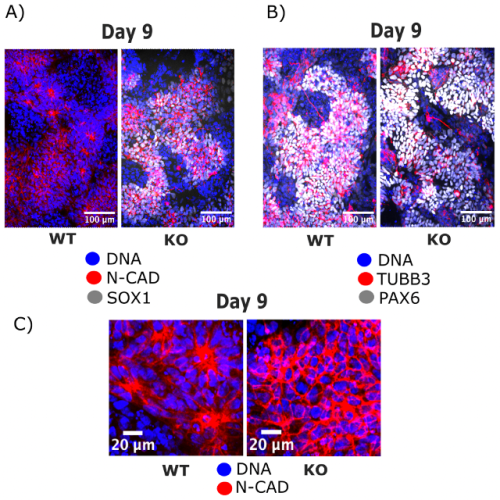
After successfully growing the iPSCs, I fixed them before differentiation and then at several stages during neural rosette formation (Fig. 1). The fixed cells were processed for immunofluorescence (IF) imaging and quantitative polymerase chain reaction (qPCR) to determine morphological changes that take place during cortical differentiation and to see its effect on gene expression, respectively. I isolated RNA using the Mini prep kit, but unfortunately, was unable to continue with the qPCR as some essential components did not arrive in time. Nevertheless, we got interesting results from the imaging alone (Fig. 2). Immunocytochemistry staining was another important lab skill I gained during the studentship. After surveying the current literature, I chose antibodies that reveal morphological features of neural rosettes: SOX1 as an early differentiation marker, PAX6 as a well-established marker of cortical neurons, N-cadherin as a rosette lumen marker, and TUBB3 as a neural cytoskeletal marker. Additionally, DAPI was used as nuclear counterstain.
Fig. 1 shows the outline and timeline of my project. iPSCs were generated by reprogramming adult somatic cells with ‘Yamanaka factors’ into pluripotent stem cells. The iPSCs were subsequently differentiated into neurons by placing them in a neural medium. The cells were fixed and subjected to IF at day 0, 4, 7 and 9 of the cortical differentiation. Neural rosettes form at day 9.
Apart from learning new lab techniques, I was introduced to image analysis softwares such as Fiji and CellProfiler. Furthermore, I was fully immersed into the activities of the research group, including weekly journal club, group meetings and workshops. I had several presentations during the group meetings which helped me improve my ability to discuss scientific concepts and results. All these skills will be useful for the rest of my MSci degree and for a future PhD.
Project outcomes
iPSCs stained with N-cadherin and SOX1 (Fig.2A) or with TUBB3 and PAX6 (Fig.2B) revealed a phenotype for the FOXG1 mutation. Zooming in on the rosette lumen stained with N-cadherin and nuclei stained with DAPI (Fig.2C) showed that neural rosettes do not form at day 9 without FOXG1 while they form in control iPSCs. Interestingly, N-cadherin, PAX6 and TUBB3 stainings reveal that FOXG1-mutant cells at day 9 look like glial progenitor cells, precursors that should form later in the organoid. Altogether, these findings provoke numerous questions about underlying mechanisms of these morphological changes, effect on gene expression and further differentiation into excitatory/inhibitory neurons, that will be researched by the lab in the future. If I had more time in the lab, I would have looked into other gene mutations significant for the comorbidity of autism and epilepsy that may affect the formation of neural rosettes, namely KMT2A and FGFR2. That would help us get a more robust understanding of the shared neurodevelopmental defects of these disorders.
Fig. 2: IF imaging of iPSCs at day 9 of cortical differentiation shows the disruption in neural rosette formation that occurs in FOXG1-mutant cells (KO) while neural rosettes develop normally in control cells (WT).
Summary
Overall, the summer studentship was an amazing experience as I designed my own experiments that I saw through from the beginning to the end. Along the way I gained valuable skills in the lab and research in general. This project introduced me to the exciting field of organoid research that is redefining the way we study neurodevelopmental disorders, and I would like to continue with it in my scientific future. I would strongly recommend applying for the Gurdon grant to students interested in delving deeper into a developmental biology topic and in experiencing what being a scientist entails.
CITED2 is a Conserved Regulator of the Uterine-Placental Interface Marija Kuna, Pramod Dhakal, Khursheed Iqbal, Esteban M. Dominguez, Lindsey N. Kent, Masanaga Muto, Ayelen Moreno-Irusta, Keisuke Kozai, Kaela M. Varberg, Hiroaki Okae, Takahiro Arima, Henry M. Sucov, Michael J. Soares
UMAP scatterplots of multiome (snRNA-ATACseq) data of invading trophoblast from Arutyunyan, et al.
Spatially resolved single-cell multiomics map of human trophoblast differentiation in early pregnancy Anna Arutyunyan, Kenny Roberts, Megan A Sheridan, Ilia Kats, Luz Garcia-Alonso, Britta Velten, Regina Hoo, Kevin Troulé Lozano, Louis-Francois Handfield, Luca Marconato, Elizabeth Tuck, Lucy Gardner, Cecilia Icoresi Mazzeo, Iva Kelava, Elena Prigmore, Sarah A Teichmann, Omer Ali Bayraktar, Ashley Moffett, Oliver Stegle, Margherita Y Turco, Roser Vento-Tormo
Cellular Maturation of Oligodendrocytes is Governed by Transient Gene Melting Kevin C. Allan, Tyler E. Miller, Andrew R. Morton, Marissa A. Scavuzzo, Matthew S. Elitt, Benjamin L.L. Clayton, Lucille R. Hu, Jost K. Vrabic, Hannah E. Olsen, Daniel C. Factor, Jonathan E. Henninger, Richard A. Young, Charles Y. Lin, Peter C. Scacheri, Paul J. Tesar doi: https://doi.org/10.1101/2022.11.17.516981
Expression bias in retinoic acid responsive genes defines variations in neural differentiation of human pluripotent stem cells Suel-Kee Kim, Seungmae Seo, Genevieve Stein-O’Brien, Amritha Jaishankar, Kazuya Ogawa, Nicola Micali, Victor Luria, Amir Karger, Yanhong Wang, Thomas M. Hyde, Joel E. Kleinman, Ty Voss, Elana J. Fertig, Joo-Heon Shin, Roland Bürli, Alan J. Cross, Nicholas J. Brandon, Daniel R. Weinberger, Joshua G. Chenoweth, Daniel J. Hoeppner, Nenad Sestan, Carlo Colantuoni, Ronald D. McKay
Adaptation to ex vivo culture drives human haematopoietic stem cell loss of repopulation capacity in a cell cycle independent manner Carys S. Johnson, Kendig Sham, Serena Belluschi, Xiaonan Wang, Winnie Lau, Kerstin B. Kaufmann, Gabriela Krivdova, Emily F. Calderbank, Nicole Mende, Jessica McLeod, Giovanna Mantica, Matthew J. Williams, Charlotte Grey-Wilson, Michael Drakopoulos, Shubhankar Sinha, Evangelia Diamanti, Christina Basford, Anthony R. Green, Nicola K. Wilson, Steven J. Howe, John E. Dick, Bertie Göttgens, Natalie Francis, Elisa Laurenti
Spatiotemporal coordination of stem cell behavior following alveolar injury Maurizio Chioccioli, Sumner Magruder, John E. McDonough, Jessica Nouws, Tao Yang, David Gonzalez, Lucia Borriello, Brian Traub, Xianjun Ye, Caroline E. Hendry, David Entenberg, Smita Krishnaswamy, Naftali Kaminski, Maor Sauler
A human mitofusin 2 mutation causes mitophagic cardiomyopathy Antonietta Franco, Jiajia Li, Daniel P. Kelly, Ray E. Hershberger, Ali J. Marian, Renate M. Lewis, Moshi Song, Xiawei Dang, Alina D. Schmidt, Mary E. Mathyer, Cristina de Guzman Strong, Gerald W. Dorn II
 (No Ratings Yet)
 (4 votes)