Behind the paper: Using OptoShroom3 to alter organoid shape with light
Posted by Guillermo Martinez-Ara, on 15 December 2022
Guillermo Martínez-Ara and colleagues from Miki Ebisuya lab have developed a new optogenetic tool that induces apical constriction in mammalian epithelia. The new tool called OptoShroom3 induces tissue folding in epithelial colonies and provokes changes in curvature and thickness in neural organoids. The authors showed that this optogenetic approach gives spatiotemporal control to manipulate the structure of mammalian tissues.
How did you get started on this project?
This project started before I got to the lab, back when my supervisor, Miki Ebisuya, was based in Japan. Two very talented postdocs spent three years exploring and developing ideas on how to create a tool to control morphogenesis.
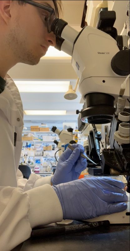
I was deeply interested in the creative approaches of synthetic biology and the development of new biological tools, so I did my master’s in systems and synthetic biology. Then, I found about Miki’s lab and thought that their application of synthetic biology to study development was fascinating. Luckily, my interests matched with Miki’s and I was invited to join the project after the lab moved to EMBL Barcelona. For my PhD project, I joined forces with Núria Taberner (one of the talented postdocs) to develop a tool to provoke apical constriction in mammalian systems. I thought this project was super exciting. Using optogenetics and synthetic biology to manipulate tissue structure – I was totally in!
Can you summarise your findings?
Briefly, we developed a new optogenetic tool (OptoShroom3) that allowed spatiotemporal control of apical constriction in mammalian tissues. To demonstrate its potential applications, we showed that OptoShroom3 could be used to alter tissue shape in epithelial cell lines, to induce folding in colonies, and to change the shape of neural organoids. We developed the first tool (to our knowledge) that could be easily applied to manipulate mammalian tissue structure through the control of biological forces.
What are the advantages of using OptoShroom3 compared with the previously existing optogenetic tools used for inducing actomyosin constriction?
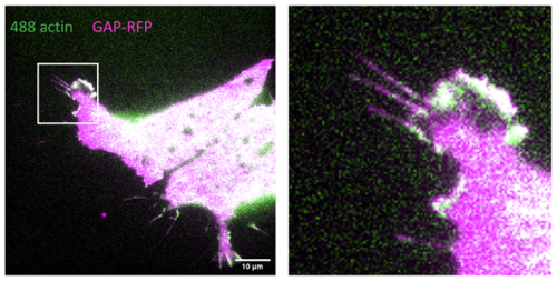
Most of the existing tools for the induction of actomyosin constriction relied on plasma membrane recruitment. This means that the factors used to induce contractility (normally related to the Rho pathway), are brought to the cell membrane upon illumination. This, in principle, gives more freedom to induce contractility in any area of the plasma membrane. However, when it comes to inducing tissue shape changes it becomes more challenging. If we want to induce constriction in one area of the cells (say apical surface) we would require very precise illumination of a single plane in the tissue, which requires two photon microscopy (as the lab of Stefano de Renzis had previously done). When the shape of tissues becomes convoluted, like we see in organoids, this becomes even more complicated, if not just impossible. We cannot easily create multiphoton stimulation patterns in 3D shapes, and even if it is achievable, this technique is not easily accessible for most scientists.
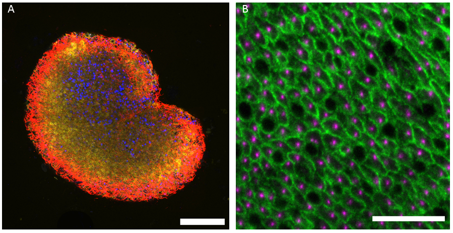
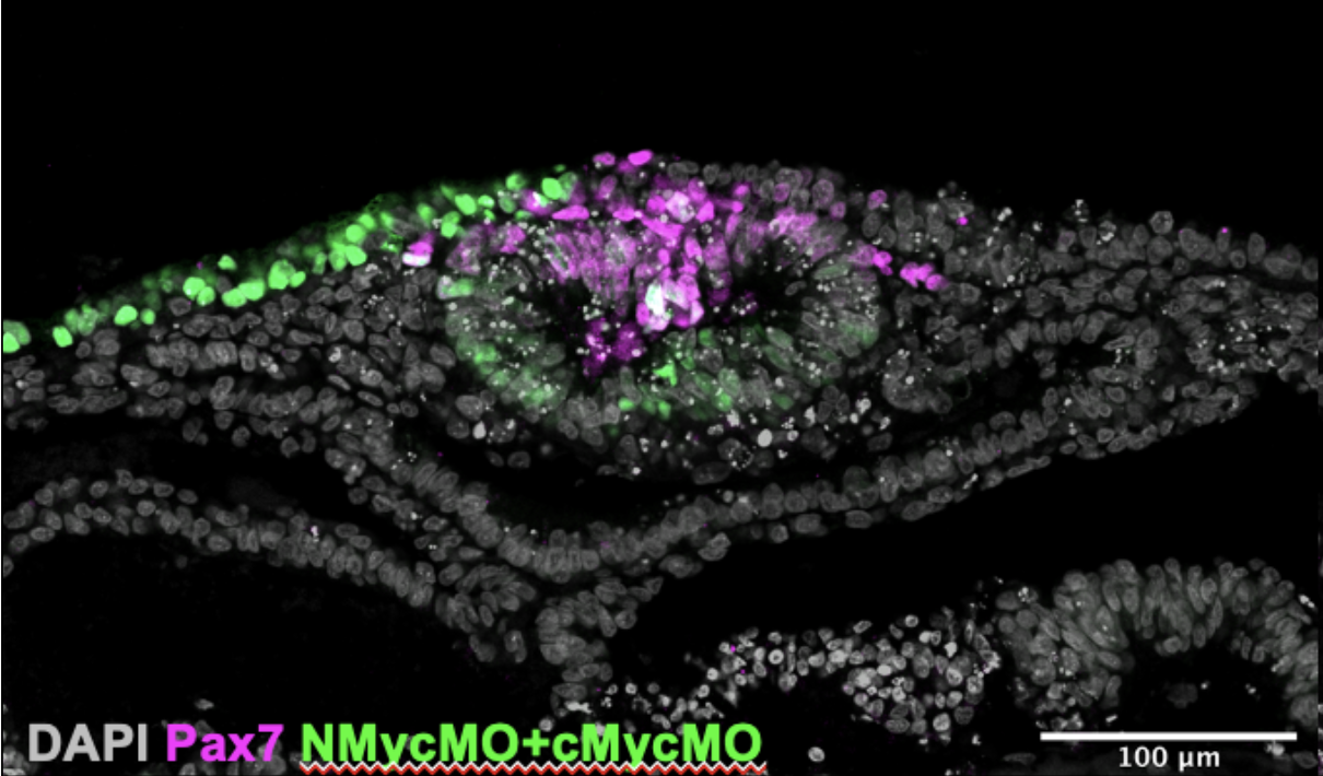
The advantage of OptoShroom3 is that it directs constriction specifically to the apical surface. Then, if all the tissue is stimulated, only the apical area of the cells will constrict, inducing selective constriction. This allowed us to induce changes in the shape of organoids with complex structures such as optic vesicle organoids. We stimulated the whole vesicle organoid to provoke localised constriction on the apical side, which led to changes in shape.
Do you have any tips or tricks for researchers using OptoShroom3 in the future?
Whenever someone contacts us with the aim of using OptoShroom3 in their own system, I normally reply something like this:
How well OptoShroom3 works in your system will depend on two things.
1. How epithelial the cell line is, because OptoShroom3 relies on the apical cytoskeletal structures, specifically on the localization of actin along the apical side of cell-cell junctions. When this structure is present, the N-terminal part of Shroom3 will localise there and then OptoShroom3 will be able to induce constriction locally on the apical side. Without epithelialization, there’s no apical constriction.
2. The ratio of expression between both components. Normally, we try to maximise the expression of both the NShroom3 component and the CShroom3 part. However, very high expression of CShroom3 seems to be somewhat toxic, so the optimal levels for the tool to work is high NShroom3-iLID and medium-low SspB-CShroom3 expression.
Finally, I would suggest people to be patient with optogenetic tools. At the beginning, they may not seem easy to use. There are several parameters to be optimised: laser intensity, intervals of stimulation and rest, temporal resolution when imaging… Once you get an idea of how much laser power you need to activate the tool on your microscope, and for how long you need to stimulate to see changes in shape, things will be much easier. I guess this applies to all optogenetic tools. You should give yourself some time to play around with these parameters!
When doing the research, did you have any particular result or eureka moment that has stuck with you?
Yes! I remember clearly the morning when I first saw the deformation of an optic vesicle in the confocal microscope room. The deformation was very clear, and it was finally a direct confirmation that we could use OptoShroom3 to alter tissue shape in organoids, which was one of our main goals! I don’t consider it an ‘eureka’ moment, but rather a happy memory of when an experiment finally works after a lot of effort.
And what about the flipside: any moments of frustration or despair?
There are always ups and downs in academic research. For us, in the first review of the OptoShroom3 publication reviewers were asking for some more experiments and controls. This would normally be ok (we knew it was a possibility), but then one month after getting these requests the confocal microscope I was using for all the experiments suddenly broke. To make it worse, it took three months to get it fixed. During that time, I tested other microscopes around in the building, but because we wanted to compare the new results to the original ones, we needed to make sure that the amount of light used for stimulation was the same. Of course, different microscopes, with different designs, optics and laser sources could not be easily compared. In the end, I spent those three months testing microscopes, planning the new experiments and, perhaps, building patience and resilience. Overall, we had a three-month delay for the project, which back then seemed catastrophic, but later on seems like it was not the end of the world.
Image analysis is a key part of your work, can you tell us about the custom-made pipeline and any tips for researchers looking to use or repurpose the code?
I tried my best to leave clear comments in the code and make it publicly available so that people can extract whatever is useful for them. However, as we mention in the GitHub page, my main aim was to make functional code for the paper, therefore it is possible that some people will struggle to understand it or use it. In that case, my main tip is to contact me! I’m always happy to help and discuss the method with researchers interested in biological image analysis.
Are you testing OptoShroom3 in vivo?
As far as I know, Miki’s lab is not planning on working directly on embryos in the near future. The lab focuses on the use of in vitro systems for synthetic developmental biology. I am also not planning to work in vivo any time soon. However, we do have some collaborators working to create some OptoShroom3 transgenic mice, which is incredibly exciting!
Where will this story take the lab?
As I said, our first aim was to make a tool that we could use to perturb tissue structure using biological forces. Now that is working the logical question is: What do we do with it?
We believe that these types of tools open the door to ask new questions, or at least to ask old questions from a different perspective. We are especially interested in how changes in tissue structure can impact other developmental processes, and we believe that organoids are a great model to ask these questions!
What is next for you after this paper?
While Miki’s lab will continue to explore how OptoShroom3 can be used to study possible feedbacks between shape and other developmental processes, I am getting ready to move to a postdoc position, in which I’m hoping to develop more exciting tools to manipulate or even program tissues!
 (2 votes)
 (No Ratings Yet)