Dr David A. Menassa and Professor Diego Gomez-Nicola at the University of Southampton, UK recently published an article in Developmental Cell where they reveal how the microglial population colonises the human frontal cortex. Dr Menassa gave us a behind the scenes look at how the story came together.
How did you get started on this project?
I joined the Gomez-Nicola lab in October 2017 as a research fellow. The funding had been granted by the Leverhulme Trust prior to my arrival, and all was in place to set the human lifespan study in motion.
The task of securing healthy human tissues from whole embryos to advanced ageing was a challenging one. It took several years to establish the tissue collection and 12 tissue resources within the UK and abroad were consulted. There was a substantial administrative load. Human tissue work in general is fraught with challenges: even when one acquires the tissues, there are no guarantees that the antigenic targets will have been preserved. The assumption is that they should be, but there are many factors that come into play making work with human tissues a good exercise in patience, focus and resilience. Once the frustrations were out of the way, the project kicked off at full speed. I was lucky to be part of a very supportive laboratory environment, working alongside a team of intramural and extramural collaborators. This paper is evidence of great teamwork and we are delighted to be able to share our findings with the scientific community.
What was already known about the developmental dynamics of microglia in the human brain prior to your work?
Two studies on adult human tissues (including one from the Gomez-Nicola lab) were published in 2017 showing that microglial turnover in humans is much faster than previously thought and that the entire population is renewed several times during a lifetime [2, 3]. Additionally, microglial proliferative dynamics increase during pathology and have been the target of therapies to limit proliferation in neurodegenerative disease particularly Alzheimer’s disease models [4]. Very little was known about microglial dynamics during the critical stages of human brain development and the early postnatal age, and this was largely due to the limited availability of tissues for research.
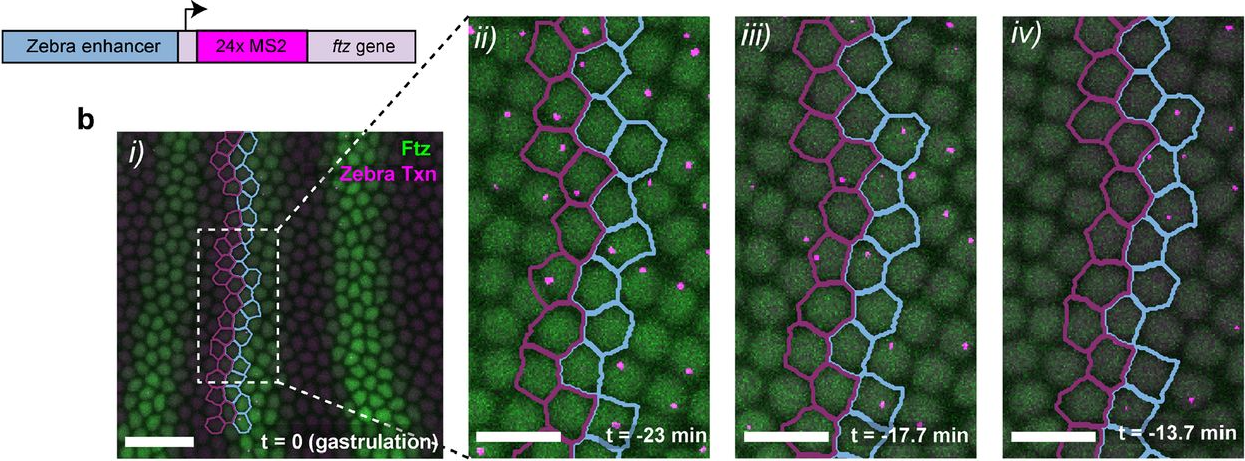
In the latter half of 2020, two papers profiled microglial developmental states during early embryonic and early fetal lives in humans [5, 6]. These studies showed that ontogenic pathways were conserved between mice and humans and that microglia became immunocompetent from as early as the 11th week of gestation. In 2022, the regional microglial transcriptome was described in humans between 8-23 weeks of gestation: this was the first study to look at specific anatomical regions such as the cortex and the cerebellum [7]. Altogether, these studies offered an important view of specific temporal windows during development. In our study, we covered the entire human lifespan, detailing the spatiotemporal dynamics of microglia from the moment these cells arrive to the human developing brain at 4 pcw up until they turnover very slowly in adult life. We profiled these cells in all layers of the cortex in the frontal lobe, the brain region responsible for higher order cognitive and executive function in humans.
Can you summarise your findings?
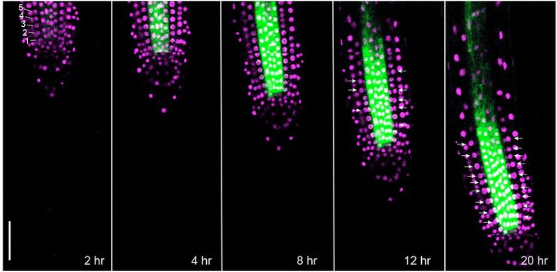
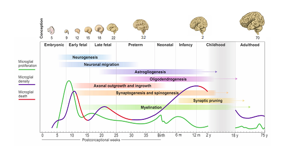
There are four important temporal windows during human brain development when we see microglial changes: the first one is when microglia arrive to the brain with the onset of circulation at 4 pcw; the second is at the transition between embryonic to fetal life at 9-10 pcw; the third is at 13-14 pcw and the fourth is during early childhood between 0.5-1 year of age. Microglial expansion in the brain is via a wave-like pattern of cycles of migration and proliferation, increasing cell number, which is further refined by selective cell-death. The changes in microglial dynamics are aligned with co-occurring neurodevelopmental processes. We found no sex differences during these developmental stages. In adult life, the population self-renews at a relatively steady rate until advanced ageing. This pattern of colonisation is unique to humans and is strikingly different than what we see in mouse.
Figure 1 Microglial dynamics across the human lifespan. This graphical abstract was modified from [1] and shows the proliferation, cell number and cell death changes of microglia between 3 postconceptional weeks and 75 years of life. The arrows represent ongoing processes.
When doing the research, did you have any particular result or eureka moment that has stuck with you?
Seeing the first fully labelled embryo for microglia, other tissue-resident macrophages and proliferative cells in the brain matter, liver, spleen, spinal cord, and heart all visible in one slide was a special moment. I remember sending Dr Gomez-Nicola the first image and we were both excited and thrilled that it was all worth the wait; the signal was very clear and from that point onwards, we were confident that the project was heading in the right direction. The second moment was when we had all the data unblinded and plotted across the lifespan for all tissues: it was a roadmap, and we could see the exact pattern that microglia followed prenatally and postnatally.
And what about the flipside? Any moment of frustration or despair?
Human tissue research is challenging, but the end-result is very rewarding. In this paper, we could not obtain samples with consent for research use from late childhood and adolescence. Therefore, we are missing part of the whole picture. As the pattern of growth and myelination of the frontal lobe is unique in humans during these stages, it will be interesting to see what happens to microglial dynamics.
Where will this story take you next?
Now that we know the temporal windows of relevance in microglial dynamics, the next step is to begin dissecting how microglial cells interact with the neurodevelopmental environment. This could be by elucidating the cues that these cells get from the brain at each step and in this way, we can start identifying why the population behaves the way it does. This is important as microglia are part of the neuropathology of neurodevelopmental disorders. To find out how these cells contribute to normal and altered neurodevelopment, we need to dissect the signalling pathways between them and developing neurons.
What is next for you after this paper?
I am currently stipendiary lecturer of neurophysiology and neuroscience at the Queen’s College, University of Oxford and visiting researcher in neurodevelopment at the Croatian Institute for Brain Research, University of Zagreb. I am continuing my research on microglial development and how this relates to neurodevelopmental disorders. I am applying for tenured positions in the UK and Europe to set up my own research group.
Bibliography
1. Menassa, D.A., et al., The spatiotemporal dynamics of microglia across the human lifespan. Dev Cell, 2022.
2. Askew, K., et al., Coupled Proliferation and Apoptosis Maintain the Rapid Turnover of Microglia in the Adult Brain. Cell Rep, 2017. 18(2): p. 391-405.
3. Réu, P., et al., The Lifespan and Turnover of Microglia in the Human Brain. Cell Rep, 2017. 20(4): p. 779-784.
4. Olmos-Alonso, A., et al., Pharmacological targeting of CSF1R inhibits microglial proliferation and prevents the progression of Alzheimer’s-like pathology. Brain, 2016. 139(Pt 3): p. 891-907.
5. Bian, Z., et al., Deciphering human macrophage development at single-cell resolution. Nature, 2020. 582(7813): p. 571-576.
6. Kracht, L., et al., Human fetal microglia acquire homeostatic immune-sensing properties early in development. Science, 2020. 369(6503): p. 530-537.
7. Li, Y., et al., Decoding the temporal and regional specification of microglia in the developing human brain. Cell Stem Cell, 2022. 29(4): p. 620-634.e6.
by Haneen Alsehli, Shrinidhi Madhusudan, Marie-Christin Leitner and Katherina Tavernini
For most of us, this was the first in-person conference since starting our PhDs, or since the COVID-19 pandemic hit. All of us were very excited to get inspired, network and talk about our work by presenting our projects, either with a poster or giving a selected talk.
First day, first lunch and we had already met new people, we started chatting and had a good time. Despite being at different stages of our PhDs, we shared the same thoughts and fun stories – we laughed a lot! From that moment on, we had a great conference group, where the friendships will last beyond the meeting.
This conference was located in a beautiful setting just a one hour train ride south of London. At Wotton House,we could explore the beautiful natural surroundings during breaks, but also study, work and listen to talks in a historic setting. Overall, we were exceptionally well taken care of during this meeting.
During the talks and poster sessions, we had the chance to listen to outstanding talks from experts in the field. We all appreciated the amount of unpublished data, and for Haneen, who is writing up at the moment, this kind of literature review was very useful. For others, hearing about new methods in talks that were outside our areas of expertise were beneficial, as we began to think about translating them to our own work.Furthermore, the industry sponsors at the event were also very accommodating and we look forward to taking their expertise and methodology back to our institutes.
Since we were all presenting our work, we all experienced the great atmosphere of this meeting. We had fruitful discussions at our posters that not only allowed us to gather ideas for future experiments, but to get feedback on our work from an international audience. We discussed our work with group leaders, representatives from industry, postdocs and of course, fellow students, which we appreciated and found very rewarding.
This meeting was highly interactive and we all acknowledge the effort that was made to foster interactions among the participants. The ‘speed-networking’ event forced some of us outside of our comfort zones, to interact with PIs and senior scientists. Each dinner, we were randomly assigned to tables and had a dedicated seat. Although being apprehensive initially about the place cards, we soon came to appreciate it, especially after having interesting conversations with participants that we might not have otherwise met. With this setting, we had the opportunity to talk to different group leaders, students, editors; to discuss science, ask questions and spin ideas about future developments.
We all really appreciated the effort the organisers took to make this conference as safe as possible. We felt that Covid measures were taken seriously, which allowed for great and almost carefree networking.
If you ever have the chance to participate in this conference, go for it! You will hear about the latest amazing research, make new friends, interact and network with experts in the field from all over the world. We would definitely recommend attending and meeting great people and potential future collaborators along the way.
Doing great science depends on teamwork, whether this is within the lab or in collaboration with other labs. However, sometimes the resources that support our work can be overlooked. Our ‘Featured resource’ series aims to shine a light on these unsung heroes of the science world. In our latest article, we hear from Tanya Z. Berardini, Ph.D (TAIR Director) and Leonore Reiser, Ph.D (Senior Scientific Curator)] who describe the work of TAIR.
The Arabidopsis Information Resource (TAIR) was established in 1999 with US National Science Foundation funding and the goal of creating a database of genetic and molecular biology data for the model higher plant Arabidopsis thaliana. Arabidopsis was the first plant genome to be sequenced (1). Since then, over twenty years of research have continually improved the sequence and functional annotation of a genome that serves as a reference for an ever-increasing number of newly sequenced plant genomes (2). TAIR integrates, interconnects, and consolidates information from peer-reviewed published literature with sequence and stock information (3) so that researchers can spend less time searching for information and more time developing and testing hypotheses guided by work that has already been done.
Who runs the resource?
TAIR was created at the Department of Plant Biology of the Carnegie Institute for Science and the National Center for Genome Resources (NCGR) and supported by the NSF from 1999 to 2013. Since 2014, it has been administered and maintained by the non-profit organization, Phoenix Bioinformatics (4). Phoenix has a small team of dedicated data curation scientists, who meticulously curate and update TAIR data, and software engineers, who maintain and update TAIR’s database and tools. Newly curated information from the literature is added weekly.
Where does funding come from?
Since 2014, TAIR’s operations have been funded by subscriptions from academic and other non-profit institutions, individual researchers, corporations and countries (e.g., China through its National Science and Technology Library). Institutional subscriptions are priced based on past year usage, so that institutions that use the resource more contribute more to its maintenance and growth. TAIR’s subscription support is as global as its user base. Phoenix Bioinformatics has additional funding from the NSF and the Sloan Foundation for other aspects of its work.
Can one access TAIR without a subscription?
Unlimited access to TAIR’s data and tools almost always requires a subscription. However, TAIR provides a limited number of free page views for occasional users each month. This is similar to the monthly allotment of complimentary articles often offered by online newspapers and magazines. After the monthly limit is reached a subscription is encouraged. Upon request, TAIR grants full access for teachers and students at non-subscribing institutions for classes that use TAIR in their curriculum. US Historically Black Colleges and Universities (HBCUs) are granted free access. Finally, we grant country-wide access to those countries that fall into the ‘low-income economies’ categorization of the World Bank.
What tools and resources are available for researchers?
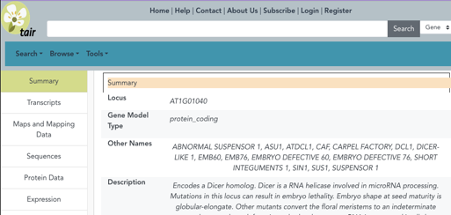
The TAIR locus page is a treasure trove that consolidates in-house data, data pulled from other resources by APIs, and links to external resources with complementary information. New articles, gene symbols and full names, gene summaries, Gene Ontology (GO) and Plant Ontology (PO) annotations, germplasms and phenotypes based on those articles are added by data curation scientists to a subset of loci on a weekly basis.
Researchers can find information for single genes or they can download data (sequences, descriptions, GO annotations) for sets of genes or even the whole genome.
There are two types of quarterly data releases: (A) Public data releases are a year old and are released for public reuse with a CC-BY reuse license. (B) Subscriber data releases reflect the most recent year’s data.
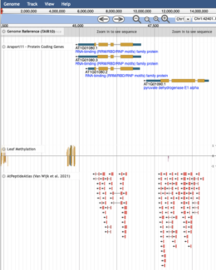
TAIR provides data search and browsing services, data analysis tools, and data visualization tools, such as genome browsers (e.g., JBrowse) and a BLAST service that includes unique datasets.
JBrowse at TAIR, 3 out of 250+ tracks shown
How can the community contribute?
Make data FAIR (5).
Use AGI identifiers for the Arabidopsis genes in papers!
Use stock identifiers from NASC and ABRC for seed and DNA stocks. They allow us to link these stocks unambiguously to locus records at TAIR.
Register gene symbols, use the symbols for genes that already exist and check that symbols you want to use are not already in use.
Beta site (in progress): New look of the TAIR Locus Page
Any hidden gems, features that are new, or that researchers might be less aware of?
The biggest hidden gem is that if you email us, we will respond within 24 hrs during a regularly scheduled work week.
Not only can one view information for one gene at a time, one can also upload a list of genes and retrieve gene descriptions, sequences, and GO annotations (and other types of data) in bulk.
The TAIR YouTube channel has tutorials and webinars of different lengths explaining various features of the resource. Watch a video and learn something new.
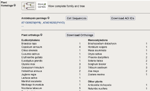
Access to PhyloGenes (www.phylogenes.org) is integrated into the TAIR Locus page. You can link to the Panther-based gene family, explore experimental and phylogenetically-inferred functional information for gene family members in Arabidopsis and 29 other plants, and take advantage of experimental annotations made in Arabidopsis and other model organisms.
Plant Homolog Section of TAIR Locus Page
The TAIR Job listings page is very active with about three new listings posted a week. Some labs recruit almost exclusively by posting their grad student and post-doc openings at https://www.arabidopsis.org/news/jobs.jsp. All openings are also shared through our Twitter account (@tair_news).
References:
The Arabidopsis Genome Initiative. Analysis of the genome sequence of the flowering plant Arabidopsis thaliana. Nature. 408, 796–815 (2000). doi.org/10.1038/35048692
Nicholas J Provart, Siobhan M Brady, Geraint Parry, Robert J Schmitz, Christine Queitsch, Dario Bonetta, Jamie Waese, Korbinian Schneeberger, Ann E Loraine. Anno genominis XX: 20 years of Arabidopsis genomics, The Plant Cell. 33(4):832–845 (2021). doi.org/10.1093/plcell/koaa038
Tanya Z. Berardini, Leonore Reiser, Donghui Li, Yarik Mezheritsky, Robert Muller, Emily Strait, Eva Huala. The Arabidopsis information resource: Making and mining the “gold standard” annotated reference plant genome. Genesis. 53(8):474-85 (2015). doi.org/10.1002/dvg.22877
Leonore Reiser, Tanya Z. Berardini, Donghui Li, Robert Muller, Emily M. Strait, Qian Li, Yarik Mezheritsky, Andrey Vetushko, Eva Huala. Sustainable funding for biocuration: The Arabidopsis Information Resource (TAIR) as a case study of a subscription-based funding model, Database. Volume 2016, (2016). doi.org/10.1093/database/baw018
Leonore Reiser, Lisa Harper, Michael Freeling, Bin Han, Sheng Luan. FAIR: A Call to Make Published Data More Findable, Accessible, Interoperable, and Reusable, Molecular Plant 11(9):1105-1108 (2018). doi.org/10.1016/j.molp.2018.07.005
Contributed by Tanya Z. Berardini, Ph.D (TAIR Director) and Leonore Reiser, Ph.D (Senior Scientific Curator)
Rnf20 shapes the endothelial control of heart morphogenesis and function Linda Kessler, Rui Gao, Nalan Tetik-Elsherbiny, Olga Lityagina, Azhar Zhailauova, Yonggang Ren, Felix A. Trogisch, Julio Cordero, Yanliang Dou, Yinuo Wang, Evgeny Chichelnitskiy, Joscha Alexander Kraske, Patricia Laura Schäfer, Chi-Chung Wu, Guillermo Barreto, Michael Potente, Thomas Wieland, Roxana Ola, Joerg Heineke, Gergana Dobreva
Universal DNA methylation age across mammalian tissues A.T. Lu, Z. Fei, A. Haghani, T.R. Robeck, J.A. Zoller, C.Z. Li, R. Lowe, Q. Yan, J. Zhang, H. Vu, J. Ablaeva, V.A. Acosta-Rodriguez, D.M. Adams, J. Almunia, A. Aloysius, R. Ardehali, A. Arneson, C.S. Baker, G. Banks, K. Belov, N.C. Bennett, P. Black, D.T. Blumstein, E.K. Bors, C.E. Breeze, R.T. Brooke, J.L. Brown, G. Carter, A. Caulton, J.M. Cavin, L. Chakrabarti, I. Chatzistamou, H. Chen, K. Cheng, P. Chiavellini, O.W. Choi, S. Clarke, L.N. Cooper, M.L. Cossette, J. Day, J. DeYoung, S. DiRocco, C. Dold, E.E. Ehmke, C.K. Emmons, S. Emmrich, E. Erbay, C. Erlacher-Reid, C.G. Faulkes, S.H. Ferguson, C.J. Finno, J.E. Flower, J.M. Gaillard, E. Garde, L. Gerber, V.N. Gladyshev, V. Gorbunova, R.G. Goya, M.J. Grant, C.B. Green, E.N. Hales, M.B. Hanson, D.W. Hart, M. Haulena, K. Herrick, A.N. Hogan, C.J. Hogg, T.A. Hore, T. Huang, J.C. Izpisua Belmonte, A.J. Jasinska, G. Jones, E. Jourdain, O. Kashpur, H. Katcher, E. Katsumata, V. Kaza, H. Kiaris, M.S. Kobor, P. Kordowitzki, W.R. Koski, M. Kruetzen, S.B. Kwon, B. Larison, S.G. Lee, M. Lehmann, J.F. Lemaitre, A.J. Levine, C. Li, X. Li, A.R. Lim, D.T.S. Lin, D.M. Lindemann, T.J. Little, N. Macoretta, D. Maddox, C.O. Matkin, J.A. Mattison, M. McClure, J. Mergl, J.J. Meudt, G.A. Montano, K. Mozhui, J. Munshi-South, A. Naderi, M. Nagy, P. Narayan, P.W. Nathanielsz, N.B. Nguyen, C. Niehrs, J.K. O’Brien, P. O’Tierney Ginn, D.T. Odom, A.G. Ophir, S. Osborn, E.A. Ostrander, K.M. Parsons, K.C. Paul, M. Pellegrini, K.J. Peters, A.B. Pedersen, J.L. Petersen, D.W. Pietersen, G.M. Pinho, J. Plassais, J.R. Poganik, N.A. Prado, P. Reddy, B. Rey, B.R. Ritz, J. Robbins, M. Rodriguez, J. Russell, E. Rydkina, L.L. Sailer, A.B. Salmon, A. Sanghavi, K.M. Schachtschneider, D. Schmitt, T. Schmitt, L. Schomacher, L.B. Schook, K.E. Sears, A.W. Seifert, A. Seluanov, A.B.A. Shafer, D. Shanmuganayagam, A.V. Shindyapina, M. Simmons, K. Singh, I. Sinha, J. Slone, R.G. Snell, E. Soltanmaohammadi, M.L. Spangler, M.C. Spriggs, L. Staggs, N. Stedman, K.J. Steinman, D.T. Stewart, V.J. Sugrue, B. Szladovits, J.S. Takahashi, M. Takasugi, E.C. Teeling, M.J. Thompson, B. Van Bonn, S.C. Vernes, D. Villar, H.V. Vinters, M.C. Wallingford, N. Wang, R.K. Wayne, G.S. Wilkinson, C.K. Williams, R.W. Williams, X.W. Yang, M. Yao, B.G. Young, B. Zhang, Z. Zhang, P. Zhao, Y. Zhao, W. Zhou, J. Zimmermann, J. Ernst, K. Raj, S. Horvath
Discovery and characterization of LNCSOX17 as an essential regulator in human endoderm formation Alexandro Landshammer, Adriano Bolondi, Helene Kretzmer, Christian Much, René Buschow, Alina Rose, Hua-Jun Wu, Sebastian Mackowiak, Bjoern Braendl, Pay Giesselmann, Rosaria Tornisiello, Krishna Mohan Parsi, Jack Huey, Thorsten Mielke, David Meierhofer, René Maehr, Denes Hnisz, Franziska Michor, John L. Rinn, Alexander Meissner
The Neuroscience Multi-Omic Archive: A BRAIN Initiative resource for single-cell transcriptomic and epigenomic data from the mammalian brain Seth A. Ament, Ricky S. Adkins, Robert Carter, Elena Chrysostomou, Carlo Colantuoni, Jonathan Crabtree, Heather H. Creasy, Kylee Degatano, Victor Felix, Peter Gandt, Gwenn A. Garden, Michelle Giglio, Brian R. Herb, Farzaneh Khajouei, Elizabeth Kiernan, Carrie McCracken, Kennedy McDaniel, Suvarna Nadendla, Lance Nickel, Dustin Olley, Joshua Orvis, Joseph P. Receveur, Mike Schor, Timothy L. Tickle, Jessica Way, Ronna Hertzano, Anup A. Mahurkar, Owen R White
Transient Polycomb activity represses developmental genes in growing oocytes Ellen G. Jarred, Zhipeng Qu, Tesha Tsai, Ruby Oberin, Sigrid Petautschnig, Heidi Bildsoe, Stephen Pederson, Qing-hua Zhang, Jessica M. Stringer, John Carroll, David K. Gardner, Maarten van den Buuse, Natalie A. Sims, William T. Gibson, David L. Adelson, Patrick S. Western
The imprinted Mir483 is a growth suppressor and metabolic regulator functioning through IGF1 Ionel Sandovici, Denise S. Fernandez-Twinn, Niamh Campbell, Wendy N. Cooper, Yoichi Sekita, Ilona Zvetkova, David Ferland-McCollough, Haydn M. Prosser, Lila M. Oyama, Danilo Cimadomo, Karina Barbosa de Queiroz, Cecilia S.K. Cheuk, Nicola M. Smith, Richard G. Kay, Katharina Hoelle, Noel H. Smith, Stefan H. Geyer, Lukas F. Reissig, Wolfgang J. Weninger, Kenneth Siddle, Anne E. Willis, Martin Bushell, Susan E. Ozanne, Miguel Constância
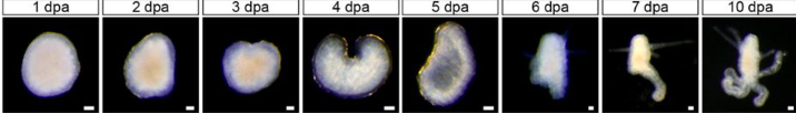
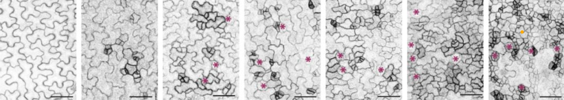
Cellular senescence modulates progenitor cell expansion during axolotl limb regeneration Qinghao Yu, Hannah E. Walters, Giovanni Pasquini, Sumeet Pal Singh, Daniel León-Periñán, Andreas Petzold, Preethi Kesavan, Cristina Subiran, Ines Garteizgogeascoa, Dunja Knapp, Anne Wagner, Andrea Bernardos, María Alfonso, Gayathri Nadar, Andreas Dahl, Volker Busskamp, Ramón Martínez-Máñez, Maximina H. Yun
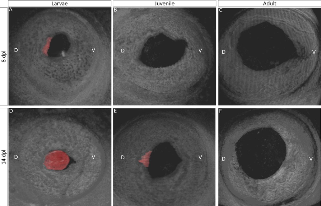
Effects of α-crystallin gene knockout on zebrafish lens development Mason Posner, Kelly L. Murray, Brandon Andrew, Stuart Brdicka, Alexis Roberts, Kirstan Franklin, Adil Hussen, Taylor Kaye, Emmaline Kepp, Mathew S. McDonald, Tyler Snodgrass, Keith Zientek, Larry L. David
Machine-learning dissection of Human Accelerated Regions in primate neurodevelopment Sean Whalen, Fumitaka Inoue, Hane Ryu, Tyler Fairr, Eirene Markenscoff-Papadimitriou, Kathleen Keough, Martin Kircher, Beth Martin, Beatriz Alvarado, Orry Elor, Dianne Laboy Cintron, Alex Williams, Md. Abul Hassan Samee, Sean Thomas, Robert Krencik, Erik M. Ullian, Arnold Kriegstein, John L. Rubenstein, Jay Shendure, Alex A. Pollen, Nadav Ahituv, Katherine S. Pollard
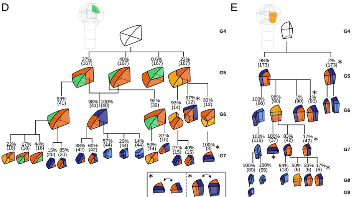
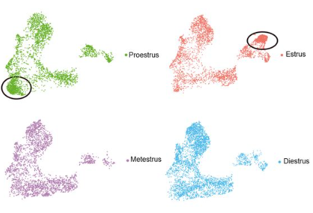
A single cell atlas of the cycling murine ovary ME Morris, MC Meinsohn, M Chauvin, HD Saatcioglu, A. Kashiwagi, NA. Sicher, NMP Nguyen, S Yuan, Rhian Stavely, M Hyun, PK Donahoe, B Sabatini, D Pépin
A CRISPR/Cas9-based enhancement of high-throughput single-cell transcriptomics Amitabh C. Pandey, Jon Bezney, Dante DeAscanis, Ethan Kirsch, Farin Ahmed, Austin Crinklaw, Kumari Sonal Choudhary, Tony Mandala, Jeffrey Deason, Jasmin Hamdi, Azeem Siddique, Sridhar Ranganathan, Phillip Ordoukhanian, Keith Brown, Jon Armstrong, Steven Head, Eric J. Topol
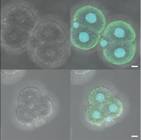
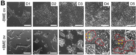
hPGCLCs in 2D culture using BME overlay method from Overeem, et al.
A human fetal lung cell atlas uncovers proximal-distal gradients of differentiation and key regulators of epithelial fates Peng He, Kyungtae Lim, Dawei Sun, Jan Patrick Pett, Quitz Jeng, Krzysztof Polanski, Ziqi Dong, Liam Bolt, Laura Richardson, Lira Mamanova, Monika Dabrowska, Anna Wilbrey-Clark, Elo Madissoon, Zewen Kelvin Tuong, Emma Dann, Chenqu Suo, Isaac Goh, Masahiro Yoshida, Marko Z Nikolić, Sam M Janes, Xiaoling He, Roger A Barker, Sarah A Teichmann, John C. Marioni, Kerstin B Meyer, Emma L Rawlins
Bat pluripotent stem cells reveal unique entanglement between host and viruses Marion Déjosez, Arturo Marin, Graham M. Hughes, Ariadna E. Morales, Carlos Godoy-Parejo, Jonathan Gray, Yiren Qin, Arun A. Singh, Hui Xu, Javier Juste, Carlos Ibáñez, Kris M. White, Romel Rosales, Nancy J. Francoeur, Robert P. Sebra, Dominic Alcock, Sébastien J. Puechmaille, Andrzej Pastusiak, Simon D.W. Frost, Michael Hiller, Richard A. Young, Emma C. Teeling, Adolfo García-Sastre, Thomas P. Zwaka
Transplantable human thyroid organoids generated from embryonic stem cells to rescue hypothyroidism Mírian Romitti, Adrien Tourneur, Barbara de Faria da Fonseca, Gilles Doumont, Pierre Gillotay, Xiao-Hui Liao, Sema Elif Eski, Gaetan Van Simaeys, Laura Chomette, Helene Lasolle, Olivier Monestier, Dominika Figini Kasprzyk, Vincent Detours, Sumeet Pal Singh, Serge Goldman, Samuel Refetoff, Sabine Costagliola
A single cell atlas of the cycling murine ovary ME Morris, MC Meinsohn, M Chauvin, HD Saatcioglu, A. Kashiwagi, NA. Sicher, NMP Nguyen, S Yuan, Rhian Stavely, M Hyun, PK Donahoe, B Sabatini, D Pépin
Initial recommendations for performing, benchmarking, and reporting single-cell proteomics experiments Laurent Gatto, Ruedi Aebersold, Juergen Cox, Vadim Demichev, Jason Derks, Edward Emmott, Alexander M. Franks, Alexander R. Ivanov, Ryan T. Kelly, Luke Khoury, Andrew Leduc, Michael J. MacCoss, Peter Nemes, David H. Perlman, Aleksandra A. Petelski, Christopher M. Rose, Erwin M. Schoof, Jennifer Van Eyk, Christophe Vanderaa, John R. Yates III, Nikolai Slavov
You may have seen on twitter that our Senior Editor, Seema Grewal, will shortly be leaving Development (sob, sob) to take up a new role as the Executive Editor of our sister journal, Journal of Cell Science (hurrah!). This means that there’s an opening for a new Reviews Editor on Development, and we’re excited to be recruiting a new colleague to join our team. Full details of the position can be found here, and if you’re potentially interested but want to find out more, you are welcome to get in touch with me for an informal chat. Obviously I’m biased, but this really is a great opportunity to contribute to the journal and hence to the community more broadly.
We also have another job opening at the moment, for a new Community Manager for FocalPlane, our platform for the microscopy community. FocalPlane operates along similar lines to the Node, and the Community Manager has a great opportunity to help shape the future of the site, support the community and build their science communication skills. To find out more, take a look at the full job advert, and again you’re welcome to reach out if you’d like more details.
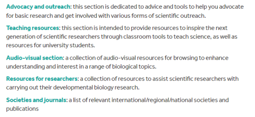
In 2017, our Node Intern, Sarah Morson, revamped our ‘Resources’ topic area on the Node. With a bigger emphasis on advocacy and outreach, we also cover teaching resources, societies and journals, audio-visual resources and resources for researchers.
More recently, we have added a ‘Featured resource’ series to the Node. In this series, we invite the ‘resources’ to showcase the services that they offer the scientific community. So far, we have heard from the following resources, with more to come in the future!
We would welcome any nominations (including self-nominations). You can contact us at thenode@biologists.com, with nominations or links to include in our Resources topic area.
“And that is one of the sort of worrying things about climate change; as we get more unpredictable weather patterns, can we actually design resilient wheat? So switching from a focus of just increasing wheat yields at any cost to having wheat that’s really robust to fluctuating weather conditions such as drought, but also flooding and unpredictable patterns basically.”
Dr Hannah Rees, Earlham Institute
In the latest episode of the Genetics Unzipped podcast, we’re looking at the future of food. With climate change making crop harvests more unpredictable and fresh water becoming a more scarce resource, what are geneticists doing to make sure we will still have food on our plates? Dr Kat Arney chats with Dr Hannah Rees about giving wheat jet lag to create a more reliable crop, and Dr Sally Le Page talks to Dr Tarang Mehta about breeding genetically improved tilapia for fish farming.
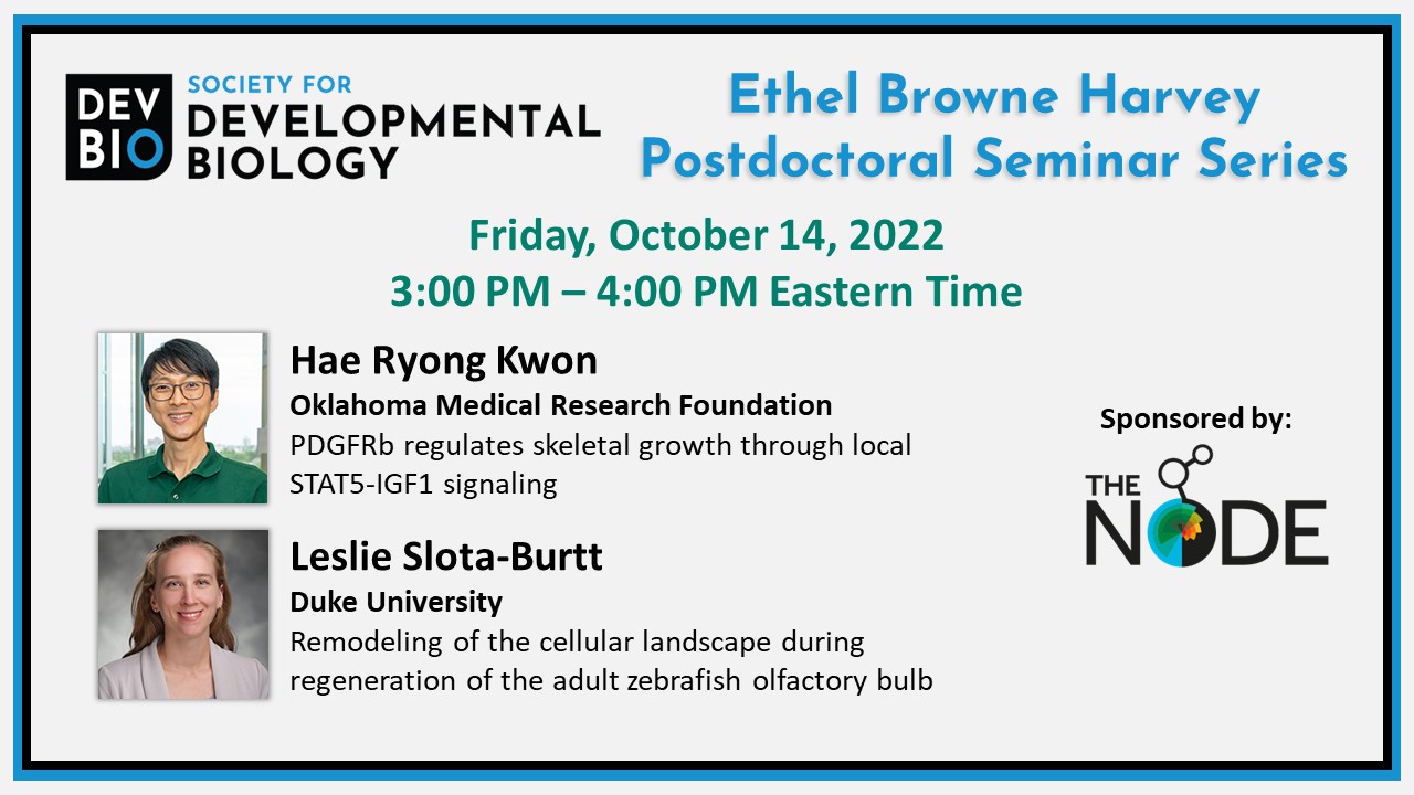
The next Society for Developmental Biology Ethel Browne Harvey Postdoctoral Seminar will be held Friday, October 14, at 3 pm ET (9 pm CEST). This seminar featuring Hae Ryong Kwon from Oklahoma Medical Research Foundation and Leslie Slota-Burtt from Duke University is generously sponsored by The Node.
Hae Ryong Kwon did his undergraduate studies in Microbiology at Chungbuk National University in South Korea. He completed a Master’s in Microbiology and Biotechnology at Chugbuk National University and a Master’s in Genome Science and Technology at the University of Tennessee. Kwon went on to earn his doctorate in Molecular, Cellular, Developmental, and Neural Biology at the University of Albany, State University of New York where he studied the function of endothelial cells in early salivary gland development in Melinda Larson’s lab. In 2016, Kwon joined Lorin E. Olson’s lab at the Oklahoma Medical Research Foundation where he studies the roles of platelet-derived growth factor signaling in human pathogenesis driving genetic diseases such as Kosaki overgrowth syndrome, Penttinen syndrome, and infantile myofibromatosis. Kwon was the recipient of an NIH National Research Service Award (F32) from the National Heart, Blood and Lung Institute.
Leslie Slota-Burtt did her undergraduate studies in Chemistry at the University of Florida. She earned her doctorate in the Developmental and Stem Cell Biology Program at Duke University where she studied cell type specification and evolution of the developing sea urchin nervous and digestive systems in Dave McClay’s lab. In 2019, Slota-Burtt joined Kenneth Poss’ lab at Duke University where she studies adult brain regeneration, specifically how genes and signaling pathways are activated after brain injury in the zebrafish. Slota-Burtt is the recipient of the NIH National Research Service Award (F32) from the Eunice Kennedy Shriver National Institute of Child Health and Human Development.
What’s in a name? From defining ‘epigenetics’, to naming nervous system organoids and assembloids, #SciTwitter has been alive with debate over the last two weeks. We bring you some of our favourite Twitter threads on these topics.
What’s in a name, part one
The lively discussion from #EMBOepigenome on what is real and hearsay in epigenetics spilled over onto Twitter. What does epigenetics mean to you and where do you sit on Zack Chiang’s epigenetics alignment chart? As always, click on the Tweets to read the full thread!
Things are getting intense! @ericmiska is leading a roundtable about what is real and what is heresy in epigenetics. As you can imagine, a lot of nuanced thoughts in this crowd. #EMBOepigenomepic.twitter.com/EqGJPReCyE
To facilitate discussion both within the scientific community and with the general public, researchers came together to produce a framework for naming neural organoids and assembloids: https://www.nature.com/articles/s41586-022-05219-6
Self-organizing systems have been one of the most exciting recent advances in stem cell research However, many names & classifications are used making it challenging to convey the science We now got together as a field to provide a nomenclature framework Out in @Nature today👇1/9 pic.twitter.com/MpBs858Wmx
Initially Hox genes were called with the species letter (mhox for mouse, Xhox for Xenopus..) until it was realized that there were more than 26 species on earth🙄 Standardisation worked pretty well in this case https://t.co/oh8SaeFWfo
During the panel discussion at our recent Development Meeting ‘From Stem Cells to Human Development’, we also discussed the importance of public perception, as well as consistency, in naming the multitude of in vitro models of human development. Go to 1hr45mins for the start of the panel discussion.
Enthusiastic about science communication and looking for a chance to broaden your writing experience alongside your research activities? The Node, our community site for developmental and stem cell biologists, is looking to appoint three correspondents who will play a key role in developing and writing content over the coming year.
As part of a small cohort, you will have the chance to engage with fellow researchers and scicomm enthusiasts as you work together to plan and generate fresh content. You will also gain insight into the publishing industry through meetings with our in-house Editors, Community Managers and Science Communications Officer, and receive regular feedback on your writing.
We will help raise your profile as a researcher and science communicator, and are also happy to support you by contributing towards conference attendance costs relating to the role, providing reference letters, or in other ways.
You will be expected to contribute around six posts over the course of the year – this could involve creating your own blog series around a theme of your choice, reporting on the latest exciting developments in developmental and stem cell biology, interviewing inspiring scientists, or writing about conferences and other events. We are also open to any other ideas you might have as we would like to shape a programme that both appeals to your interests and benefits the research community.
Please note, we are also recruiting correspondents for FocalPlane, so when applying you will have the option of choosing to apply for the Node, FocalPlane or both.
We encourage applications from all individuals regardless of sexual orientation, gender identity or expression, religion, ethnicity, age, neurodiversity or disability status. We also welcome applicants from a range of geographic locations.
Please get in touch with us if you have any questions about the programme at thenode@biologists.com
 (1 votes)
 (No Ratings Yet)