Debate on the status of the field of developmental biology continue to rumble on, including an open letter to Claudio Stern from Peter Lawrence published on the Node. You can read the letter here, as well as a reply from Alfonso Martinez Arias. Remember, the Node is your site and if you would like to share your hope and fears for the future of developmental biology all you need to do is register and then you are free to post. Please get it contact if you need any support doing this, or if you would like to collaborate with others on a post.
If you would like to write for the Node, check out our recent list of writing ideas. If you would like to contribute to our ‘Developing news’ blog, please get in touch at thenode@biologists.com
At the end of 2021 and beginning of 2022, Journal of Cell Science (JCS) put out a call for essays on ‘Equity, diversity and inclusion in cell biology’. JCS introduced the series to amplify voices that are not always heard in this space. After receiving some fantastic essays on a range of different topics, they selected three winners and have now published these essays, as well as those from the runners-up.
If you have a story about equity, diversity and inclusion that you’d like to share, we’d love for you to post it on the Node. Get in contact (thenode@biologists.com) if you have any questions or would like help posting your article.
Birds do it. Bees do it. We even have evidence that fleas do it, although whether or not those fleas are educated remains to be determined. And by ‘it’, I of course mean cannibalism.
In the latest Genetics Unzipped podcast, Dr Sally Le Page explores the gruesome side of family life in the natural world, getting stuck into a spot of cannibalism and asking: “When exactly should you eat your relatives?”
Sally takes a look at different examples of cannibalism across the animal kingdom, from offspring eating their mothers to males giving up their lives for sex and siblings devouring each other in the womb.
We meet the Taita Hills caecilian, an amphibian species where mothers produce extra thick skin that is eaten by their young, and spiders who digest their own internal organs to feed their spiderlings. We discover how male spiders give up everything, including their lives, for a chance at mating, why barn owls employ a ‘lifeboat’ strategy to eat their siblings, and why you might not want to put your hand inside a sand tiger shark’s uterus…
The first LMCB retreat in 10 years provided a much-needed rejuvenation of scientific connections and discussions after two years of Covid restrictions.
Hinxton Hall, on the Wellcome Genome Campus near Cambridge, was a perfect scenic location for what the next two days had in store for us. The retreat began with a talk from the LMCB director, Alison Lloyd, who reminded us about the importance and wonders of tissue biology from the angle of peripheral nerve homeostasis and repair. This introduction set the stage for a range of exciting talks throughout the next two days, presented by LMCB PhD students, postdocs, and group leaders, as well as invited external speakers.
The internal talks provided an opportunity to sample the range of cutting-edge research from across the institute. The topics went across scales from identifying enhancers in neurons to collective cell dynamics and mechanobiology and understanding how morphogenesis shapes an organ through development.
One of the many highlights of the retreat was the flash talk session where selected PhD students and postdocs explained their research in three minutes. This was a friendly competition with the winning prize going to Giulia Paci from the Mao lab, who presented her work on microfabricated tools to apply mechanical forces in developing tissues. This session highlighted the range of techniques and approaches used across the institute including mathematical modelling, electron microscopy, and state of the art light microscopy.
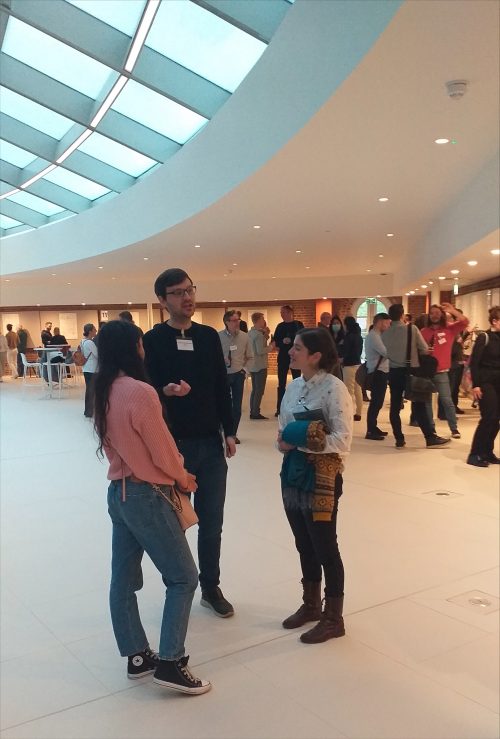
If you didn’t think the flash talks were challenging enough, every student had the chance to draw their own research posters at the poster sessions. This saw a range of artistic attempts to explain research projects with pen and paper. I personally found the poster session to be so much fun, and I could now put names to faces and faces to exciting research projects. It was also a good opportunity to identify converging interests and to think about future collaborations and discussions.
In depth discussions at the well prepared poster session
In addition to talks from researchers within the LMCB, we were lucky to have excellent talks from three external scientists. The first of these was Soyon Hong from the UK Dementia Research Institute at UCL, who talked about the role of microglia in Alzheimer’s disease. We also had an entertaining talk from Mark Miodownik on the use and potential of animate materials to create a more sustainable planet for the future. The final external speaker was Rachel McKendry, who spoke about using quantum materials for early disease diagnosis. This was particularly relevant in the context of the Covid pandemic, as she highlighted technology that enabled the detection of low vial loads.
Now onto the social events, which lived up to their high expectations. The retreat started with a speed-dating session where we moved around small groups and gave a 30 second introduction. It was a great way to hear about the range of interests from colleagues and to get a hint of their hobbies away from science.
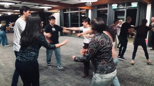
One general aspect I have noticed about scientists is their humour, perhaps because research requires a creative quirk. This was certainly confirmed in the pantomimes. Students and postdocs had filmed a pantomime prior to the retreat which was shown as a short film after dinner. Our pantomime was scripted and filmed by the very talented Jimmy Van Hear, a PhD student in the Mao lab, and it was called the Cephalopod Contest, taking inspiration from the popular Netflix series ‘Squid Game’. As you can imagine, this was great light-hearted fun about the institute, the life of a scientist, and of course we also mocked our group leaders. Don’t worry…. the group leaders got their revenge on our pantomime with a LIVE performance of their own! After the endless laughs, the first night ended with a Ceilidh. Yes, that’s a bunch of energetic scientists being taught Scottish dance moves to fast paced music. This was a great chance to chill out and have fun outside of the scientific activities.
First night fun with a Ceilidh
It wouldn’t have been a complete scientific retreat without a biology-themed Dragons’ Den competition! Over the two days, we got together into designated groups and brainstormed pitches and project names for the Dragons (LMCB group leaders). Group members then presented to the Dragons, who were willing to invest their own chocolate coins for the most promising projects. The projects were humorous, creative, and often inspired by our external speakers. We heard about a proposal to grow meat from your own cells and therefore bypassing any issues around animal consent (although as one Dragon pointed out, you could call this cannibalism). Another project pitched to speed up wound healing with a bioactive spray. But the winner of the audience vote, perhaps inspired by Mark Miodownik’s talk on animate materials, was the project ‘FungalFind’ which aimed to identify pollutants with genetically engineered fungi.
An exciting aspect of scientific training involves interacting with the community – gaining the social skills to spark collaboration, start new conversations, ask relevant questions and communicate with scientists from a range of different disciplines. Starting my PhD journey in a pandemic and joining a new research institute meant that the community aspect of science had faded, and my social skills felt rusty. During the retreat, I have met the faces I pass in the corridor, and I feel well connected to different research groups across my institute. I also feel much more confident about approaching colleagues and getting my ideas across. This experience has stressed the importance of in-person interactions and highlighted how ideas and creativity are much more fruitful in real life.
The survey should take around 6 minutes to complete and we encourage everyone to participate – even if you have not attended a Development presents… webinar before. Your feedback will help to determine the future direction of the series. Please complete the survey before Wednesday 6 July.
The Centre for Trophoblast Research will again run its popular course in placental biology, online, from Monday 11th to Friday 15th July 2022.
This online course is aimed at students, post-docs, established researchers, medical & veterinary healthcare professionals and industry colleagues interested in cutting-edge placental biology and research.
Most content will be pre-recorded lectures and practical sessions given by investigators at the forefront of the placental biology field. There will be daily live Q&A sessions with the speakers, and opportunities to give talks and network.
Lectures and practical topics include:
Human & mouse placental development Organoids, explant and trophoblast cultures Materno-fetal interactions Immunology Placental transport Angiogenesis Clinical study design Placental metabolism Stem cell embryo models Genome editing Grant/fellowship writing History of human embryos
We hope you will join us!
Limited places. Register here : https://www.trophoblast.cam.ac.uk/placentalbiologycourse/registration
The Fossils, Phylogenies, Genomes, Embryos & the Evolution of the Deuterostomes symposium took place at the Natural History Museum in London to honour the work and contributions of the late palaeontologist R.P.S. ‘Dick’ Jefferies. In a field that is often looking to the future, it can be easy to take for granted the work that has come before or to overlook the early iterations of hypotheses we investigate today. This brilliant symposium, featuring speakers working across the scope of deuterostome evolution, was a fitting celebration of the pioneering work carried out by Dick Jefferies.
Joining the British Museum in the 1960s, Dick’s research focused on deuterostome fossils. Although many of his predictions have subsequently been disproved, some of his then-radical ideas have in fact been confirmed by more modern techniques. For example, his perhaps unexpected hypothesis of a clade comprising the tunicates and vertebrates (the Olfactores), has been subsequently supported by molecular data. Modern imaging approaches have also been key to confirming the validity of some of his ideas. A brilliant talk by Imran Rahman showed beautiful high-resolution X-ray scans supporting Jefferies’ idea of gill slits in early echinoderm fossils called the Stylophora. A particularly entertaining talk by Bertrand Lefebvre to round out the day detailed the painstakingly slow fossil sectioning-and-tracing technique employed by Jefferies’ to draw the same hypotheses now reached with very rapid microscopy approaches.
A particular highlight of the symposium was the breadth of research represented in the talks. Trends in developmental biology might sometimes lean towards developmental genetics and -omics, but the ‘evo’ side of evo-devo holds valuable contributions for the field. An excellent talk by Elizabeth Clark detailed the modelling of locomotion from fossil traces, and several talks (Paschalia Kapli, Graham Budd, Rachel Warnock) discussed persistent problems in phylogenetics. It was great to see the representation of such diverse research, and clear that there are still many open questions in the field – some of them pondered by Jefferies himself – remaining to be answered.
The Fossils, Phylogenies, Embryos & the Evolution of the Deuterostomes meeting was held on 12 May. It was supported by The Company of Biologists and The Palaeontological Association and organised by Max Telford, Jeffrey Thompson, Tim Ewin, Tim Littlewood, Greg Edgecombe, and Paul Barrett.
I read your article just now, it is so perceptive of where things have gone in ‘Embryology’. The problem with us oldies is that we compare approaches, but the new generations can ignore us saying we have rose-tinted glasses about the past. And that may be true, but it is not an argument and your article provides real arguments. One core theme running through the article is that the balance between data collection and experiments that are designed to understand has gone completely wrong over the last few decades.
It is so comforting to find someone else who sees this as clearly as you do. When we began our work, Wigglesworth was my mentor. His style was careful observation, then a question (how? why?), then a series of simple and direct experiments to answer it. I have always tried to follow this approach, and with genetics, specifically genetic mosaics, we have had powerful (although not so simple) methods to do this.
I don’t think our papers have changed fundamentally in this long time, we still try to get at mechanisms by carefully designed experiments and I submit that they meet high standards of technique and rigour. But we can’t publish them in major journals as we used to, indeed we don’t even try. I submit this is because they don’t meet the new criteria. You explain what these criteria are in your article. To put it cynically, I believe that success goes to those who put in so much fashionable data that no reviewer can fault it, they are almost ‘drowned’ into submission. When we showed that a cell could have two opposite polarities, in vivo and in situ, depending on inputs from its different neighbours, I naively thought it would be of great interest as it challenges many of the decades-old perceptions of planar cell polarity. It was published in eLife and the evidence is so clear. But it created not even a ripple in the field. That, more than anything, showed me that developmental biology has moved into a new landscape where slag heaps of fashionable data interfere with sight lines (and thought). Just as you put so nicely in your article, thank you for writing it.
Postdoc position immediately available in Guillermo Oliver’s lab at Northwestern University in Chicago to join our team to further characterise the novel functional roles of the lymphatic vasculature in organ development and regeneration in health and disease using available mouse models. Candidates should have recently obtained a PhD or MD degree and have experience working with mice. Interested individuals should email their CV and brief description of research interests to:
Guillermo Oliver, Ph,D,
Director Center for Vascular and Developmental Biology,
In the past week, #devbio twitter has been discussing an opinion article from Claudio Stern, published in Developmental Biology. In the article, Claudio discusses progress in developmental biology from a historical perspective before focussing on the present-day and future challenges for the field. In response the article, starting with a twitter thread from Alex Schier, many in the community disagreed with Claudio’s bleak outlook, some even going so far as to say that it is a golden age for developmental biology. We pick out a few of our favourite tweets (click on the link for the full thread). Let us know what you think about the current outlook for #devbio.
A thought: The problem isn't that mind-blowing dev biol isn’t being done; it's that Cell/Science/Nature won't publish it without some ridicul-omics. Why? The field's weak storytelling in recent years? Our failure to nurture and promote a new generation of stars? Both? Or?
amazing discussion – both Claudio's and Alex's points. I find DevBio exciting and VERY painful to leave behind. But why are there so few applications to the ERC? It's now one of the smallest fields in LifeScience – not in terms of awards but in applications. https://t.co/ECLqFqmeZ0
If you would like to write for the Node, check out our recent list of writing ideas. If you would like to contribute to our ‘Developing news’ blog, please get in touch at thenode@biologists.com
 (No Ratings Yet)


 (5 votes)