Basement membrane mechanical properties
Posted by Uwe Töpfer, on 20 May 2022
Extrinsic forces have an underestimated impact on shaping tissues and organs. Extracellular matrixes surround tissues and provide the cells with mechanical stimuli to drive morphogenetic processes. A specialized extracellular matrix (ECM) that is crucial for the development of epithelial tissues and organs is the basement membrane. The composition of this matrix varies from tissue to tissue and can consist of hundreds of distinct proteins. However, four main components are present in nearly every basement membrane across metazoan life: Laminins, a family of heterotrimeric (α,β,γ) glycoproteins, that self-assemble into networks and interact with cell surface receptors like integrins or dystroglycans. Collagen IV is a triple-stranded helical structure forming networks by covalent interactions. Nidogen is a sulfated glycoprotein that is supposed to connect Laminin and Collagen IV and Perlecan, a heparan sulfate proteoglycan that is involved in cell-signaling by binding of multiple growth factors.
To what extent the presence of these main components is interdependent during the maintenance of basement membranes and to what extent individual components contribute to the mechanical properties of basement membranes is not well understood.
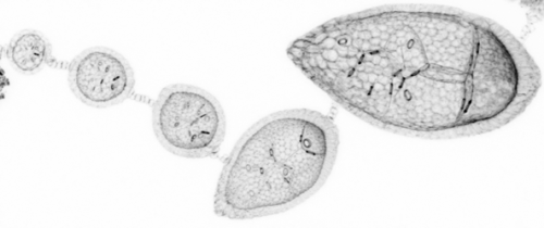
In this Issue of Development (Development (2022) 149 (10)) we address these questions and show that Laminin and Collagen IV networks partially persist independently from each other and measured the mechanical properties of the basement membrane after knock-down of single main components.
https://doi.org/10.1242/dev.200456
Reference
Uwe Töpfer, Karla Yanín Guerra Santillán, Elisabeth Fischer-Friedrich, Christian Dahmann; Distinct contributions of ECM proteins to basement membrane mechanical properties in Drosophila. Development 15 May 2022; 149 (10): dev200456. doi: https://doi.org/10.1242/dev.200456
 (No Ratings Yet)
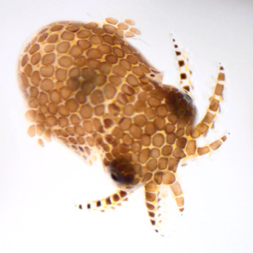
 (4 votes)