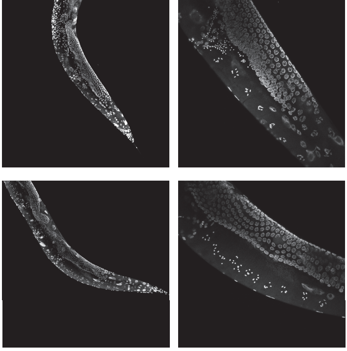
1. Choroid plexus/CSF signaling niche: development, stem cells and plasticity
2. Barrier and neuroimmune function
3. Propelling and sensing the CSF: ependyma and cilia
4. Novel signaling sources: extra-axial CSF, Interstitial fluid, meninges
5. Choroid plexus/CSF and disease
The Company of Biologists Workshops provide leading experts and early career scientists from a diverse range of scientific backgrounds with a stimulating environment for the cross-fertilisation of interdisciplinary ideas. The programmes are carefully developed and are intended to champion the novel techniques and innovations that will underpin important scientific advances.
There are currently multiple funded spaces for early-career researchers to attend this exciting event (deadline = 15 March). To find out more and apply online please visit
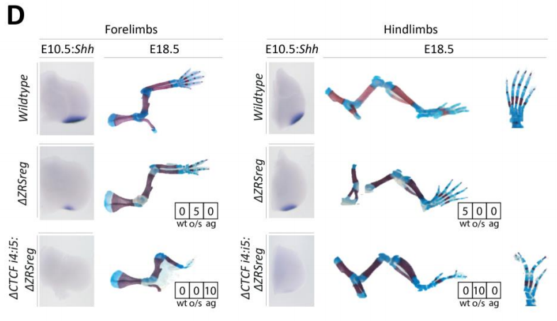
In our recent paper published in Current Biology, we unravel the direct and early role for Hox genes in the regulation and natural variation of the forelimb position in birds. Here I will share with you the story behind this paper.
I have always been fascinated by the question of how, from a single cell, a multicellular organism with a complex, highly organized three-dimensional structure can arise. Therefore, when I started my PhD in Dr. J. Gros’ lab and got the opportunity to work on the question of limb position in chicken embryos, I was thrilled. How limbs reproducibly form along the vertebrate body is a fascinating, long standing question for developmental but also for evolutionary biologists. Indeed, this question has two main aspects:
one purely developmental aspect – in trying to understand the cellular and molecular mechanisms establishing limb position during the embryo development
one evolutionary aspect – in tackling the question of diversity in limb position amongst tetrapod species.
The context
In tetrapods, while limbs always position at the level of the cervico-thoracic (for the forelimb) or lumbo-sacral (for the hindlimb) vertebral transitions, the position of these vertebral frontiers – and hence the limb position – is highly variable between species. Avian species especially, display a wide variation in the position of their forelimb – from the sparrow that form forelimbs at the level of the 10th vertebra up to the swan in which forelimbs form at the level of the 25th vertebra – a difference of no less than 15 vertebrae!
Despite major advances in our understanding of limb patterning in three dimensions, how limbs reproducibly form along the anteroposterior axis and how variations in these positions arise remained largely unresolved. Hox genes were long suspected to regulate limb position (Tanaka, 2013; Tickle, 2015). This assumption first arose because of their well-demonstrated role in patterning the vertebrae and was further supported by cross-species comparative studies which showed that Hox gene expression domains correlate with limb position in different species (Burke et al., 1995). But the different supportive evidences were mostly correlative (Burke et al., 1995; Cohn et al., 1997; Minguillon et al., 2012; Nishimoto et al., 2014). In addition, whereas Hox gene mutant mice display vertebral identity transformations (Mallo et al., 2010), functional studies in support for a role of Hox genes in regulating limb position were lacking. Therefore, whether Hox genes would control limb initiation and position was clearly unresolved at the time we started this work.
Our Results
Where do the limbs come from? They originate from the Lateral Plate Mesoderm (LPM), a mesodermal tissue that flanks axial embryonic structures (i.e. notochord, neural tube and somites), and will emerge from this tissue at their stereotypical position, at three days of development (Hamburger and Hamilton stage 15). First, we wanted to determine when the forelimb position is primarily established. We took advantage of the possibility to do grafting experiments in chicken and quail embryos, combined with the use of transgenic quail lines generated in the lab, to establish that the forelimb position is determined very early, 24h before limb initiation (i.e. at stage 11, 2 days of development).
The finding that the forelimb position is already established by stage 11 led us to think that this positional information could be established earlier – during the process of gastrulation. Gastrulation is this key morphological process during which the three germ layers – ectoderm, mesoderm and endoderm – are formed. The LPM is generated during this process and we wondered whether it was also patterned into limb- and non-limb domains while being generated. Whereas LPM precursor cells in the epiblast had been identified through lineage-tracing experiments (Psychoyos and Stern, 1996), how the forelimb, interlimb and hindlimb cells are generated was not characterized. The process of gastrulation is really dynamic and spans for about 24h. Therefore, in order to precisely catch the dynamic behaviors of LPM precursor cells, we used yet again another advantage of the chicken embryo, which is the possibility to do live-imaging, especially in the early stages of development as the embryo is flat and can be cultured ex ovo. We therefore performed a dynamic lineage analysis of the LPM formation, and could nicely characterize that the forelimb, interlimb and hindlimb domains are sequentially generated during gastrulation.
At this point, we had described how the LPM is formed and patterned into limb and non-limb domains at the cellular level. The next step was now to investigate the molecular mechanism underlying this process.
As I mentioned earlier, our top candidates for the regulation of limb position were the Hox genes, well-known for their role in patterning vertebrae along the main body axis (Mallo et al., 2010). For our study, it is important to remember that these genes are arranged in four different clusters and display a specific chromosomal organization that reflects their sequence of activation (i.e. temporal collinearity) and their successive domains of expression along the antero-posterior axis (i.e. spatial collinearity) (Izpisúa-Belmonte et al., 1991). Interestingly, we noticed that the collinear sequence of Hoxb genes activation during gastrulation correlates with the temporal sequence of forelimb (e.g. Hoxb4) and interlimb (e.g. Hoxb7and Hoxb9) formation we had just identified, suggesting that they could play a role in the formation of these domains. To test it, we used the electroporation technique that allowed us to do functional perturbations precisely controlled in time and space. We performed overexpression and loss-of-function of different Hoxb genes in LPM precursor cells to perturb their activation during gastrulation. We could show that – as previously identified in the paraxial mesoderm (Iimura and Pourquié, 2006) – Hox genes, collinearly activated during gastrulation, establish their own stereotypical sequential expression domains in the LPM – domains that correlate with the forelimb (e.g. Hoxb4) and interlimb fields (e.g. Hoxb7 and Hoxb9).
But are these Hox expression domains important to position the forelimb?
One year before I started my PhD, a study showed that Hox4/5 genes could bind to a regulatory sequence of Tbx5 – a transcription factor essential for forelimb initiation – and activate the expression of a reporter gene under the control of this regulatory sequence (Minguillon et al., 2012). These results further supported a role for Hox genes in positioning the forelimb along the antero-posterior axis. We therefore decided to ectopically express Hoxb4 in the interlimb at stage 14 – when Hox domains are well-established – to see if we could perturb the forelimb position. We first screened embryos for an ectopic expression of Tbx5 in the interlimb, naively thinking that Hoxb4 overexpression should first induce Tbx5 expression. But that would have been too simple, Hoxb4 alone was not able to induce Tbx5 ectopic expression in the interlimb…
We were initially quite puzzled by this result. But the story could not stop there, we were missing something and we had to find out what. Then became a long journey during which we electroporated many different combinations of genes in the interlimb – e.g. Hoxb4 fused to VP16; combined Hox4/Hox5 genes; Hoxb4 plus activators of Retinoic Acid or Wnt/β catenin pathways, both involved in Tbx5 expression and forelimb initiation (Nishimoto et al., 2015); and several others – in an attempt to induce Tbx5 expression in the interlimb and a shift in limb position. Unfortunately, none of these combinations had an effect on Tbx5 expression nor limb position. At this point, we started to be somewhat desperate and were about to give up as the end of my PhD was getting closer. But reading again some of the literature, we came back to one paper in which it was shown that Hoxc9 – which is expressed in the interlimb – could repress Tbx5 expression (Nishimoto et al., 2014). Maybe the solution was there, this repressive activity was preventing all our attempts to posteriorly extend the Tbx5-positive forelimb field. This was it, the last-chance experiment! We decided to construct a dominant-negative form of Hoxc9 using a strategy others had established to generate Hox dominant-negative constructs (Denans et al., 2015). Then, we combined overexpression of Hoxb4 together with the repression of Hoxc9 in the interlimb and, not only we could nicely extend the Tbx5-positive forelimb domain in the interlimb, but we could displace the final forelimb position (Figure 1). There it was, many years after Hox genes where first suspected, we finally had the functional evidence of their direct role in regulating the forelimb position!
Figure 1: Electroporation of Hoxb4+Hoxc9dn in the right interlimb domain of stage 14 chicken embryos induces a posterior extension of the Tbx5-positive forelimb field (st14+24h), a posteriorly extended forelimb bud (st14+48h) and ultimately posteriorly shifted forelimb (st14+4 days – Alcian Blue staining)
Importantly, removing Hoxc9 repression from the interlimb alone was not sufficient to induce Tbx5 expression and limb initiation. Therefore, showing that to change the forelimb position, both a shift of the forelimb field (e.g. Hoxb4 expression) and the interlimb field (e.g. Hox9 expression) is necessary. These results brought to us one potential explanation to why the vast majority of Hox genes mutant mice do not show major perturbations in limb position. Indeed, our data argue that, to induce a shift in the forelimb position in mouse, a combination of gain-of-function for forelimb activator (e.g. Hoxb4) and loss-of-function for forelimb-repressor (e.g. Hox9) should be performed. Another interesting point worth commenting is that, while Hoxb4 and Hox9 activate and repress Tbx5 expression, eventually leading to the establishment of the definitive forelimb position, Tbx5 itself was shown to be not sufficient to induce limb initiation (Nishimoto et al., 2015). Therefore, implying that, to regulate forelimb initiation at its specific position, Hox genes do not solely act upon activation/repression of Tbx5. This point remains to be explored but one can speculate that Hox genes could activate/repress other regulators of limb initiation or act cooperatively with Tbx5 to activate the limb initiation program, as recently shown for Hoxc10 and Tbx4 in the context of hindlimb development (Jain et al., 2018).
At this point, we had now unraveled the developmental mechanism behind limb position establishment. But what about limb position diversity, how can variations in limb position arise?
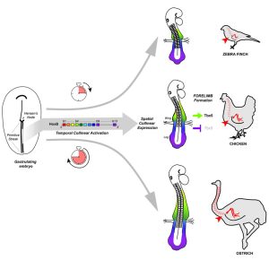
Our results suggested that natural variations in the forelimb position should be traced back to changes in Hox genes activation during gastrulation. To test this hypothesis, we took advantage of the bird natural variation in limb position. We selected three different bird species: zebra finch, chicken and ostrich as they form forelimbs at the level of the 13th, 15th and 18th vertebra, respectively, to perform a cross-species comparative analysis of Hoxb genes temporal and spatial dynamics of expression during early development. We then could provide evidence that, as predicted, changes in the timing of Hox activation during gastrulation prefigure variation in the spatial organization of these genes – i.e. in the spatial position of Hoxb4/Hoxb9 border of expression – in the LPM and ultimately, natural variation in forelimb position in birds. Working with non-conventional model organisms such as the ostrich was not trivial and required some troubleshooting and optimization at every step of the process, but it brought us with some very exciting outcomes and I think, shows the importance of using non-traditional model organisms when it comes to challenge and validate the models and predictions we establish using our favorite model organisms.
Finally, now that we had established the role of Hox genes in regulating limb position and variation, the next logical step was to investigate how such variation in Hox activation timing could be controlled. We got interested in the Retinoic Acid (RA) signaling pathway, and especially the RA catabolizing enzyme Cyp26a1, as it was already shown to be involved in hindlimb position regulation (Lee et al., 2010). We observed that Cyp26a1 onset of expression differs between the three avian species – i.e. its expression is premature and delayed in zebra finch and ostrich embryos, respectively, compared to chicken – consistent with a role for RA signaling in regulating Hox genes activation. Finally, modulating RA signaling during gastrulation in chicken embryos, provoked changes in Hox genes spatial organization in the LPM and in the Tbx5-positive forelimb field position, further suggesting that RA signaling might regulate the forelimb position through the regulation of Hox genes activation during gastrulation. This is an interesting point that will definitely need to be further investigated. Indeed, the possibility that Cyp26a1, already shown to regulate hindlimb position (Lee et al., 2010) could also regulate the forelimb position is particularly interesting. Especially, as it was recently proposed that differences in the onset of Gdf11 expression – i.e. the signaling molecule that induces Cyp26a1 expression – account for variations in hindlimb position amongst tetrapods (Matsubara et al., 2017). Therefore, one single signaling pathway would then be responsible for the regulation and natural variation of both fore- and hindlimb position.
Conclusion
From all these different results, the major conclusion of our work is that the forelimb position is determined very early, during gastrulation: it is the temporally controlled activation of Hox genes that progressively patterns the LPM into limb- and non-limb forming domains, as the main axis is being formed. And relative changes in this timed collinear activation underlie natural variation in forelimb position in birds (Figure 2).
One of the important points brought by our study is that we show the LPM is patterned by Hox genes during gastrulation following a similar mechanism as the one identified to pattern the somites (Iimura and Pourquié, 2006). These two tissues – that respectively give rise to the limb and vertebrae – both being generated and patterned during gastrulation by Hox genes, offers a simple mechanism to pattern the cervico-thoracic frontier in the somites and the forelimb position in the LPM, concomitantly therefore maintaining their tight association observed in all tetrapod species.
Figure 2: Hox genes, collinearly activated during gastrulation, establish their collinear spatial expression domains in the LPM and, within these domains, Hoxb4 anteriorly and Hox9 posteriorly, respectively activate and repress limb initiation (i.e. Tbx5 expression) therefore defining the definitive limb position. Relative changes in the temporal sequence of Hox activation underly natural variations in limb position in birds.
As a conclusion, our work that combines experimental embryology, state-of-the-art live imaging and cross-species comparative studies, addresses the major question of how the forelimb position is determined and solves a 20-year long controversy on the role of Hox genes in regulating limb position. It also provides a general mechanism for generating variation in body plan organization in vertebrates and reinforces the importance of Hox genes in shaping animal body plans.
Developmental biology is a prominent field that has captured the imagination of many scientists. Over the years, research in the area has seen a steady number of amazing accomplishments, with peaks in activity following the development and application of new technologies. Although the field continues to flourish and produce excellent work, I have recently noticed difficulty with its perception and visibility. Having joined the developmental biology community during the early 1990s, and contributing since as a stem cell researcher, cancer biologist and an MD, I have a unique perspective on these challenges. Here, I discuss these issues and challenges and offer potential solutions for a field that is very important to me.
I recently had a discussion with some colleagues at a stem cell meeting about impact factors and the conversation morphed to the topic of developmental biology journals. It appears that the impact factors of all classical developmental biology journals (Development, Developmental Biology, Genes and Development, Developmental Cell, etc.) have been declining over the years. Thus, although the field continues to publish excellent studies in top tier journals, the general area of developmental biology may need help. Of course, impact factor is not a great measure of real importance, and authors may be artificially infatuated with these numbers, as they are thought to impact promotion and other academic criteria (discussed by Pourquié, 2018). However, to me, the numbers are consistent with my perception that much of developmental biology research has not been as appreciated by other fields or the public as it used to be. I left the table feeling pretty depressed and wanted to figure out what could be done to improve the general visibility of developmental biology to scientists.
So how did this happen? Historically, developmental biology has been very successful, spawning a remarkable number of new fields such as stem cell biology, single cell genomics and chemical genetics (reviewed by Gilbert, 2017). Strangely, it appears that these new areas were not fully embraced and I believe it is this lack of inclusiveness that is the key to the problem. For example, stem cell research came from developmental biology but then, as the stem cell community grew, it was only welcomed by a few of the classical developmental biologists and journals. Stem cell journals were established and became successful and, although developmental biology journals tried to recover the stem cell field, the damage was already done. Organoids are also becoming very popular models, but are often not considered as a major area of developmental biology, or even stem cell biology. Will there be separate journals or societies for organoids? Given that all of these fields are close to each other, it would make sense – with regard to outside perception – to be as inclusive as possible by pooling them together and trumpeting all of their successes.
Notably, the stem cell field was able to grow very quickly. This was because of excellent science and, of course, the medical potential of stem cells, but it was also because of inclusiveness and clever marketing. Stem cell biologists figured out how to market developmental biology. When I started the International Society of Stem Cell Research (ISSCR) as the first President, I invited many well-known developmental biologists to join the board. At our first meeting, several of them felt that we should not talk to the press about our work. This group was worried that the press might misrepresent their views and this could affect public perception. I made the point that if we didn’t provide the public with accurate information, then there would be misinformation in the public eye and this could instead create long-term problems. The decision was made to have all of the board talk about stem cell science to as many groups as possible, including the public, government officials and other scientific groups. When creating our first meetings, we were as inclusive as possible, including many talks on animal model systems and plants. I was very happy to include one talk on stem cells in trees at the first meeting. We formed an alliance with Cell Press so that we could establish a venue for publishing our top papers (in the journal Cell Stem Cell) and the field was very supportive of progress. We engaged funding bodies and governments, and included members of various foundations on panels and committees. Ethical guidelines were put in place. Lastly, we involved physicians who might be able to translate our basic research into the clinic. Overall, we tried to be as inclusive as possible and developed an educational strategy for the public, funders, governments and scientists.
Based on my experience, I think there are a number of steps that could be taken to improve visibility of the developmental biology field:
As a field, we should consider marketing the attributes of developmental biology. There are several different audiences – potential students, post-docs and other researchers, grant funders, editors, general scientists, and the public – and we need to develop a different message for each audience. We also need to point out the successes and technologies of the field and say why it is so important for everyone to know about developmental biology and why it is a great area to be part of. Perhaps a video series could be used to illustrate great examples of success or future directions. These should also clearly state what the field is trying to accomplish now. Is there medical relevance? If so, we need to say it loudly. Overall, better marketing will make developmental biology more attractive, which will hopefully lead to more papers being published, and this will expand the community’s impact.
We also need to think more carefully about the scope and aims of developmental biology meetings. Although the meetings are highly attended by developmental biologists, it would be beneficial to aim higher and reach out to those who are at the periphery of the field, or those in the newer disciplines that run the risk of splitting off from the core community. Consider that many researchers who study human disease might rather attend a meeting about their tissue, technology or cell biology than go to a broad meeting about developmental biology. Maybe we could create a new meeting or retrofit an old meeting to be more inclusive. Perhaps this could be centered on developmental biology techniques. We have seen huge advances in bar coding and single cell techniques, for example, and many of the questions that are being tackled using these techniques are now about organ development and function. This is an opportunity to bring interesting papers into the field. The term ‘applied developmental biology’ has been used in the past (see Maartens, 2017), but perhaps we should re-visit this idea and do a better job of marketing ‘applied developmental biology’ and including it in meetings. In general, meetings should be used to show off: invite every editor possible, invite every program officer for funding, get the heavyweights of the field to attend as well as the newcomers…and do not be scared to have 2000 people there.
The field of developmental biology could also benefit from reaching out and inviting the medical community into the group. Twenty-five years ago, I was invited into the developmental biology community by some really excellent scientists. It made me want to become part of a group that included rigorous scientists with bold ideas who wanted to extend their concepts to a young doctor. I am now very proud to be part of the community. So, moving forward, we should make sure we invite more medics and translational researchers to developmental biology meetings. Companies should also be invited to participate: there is a lot of developmental biology in companies right now and they are increasingly recognizing its importance. For example, one company that I founded (Scholar Rock) works on making antibodies to members of the TGF-β family for therapeutics, and it often discusses the developmental biology that is regulated by these ligands. Developmental biology is clearly relevant to companies, so it will be important to interact with them and foster good relationships.
The ‘journal experience’ is becoming very important when deciding where to submit a paper, so this is also something that, as a community, we need to improve. Much of developmental biology work is inherently harder to do in vivo, and experiments take a long time. The field, as a whole, needs to be more wary of this. Reviewers should recognize that, sometimes, descriptive in vivo work is just as important, revealing and fascinating as complicated molecular manipulations. As such, the same level of experiments that would be requested for a more accessible and/or manipulatable system may not be appropriate. As an author, you may need to remind your editors of this and liaise with them to streamline the crucial experiments that are requested before a paper is published.
Finally, we also need to educate funding bodies and the general public about the value of basic research in developmental biology. Pointing out the impact of basic biology on the development of clinical therapies has great effect. Checkpoint blockade for cancer therapy, for example, could not have been initiated without a significant literature on T-cell developmental biology. Anti-cancer therapies that target the Hedgehog pathway would not have been possible without basic research into how this pathway functions in normal development (discussed by Ingham, 2018). Collecting a number of these anecdotes works, but researchers should also consider how their own studies add to this impact, establish novelty and push the field forward. Indeed, in a recent commentary it was pointed out by Claude Desplan that the reuse of the same signaling pathways in most developmental processes created reader and reviewer fatigue (discussed by Desplan, 2017). The fact that NOTCH mutations cause lymphoma as well as congenital heart defects may be less exciting than finding a new pathway that causes cancer, but if you are the patient with a NOTCH mutation, you will no doubt be thanking the developmental biologists for bringing some understanding of its action. Driving research forward in this way, and highlighting the importance of this progression, may help with how the field is perceived. The newly developed single cell profiling approaches open up huge research, diagnostic and therapeutic avenues to study stem cells, organ development, regeneration and cancer. We should be telling the public about this work now and claim victory when new mechanisms are found or when new applications occur. This is something that everyone can do; we all need to be advocates for the field.
In summary, it is clear to many of us that developmental biology continues to establish new principles and techniques that are helpful to many other fields. It spawns areas of research that become fields themselves. My perception is that the community could do better to enhance its visibility to other researchers and to the public. Inclusivity is very important and enhanced marketing strategies could be helpful in sending out the right signal. Much like Spemann’s organizer, we need this signal to ‘induce’ change, to ‘specify’ more developmental biologists, and to invoke a ‘community effect’ to bring as many groups as possible together to show how exciting the field is.
Competing interests
L.Z. is a founder and stockholder of Fate Therapeutics, CAMP4 Therapeutics and Scholar Rock, and a consultant for Celularity.
Tills O, Spicer JI, Grimmer A, Marini S, Jie VW, Tully E, Rundle SD. 2018. A high-throughput and open-source platform for embryo phenomics. PLOS Biology, 16:1-19.
A seasoned graduate student gave me some valuable advice at the outset of my PhD. She told me to FIND A BETTER WAY! We were both working on heterochrony – changes in the timing of developmental events. She had worked on between-species comparisons – I was about to start investigating within-species differences. The challenge in both research areas lies in its temporal nature – a poorly-timed lunch, or rest-room break and the developmental events of interest are literally a thing of the past. Development shows little regard for the diurnal rhythms of scientists either. Her advice drove me to develop a technology that would enable me to (occasionally) leave the lab. As a by-product, it produced a step change in the quality of the developmental data we were generating and, in turn, our understanding of development at the level of the whole-organism.
OpenVIM
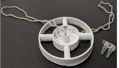
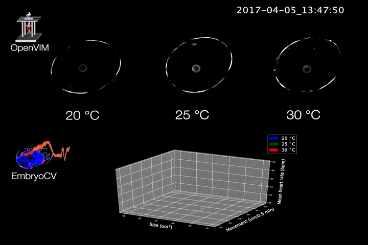
OpenVIM is the result – a modular open-source video light microscope (Fig.1). Specifically tailored for automated time-lapse imaging of large numbers of aquatic embryos, for the duration of their development, or the experiment. The key with OpenVIM is that we can capture the dynamic process of embryonic development by generating realtime image sequences of each embryo. These acquisitions are repeated over the duration of the experiment (or their development). I started my career as a marine biologist, and despite my growing interest in development, my research continues to be set in the context of understanding how animals respond to changes in their environment. Therefore, environmental control was central and so this is also integrated and we can heat, cool and provide mixtures of gases during the course of an experiment.
Fig.1. OpenVIM – Open-source video microscope, www.openvim.org.
OpenVIM produces many Terabytes (and hours) of video potentially leading to a serious bottleneck in its use. Fortunately a NERC Technology grant provided us the support to develop some complimentary analytical software that would relieve this bottleneck. We called this software EmbryoCV and it sits alongside OpenVIM and extracts as much biological information from these rich video time series as possible. The powerful combination of hardware and software we call EmbryoPhenomics – an open-source platform for phenomics in embryos.
EmbryoCV
EmbryoCV is a Python-based, high-throughput image analysis software. It sits alongside OpenVIM and automates the process of identifying embryos, measuring size, shape and movements all the way through to measuring physiological traits such as cardiac activity. This is performed on millions of frames, and hours of video, capturing both the real-time and developmental responses of embryos.
Fig.2. Video generated by OpenVIM of three Radix balthica embryos cultured at different temperatures. EmbryoCV generated measures of embryo size, movement and heart rate produced from 40 embryos at each temperature are shown in an XYZ plot.
A core component of EmbryoCV is the use of temporal fluctuations in pixel intensities in image sequences at different spatial resolutions. These signals can be used to make measurements of physiological traits such as heart rate. They can also be used to produce holistic measures such as lethal end points. to be identified.
EmbryoPhenomics
The combination of OpenVIM hardware and EmbryoCV software is called EmbryoPhenomics – an open-source platform for phenomics in embryos (Fig.2). Our recent paper describes the application of EmbryoPhenomics to perform four distinct climate-change themed experiments, using a freshwater gastropod and a marine amphipod – two species with very different types of development. In combination these experiments generated > 30 M images (approx 50 TB of image data). The EmbryoCV software was able to analyse > 93 % of these images.
Why phenomics?
David Houle comments ‘Organisms are fantastically complicated. Traditional biological intuition comes down to choosing an interesting character as the the object of study, from the essentially infinite number of characters that could be identified. Scientists who pick characters that are both biologically important and readily measured can obtain interesting results that resonate with others; those that do not pick so fortunately struggle‘.
Phenomics can be defined as the acquisition of high-dimensional phenotypic data on an organism-wide scale Houle et al 2010. Technologies for high-throughput screening in phenomics address the challenge of data acquisition in much the same way that Next Generation Sequencing did in genomics – it reduces the limitations of traditional approaches, by applying high-throughput technologies to capture the complexity and interconnectedness of biological responses. Phenomics can provide a quality and quantity of data supportive of new approaches to understanding responses.
Biology is complex. Capturing the dynamic process of embryonic development using a handful of measures, perhaps chosen as much for their ease of measurement as their biological relevance is never going to be an optimal strategy. Nonetheless, this is the challenge faced by the majority of biologists, who do not possess the means to perform experiments to the scale that they would like.
Where next?
EmbryoPhenomics is open-source – both OpenVIM hardware and EmbryoCV software are freely accessible to people to adapt and modify to their own needs. The downstream data produced by EmbryoCV are vast and we are actively exploring different approaches to interrogate these, including deep learning. We are also streamlining the workflow of EmbryoPhenomics so that data are produced during, rather than after, an experiment.
Check out our Vimeo Channel for some examples of the types of video and diversity of organisms that we work with.
More information on EmbryoPhenomics can be found in the paper or on these websites:
A PhD position is open in the QARMA team (LIS – Marseille, France). The recruited student will join Paul Villoutreix’s group ( @paulvilloutreix ): Data Science and Developmental Biology. PhD students will also be part of the Turing Centre for Living Systems (CENTURI), an interdisciplinary research centre located in Marseille.
PhD project: The intrinsic geometry of a developing embryo – More info
Deadline: March 01, 2019
PhD duration: 3 years
Expected profile – selection criteria
The team is expecting students with a background in machine learning and a good knowledge of biology as well as a will to open new avenues at the intersection of biology and computer science. Candidates will be evaluated based on the following criteria:
Academic achievements
Past research experience (internships, master thesis)
Interest to work in a multidisciplinary research environment
Enthusiasm and communication skills
How to apply: Students are required to apply on CENTURI’s website. Applications must include the following documents (compiled into a single PDF file):
CV
cover letter
transcript of your MSc’s grades (M1 and M2 if available)
Last year, I started to experiment with signing my reports for peer review of manuscripts, inspired by other people on twitter (@kaymtye, @AndrewPlested who in turn were inspired by Leslie Voshall). This year, the experiment is a bit different. I will only review for journals that allow non-anonymous peer-review.
Why?
That was the question raised by an editor. At first the editor did not want me to sign my review, since that was the default. However, after some back-and-forth over email, permission was granted. My main argument to sign was that I think it makes me a better reviewer (that’s right “I think”, these things are difficult to quantify, you know), since I will be less sloppy more precise, more constructive and more realistic in terms of requesting new experiments. Another advantage is that the authors have a better idea of who they are dealing with. They can better assess the expertise of the referee and respond accordingly. Recently, I received non-anonymous reviews for a submitted manuscript for the first time. This was a very positive experience and it strengthens my opinion that signed reviews make the peer review process more human.
I realize that I can sign my reviews and reveal my identity because I am privileged. I have a permanent position at a well-regarded university in a research group with a solid track record. However, being privileged should not stop me. And I think that the privileged have an important role in improving the peer review system. Signed peer reviews are not necessarily a magic bullet, but a good start would be to move away from the anonymous review as a standard. Journals that allow signed reviews should make that clear to reviewers during the peer review process.
What’s next?
The debate around signed reviews is not new. Similar issues with disclosing one’s identity apply to commenting on preprints. Signing reviews or other type of comments is not without risk for early stage career researchers or other researchers in vulnerable positions. We have discussed this in our preprint journal club and in my opinion early stage career researchers (PhD candidates, post-docs) should not sign public comments by default. If they want to disclose their identity, I’d recommend to directly contact the authors with their feedback by email.
One way to protect young researchers would be to co-review and co-sign with a senior scientist. Another opportunity is the cross-commenting on peer review reports that several journals are implementing. One could imagine that multiple reviewers draft a single review report and sign this together. This generates a review report with an author list, which has the advantage that the comments cannot be traced back to a single person. The downside is that such a collaborative review may require substantially more effort and time.
Finally…
As said in the intro, signing peer review reports is an experiment. So far, I am pleased with the results and I will continue. There may, however, be some unwanted side-effects that will stop my experiment. In the meantime, I hope that reviewers realize that signing review reports is often an option and that they give it some serious thoughts.
We offer one fully-funded postdoctoral position up to five years in the Laboratory of Genome Integrity located at the main campus of the National Institutes of Health (NIH/NCI, Bethesda, MD).Our laboratory uses human and mouse embryonic stem cells (ESCs) as well as mouse embryos to understand the molecular mechanisms underlying the maintenance/exit of pluripotency and self-renewal. Understanding cell plasticity, pluripotency and differentiation to get a better comprehension of embryonic development, cell transformation and cancer are our scientific interests.
The applicant should have or about to have a PhD in Developmental Biology, Genetics or similar, and must have demonstrated expertise in mouse embryology and in vitro embryo manipulation. Knowledge on mammalian tissue culture, molecular biology and/or next generation sequencing technologies and computational biology will be considered as an advantage. The applicant will have the opportunity to develop his/her research program or lead ongoing projects.
We seek a highly motivated, interactive, creative individual, eager to learn and develop new technologies and complex cell systems based on live cell/embryo imaging, 3D modelling and CRISPR-based editing interested in understanding how a single cell can develop into a complex multicellular organism in vitro and in vivo.
Please send a brief cover letter, CV and at least two reference letters via e-mail to:
Welcome to our monthly trawl for developmental biology (and related) preprints.
January was notable for the number of preprints on Xenopus development, plus a trio on Piezo channels, two on ctenophores, and a preprint on preprints that has also been preLighted (very meta).
The preprints were hosted on bioRxiv, PeerJ, andarXiv. Let us know if we missed anything, and use these links to get to the section you want:
Hedgehog signaling controls progenitor differentiation timing
Megan Rowton, Andrew D. Hoffmann, Jeffrey D. Steimle, Suzy Hur, Xinan Holly Yang, Alexander Guzzetta, Sonja Lazarevic, Chul Kim, Nikita Deng, Emery Lu, Jessica Jacobs-Li, Shuhan Yu, Mervenaz Koska, Erika Hanson, Carlos Perez-Cervantes, Sunny Sun-Kin Chan, Kohta Ikegami, Daniel J. Garry, Michael Kyba, Ivan P. Moskowitz
A fetus and a placenta from Sandovici, et al.’s preprint
Fetus-derived IGF2 matches placental development to fetal demand
Ionel Sandovici, Aikaterini Georgopoulou, Antonia S Hufnagel, Samira N Schiefer, Fatima Santos, Katharina Hoelle, Brian Y.H. Lam, Giles S.H. Yeo, Keith Burling, Jorge Lopez-Tello, Moritz Reiterer, Abigail L. Fowden, Graham J. Burton, Amanda N. Sferruzzi-Perri, Cristina M. Branco, Miguel Constancia
Piezo1 is required for outflow tract and aortic valve development.
Adele Faucherre, Hamid Moha ou Maati, Nathalie Nasr, Amelie Pinard, Alexis Theron, Gaelle Odelin, Jean Pierre Desvignes, David Salgado, Gwenaelle Collod Beroud, Jean Francois Avierinos, Guillaume Lebon, Stephane Zaffran, Chris Jopling
Preformed Chromatin Topology Assists Transcriptional Robustness of Shh during Limb Development
Christina Paliou, Philine Guckelberger, Robert Schöpflin, Verena Heinrich, Andrea Esposito, Andrea Maria Maria Chiariello, Simona Bianco, Carlo Annunziatella, Johannes Helmuth, Stefan Haas, Ivana Jerković, Norbert Brieske, Lars Wittler, Bernd Timmermann, Mario Nicodemi, Martin Vingron, Stefan Mundlos, Guillaume Andrey
The phylogenetically distinct early human embryo
Manvendra Singh, Thomas J Widmann, Vikas Bansal, Jose L Cortes, Gerald G Schumann, Stephanie Wunderlich, Ulrich Martin, Jose L Garcia-Perez, Laurence D Hurst, Zsuzsanna Izsvak
N6-methyladenosine dynamics during early vertebrate embryogenesis
Havard Aanes, Dominique Engelsen, Adeel Manaf, Endalkachew Ashenafi Alemu, Cathrine Broberg Vagbo, Leonardo Martin, Mads Lerdrup, Klaus Hansen, Sinnakaruppan Mathavan, Cecilia Winata, Robert B. Darnell, Peter Alestrom, Arne Klungland
LINE-1 retrotransposition impacts the genome of human pre implantation embryos and extraembryonic tissues
Martin Munoz-Lopez, Raquel Vilar, Claude Philippe, Raheleh Rahbari, Sandra R. Richardson, Miguel Andres-Anton, Thomas Widmann, David Cano, Jose L. Cortes, Alejandro Rubio-Roldan, Etienne Guichard, Sara R. Heras, Francisco J. Sanchez-Luque, Maria Morell, Elisabet Aguilar, Marta Garcia-Canadas, Laura Sanchez, Angela Macia, Pedro Vilches, Maria Concepcion Nieto-Perez, Antonio Gomez-Martin, Beatriz Gonzalez-Alzaga, Clemente Aguilar- Garduno, Adam D. Ewing, Marina Lacasana, Ignacio S. Alvarez, Richard Badge, Geoffrey J. Faulkner, Gael Cristofari, Jose L. Garcia-Perez
The UTX Tumor Suppressor Directly Senses Oxygen to Control Chromatin and Cell Fate
Abhishek Chakraborty, Tuomas Laukka, Matti Myllykoski, Alison Ringel, Matthew Booker, Michael Tolstorukov, Yuzhong Meng, Sam Meier, Rebecca Jennings, Amanda Creech, Zachary Herbert, Jessica Spinelli, Samuel McBrayer, Benjamin Olenchock, Jacob Jaffe, Marcia Haigis, Rameen Beroukhim, Sabina Signoretti, Peppi Koivunen, William G. Kaelin Jr.
| Stem cells, regeneration & disease modelling
Micropatterned hESC colonies from Britton, et al.’s preprint
Tetraploidy in rodent cardiac stem cells confers enhanced biological properties
Kathleen Broughton, Tiffany Khieu, Nicky Nguyen, Michael Rosa, Sadia Mohsin, Pearl Quijada, Jessica Wang, Oscar Echeagaray, Dieter Kubli, Taeyong Kim, Fareheh Firouzi, Megan Monsanto, Natalie Gude, Robert Adamson, Walter Dembitsky, Michael Davis, Mark Sussman
The Evolution of Placental Invasion and Cancer Metastasis are Causally Linked
Kshitiz Gupta, Junaid Afzal, Jamie D. Maziarz, Archer Hamidzadeh, Cong Liang, Eric M. Erkenbrack, Hong Nam, Jan-Dirk Haeger, Christiane Pfarrer, Thomas Hoang, Troy Ott, Thomas Spencer, Mihaela Pavlicev, Doug Antczak, Andre Levchenko, Gunter P. Wagner
YAP/TAZ as a Novel Regulator of cell volume
Nicolas Andres Perez Gonzalez, Nash Delta Rochman, Kai Yao, Jiaxiang Tao, Mihn-Tam Tran Le, Shannon Flanary, Lucia Sablich, Ben Toler, Eliana Crentsil, Felipe Takaesu, Bram Lambrus, Jessie Huang, Vivian Fu, Andrew Holland, Steven An, Denis Wirtz, Kun-Liang Guan, Sean Sun
Multimodal cell type correspondence by intersectional mFISH in intact tissues
Philip R Nicovich, Michael J Taormina, Christopher A Baker, Thuc Nghi Nguyen, Elliot R Thomsen, Emma Garren, Brian Long, Melissa Gorham, Jeremy Miller, Travis Hage, Alice Bosma-Moody, Gabe J Murphy, Boaz P Levi, Jennie L Close, Bosiljka Tasic, Ed S Lein, Hongkui Zeng
The genome of C57BL/6J “Eve”, the mother of the laboratory mouse genome reference strain
Vishal Kumar Sarsani, Narayanan Raghupathy, Ian T Fiddes, Joel Armstrong, Francoise Thibaud-Nissen, Oraya Zinder, Mohan Bolisetty, Kerstin Howe, Doug Hinerfeld, Xiaoan Ruan, Lucy Rowe, Mary Barter, Guruprasad Ananda, Benedict Paten, George M. Weinstock, Gary A. Churchill, Michael V. Wiles, Valerie A. Schneider, Anuj Srivastava, Laura Reinholdt
Splice donor site sgRNAs enhance CRISPR/Cas9-mediated knockout efficiency
Ignacio Garcia-Tunon, Veronica Alonso-Perez, Elena Vuelta, Sandra Perez- Ramos, Maria Herrero, Lucia Mendez, Jesus Maria Hernandez-Sanchez, Marta Martin-Izquierdo, Raquel Saldana, Julian Sevilla, Fermin Sanchez-Guijo, Jesus Maria Hernandez-Rivas, Manuel Adolfo Sanchez-Martin
Perceptions and Prospects in Life Sciences in a Heterogenous Latin American Population
Leonardo M.R. Ferreira, Giovanni A. Carosso, Bruno Lopez-Videla, Gustavo Vaca Diez, Laura Ines Rivera-Betancourt, Yara Rodriguez, Dalila G. Ordonez, Natalia Montellano Duran, Diana K. Alatriste-Gonzalez, Aldo Vacaflores, Soad Bohorquez, Lilian Gonzalez Auza, Christian Schuetz, Carolina Alexander-Savino, Omar Gandarilla Cuellar, Mohammed Andres Mostajo Radji
*all authors contributed equally; cross-posted from here.
The growing adoption of preprints over the last five years in the biological sciences has driven discussion within the academic community about the merits, goals, and potential downsides of disseminating work prior to peer review. However, the community has lacked a systematic bibliometric analysis (Figure 1) of preprints in which to root these discussions. Abdill and Blekhman1 have generated an analysis of the data to identify trends in preprint usage, popularity, and outcomes, and created the website rxivist.org to facilitate future analysis and to provide an alternative platform for discovering actively discussed preprints.
As members of the preLights community who support the increased uptake of – and discussion about – preprints in the life sciences, we have taken this opportunity to reflect on how preprint success can be measured, and what the data provided by Abdill and Blekhman tell us about preprints in the life sciences.
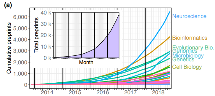
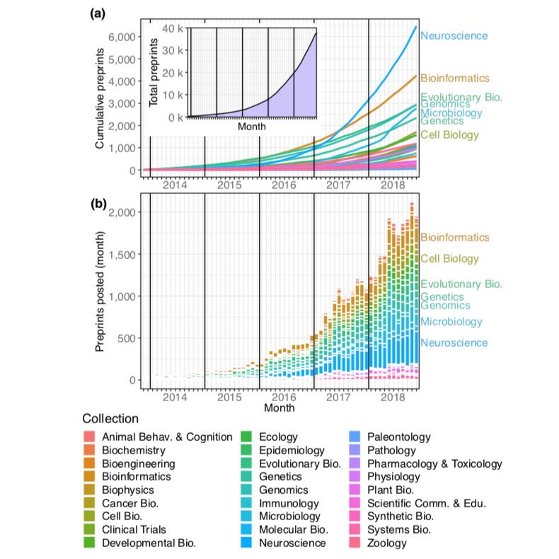
Figure 1, reproduced from Figure 1, Abdill and Blekhman 2019, under a CC-BY-ND-4.0 international license. Total preprints posted to bioRxiv over a 61-month period from November 2013 through November 2018. (a) The number of preprints (y- axis) at each month (x-axis), with each category depicted as a line in a different color. (a, inset) The overall number of preprints on bioRxiv in each month. (b) The number of preprints posted (y-axis) in each month (x-axis) by category. The category color key is provided below the figure.
What makes a preprint successful?
Citation rates have long served as the bibliometric gold standard for measuring the scientific impact of publications. However, follow-up studies and reviews can take years to make their own appearance in the literature, meaning that the impact of any one study based on citations can only be assessed in the long term (read: years to decades).
Standing in sharp contrast to this slow, cumulative view of scientific impact, the core goal of preprints is to accelerate the dissemination of scientific observations – to promote discussion, collaboration, and quick follow-up. In this sense, the ability of a preprint to reach the scientific community quickly and effectively is perhaps the ultimate measure of its success.
This alternate perspective justifies Abdill and Blekhman’s use of preprint downloads, Twitter activity and eventual publication outcomes as key quantitative metrics of preprint success, but these naturally raise some questions as well. Is using Twitter to define “popularity” acceptable? Our experience suggests Twitter is indeed the dominant social media platform for spreading preprints – but this does raise the possibility of excluding communities of scientists, and their opinions, based on a lack of Twitter presence. Our hope is that preprint curation platforms (such as preLights) will play an ever more important role in dissemination.
As with all bibliometric methods, it is important to keep in mind the potential for manipulation or misuse – as summarised in Goodhart’s Law, when a measure becomes a target, it ceases to be a good measure2. While we applaud the introduction of quantitative measures for preprint “success” in the rxivist project and the enablement of detailed analyses conducted by the authors, we are mindful that a small number of metrics should not be used to define a preprint without critical engagement and evaluation.
Social media and the dissemination of science
The use of Twitter activity as a metric for popularity of preprints on the rxivist website helps flag some issues of interest. First, biases in research communication using social media are poorly understood. For example, among scientists active on Twitter, there is widespread variation in the number of their Twitter followers3. As such, the interests of a single popular scientific influencer could potentially drive far greater disparities than are reflected in quality.
Second, preprinting opens up a space – until recently unexplored – for community engagement, spanning the gap between the dissemination of the authors’ unfiltered data and ideas, and the final peer-approved version. The bulk of this engagement takes place on social media platforms like Twitter. While the informal, loose, and relatively egalitarian structure of communicating this way can be immensely liberating, it is also easy to get lost in the noise, or to simply be overwhelmed by information overload. It is possible to go to sleep in the UK and miss a wide-ranging and critical discussion about a preprint taking place in US time zones – by the time you wake up, Twitter has moved on to the next big thing. In a world of science dissemination via social media, preprint curation and journal club initiatives (and the biorXiv comments section) must take on a critical role – that of providing a stable platform for sustained, publicly recorded engagement while remaining responsive to the abbreviated timescales driven by social media.
What happens, then, after the first few weeks, when the tweetstorms have settled, and the commentaries have been posted? We explore this in more detail below.
Time to publication
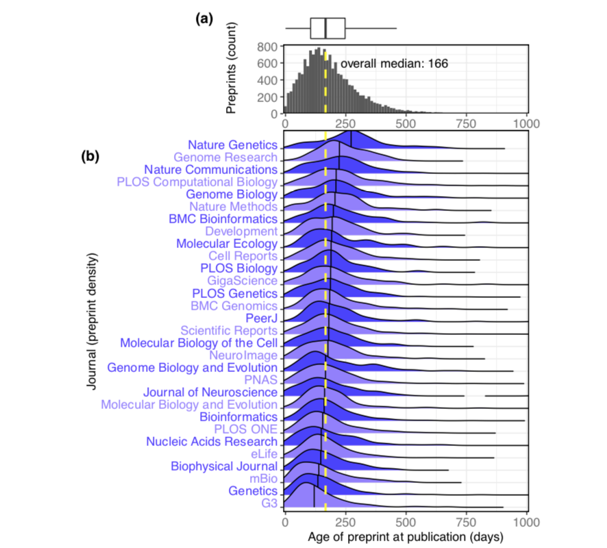
The eventual publication outcomes of preprints – their appearance in some peer-reviewed journal – feature heavily in the metrics utilised by Abdill and Blekhman. The authors found that two-thirds of bioRxiv preprints posted between 2013 and the end of 2016 were eventually published in peer-reviewed form. This high rate indicates that authors tend to post quality preprints, therefore initial fears that work not meeting certain scientific standards might ‘clog’ bioRxiv appear to be unfounded. The median time from the posting of a preprint on bioRxiv to its final publication in a peer-reviewed journal was just under six months, although this varied substantially, depending on the eventual journal of publication (Figure 2). In terms of accelerating science, pushing forward the public dissemination of new information by an average of six months could be considered a significant success- especially for early-career researchers, for whom six months represents a large fraction of their total career progress. Having their work and ideas publicly available enables them to receive feedback earlier and prepare effectively for career transitions.
Figure 2, reproduced from Figure 6 of Abdill and Blekhman, 2019, under a CC-BY-ND 4.0 international license. The interval between the date a preprint is posted to bioRxiv and the date it is first published elsewhere. (a) A histogram showing the distribution of publication intervals—the x axis indicates the time between preprint posting and journal publication; the y axis indicates how many preprints fall within the limits of each bin. The yellow line indicates the median; the same data is also visualized using a boxplot above the histogram. (b) The publication intervals of preprints, broken down by the journal in which each appeared. The journals in this list are the 30 journals that have published the most total bioRxiv preprints; the plot for each journal indicates the density distribution of the preprints published by that journal, excluding any papers that were posted to bioRxiv after publication. Portions of the distributions beyond 1,000 days are not displayed.
However, as Abdill and Blekhman point out, “time-to-publication” is influenced by a plethora of factors, including journal behavior, when preprints are posted in the publication process, and whether preprints are ever published at all.
It’s worth noting that the two “slowest” and the two “fastest” journals (in terms of time from bioRxiv posting to final publication) both fall within the field of genetics and genomics – Genetics and G3 on the “fast” end and Nature Genetics and Genome Research on the “slow” end. This suggests that field-specific attitudes and norms about preprint usage do not drive the difference in publication times by journal.
Preprints can be posted at many stages of the publication process. More field-specific data on when authors typically post preprints would help us understand how authors are utilising preprints – are they sharing work as close to publication as possible, or aiming to get feedback on their work prior to submitting it to a journal? Although the total number of infractions appears to be small, it’s worth noting that some authors appear to be flaunting the bioRxiv guidelines that state that preprints must be uploaded prior to acceptance in a journal.
Publication as a readout of preprint quality
The authors find a significant correlation between preprint download counts and the impact factor of the eventual journal of publication. This suggests that the popularity of a preprint reflects the eventual perceived impact and quality of the work, and that there is some informal consensus among the scientific community about the scientific quality of both published and unpublished preprints. However, the authors rightly point out that publication, particularly in a high profile journal, may actually drive further downloads. Additionally, both metrics are susceptible to biases that distort the connection between popularity and quality – name recognition of the principal investigator, or “hot” topics within a field, for example. Therefore, further validation will be needed to determine how precisely this measure correlates with the scientific utility of a preprint. For instance, it will be interesting to examine whether the number of downloads a preprint receives in its first month holds predictive value for eventual publication.
On the other end of the scale, what about preprints that are never published in peer-reviewed form? Missing links to final publications in the rxivist website and analysis may be partly a technical issue – changes in authors or preprint title make it difficult for automated formats to match preprints to their final publications – but this is unlikely to account for many preprints. Instead, of greater concern is whether these unpublished preprints are of lower quality and are constructed on the back of poor science that would not pass the stringency of peer review. We argue that a lack of final publication should not be held as an indictment of the quality of a preprint. First, preprints help to communicate results more rapidly, particularly in instances where matching a manuscript to typical journal expectations may be difficult or impossible – for example, following the departure of the primary author from a lab. Second, preprints can serve as useful repositories of negative results, which often remain unpublished – after all, “a negative result is still a result”. Therefore, preprints communicating preliminary or shorter stories can prompt discussion and study in the field and be as successful as those that lead to full publication, especially if they invalidate previous hypotheses or drive changes in research that lead in different directions.
Conclusion
The explosive rise of bioRxiv preprints in the life sciences since 2013 has clearly demonstrated the importance of increased speed and visibility through the publishing process. Leveraging social media has also been key to this process. The rxivist project is a laudable effort to collect and collate data on preprint usage, which can in turn be used to measure the influence and uptake of preprints in the life sciences, while he rxivist website makes this data easily accessible and allows others to easily interact with the metadata. We believe the data indicate that preprints are generally successful and that their increasing adoption is a positive trend for not just the life sciences, but science as a whole. While the metrics for preprints diverge from those used for standard peer-reviewed publications, this appropriately reflects the difference between the standards and goals for preprints and that of peer-reviewed journal articles. We thank the authors of the rxivist project for their work, and are excited to watch as our understanding of preprint publishing continues to grow in the future.
References
Abdill, R. J. & Blekhman, R. Tracking the popularity and outcomes of all bioRxiv preprints. bioRxiv 515643 (2019). doi:10.1101/515643
Biagioli, M. Watch out for cheats in citation game. Nature535, 201–201 (2016).
Côté, I. M. & Darling, E. S. Scientists on Twitter: Preaching to the choir or singing from the rooftops? FACETS3, 682–694 (2018).
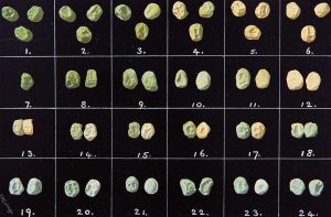
Picture: Photographic plate from Raphael Weldon’s 1902 paper in Biometrika.
In the latest episode of Genetics Unzipped, Kat Arney is exploring some more of the leading 100 ideas in genetics. She’s been digging around in the genetic vegetable patch in search of flavourful GM tomatoes, chunky onion genomes and Mendelian peas.
If you enjoy the show, please do rate and review and spread the word. And you can always send feedback and suggestions for future episodes and guests to podcast@geneticsunzipped.com

 (No Ratings Yet)
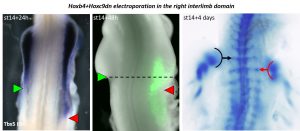
 (2 votes)
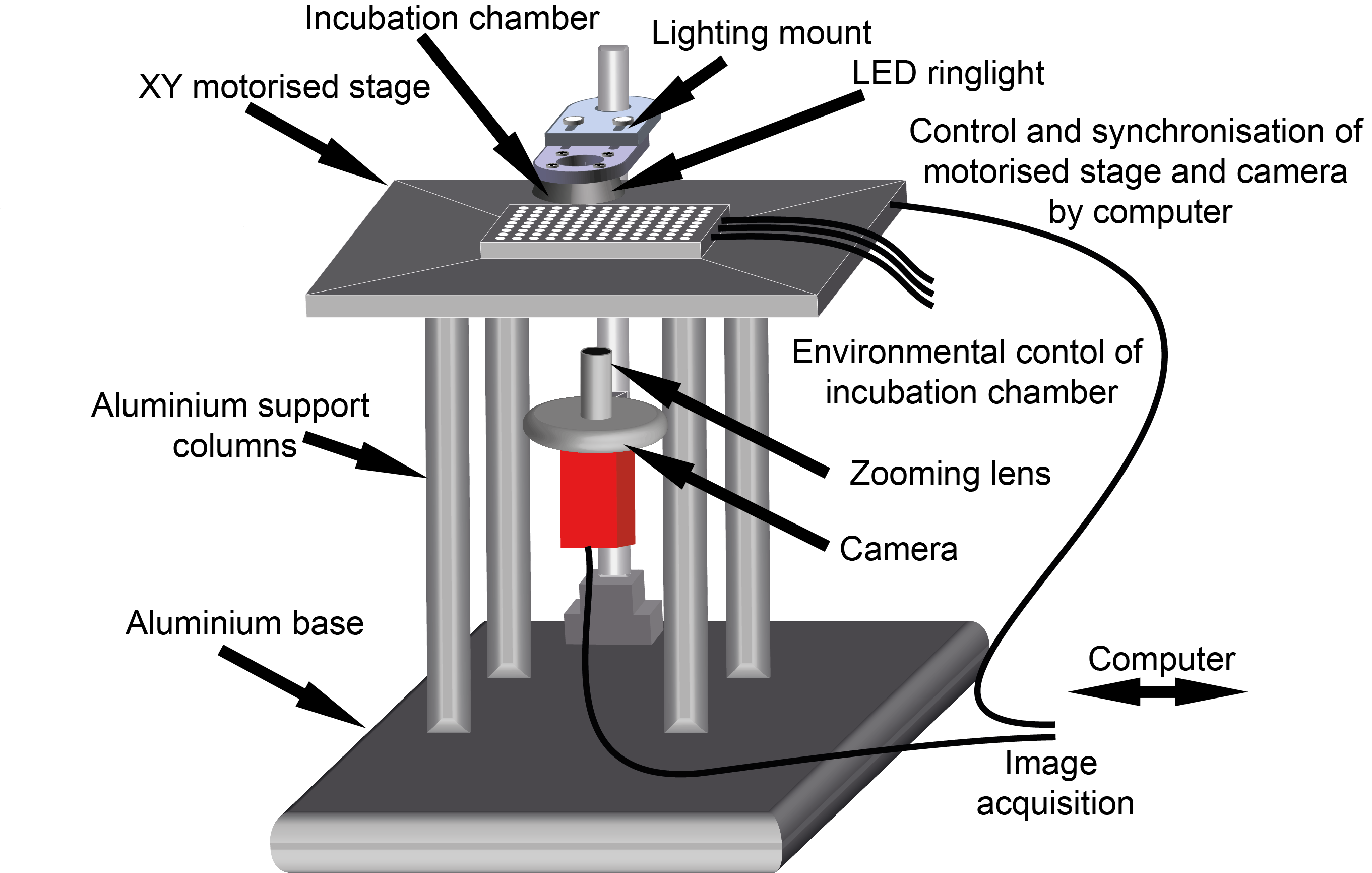
 (3 votes)