This interview, the 52nd in our series, was the first to be published in Development. We’re aiming for one interview per issue, and will continue to put them up here (once the issue has closed).
During teleost fertilisation, sperm fertilises the oocyte through the micropyle, a channel traversing the vitelline membrane at the animal pole. This crucial structure is formed by a specialised micropylar cell (MC) in the follicular epithelium that surrounds the oocyte, but many aspects of MC specification and differentiation remain incompletely understood. A paper from the current issue of Development reveals an unexpected role for the Hippo pathway effector Taz in this process, and hence in fertilisation, in zebrafish. First author and PhD student Chaitanya Dingare and his supervisor Virginie Lecaudey, Professor for Developmental Biology of Vertebrates at the Goethe University of Frankfurt in Germany, told us more about the story.
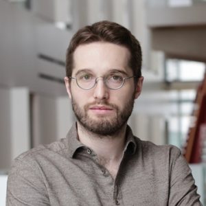
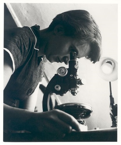
Chaitanya (L) and Virginie (R)
Virginie, can you give us your scientific biography and the questions your lab is trying to answer?
VL I studied Biology in Paris and did my PhD at the Ecole Normale Supérieure in the lab of Sylvie Schneider-Maunoury. We were interested in the mechanisms underlying the segmentation of the vertebrate hindbrain; this is when I started to work with zebrafish. During this time, I developed a solid background in molecular biology and a passion for developmental biology. Then in 2005 I joined Darren Gilmour at the European Molecular Biology Laboratory (EMBL) in Heidelberg as his lab was just starting. There I started to use the zebrafish lateral line primordium as a model of dynamically remodelling epithelium, and truly enjoyed the extraordinary scientific and international environment of the EMBL. These years were influential in my career, not only because of the amount of things I learned, but also because of the amazing people I met there, and who are now my colleagues, my collaborators and my friends. In 2009, I got the opportunity to start my own lab as a Junior Professor at the University of Freiburg, within the BIOSS Centre for Biological Signalling Studies excellence cluster. We started to focus on the mechanisms that underlie cell shape changes in epithelia and how this drives morphogenesis and organogenesis. After spending 5 years in Freiburg, in 2015 I was appointed as a full professor at the Goethe University of Frankfurt.
My lab is still focusing on how cells coordinate their behaviours to assemble coherent and functional tissues and organs during development. For this purpose, we have mainly been using the lateral line primordium, which is a beautiful system to understand how cell proliferation, cell migration, cell shape changes and cell differentiation are orchestrated in a tissue that is undergoing morphogenesis. In addition, as a group of cells that migrate superficially in a transparent embryo, this is also an ideal system to use to follow these processes in real time within a living organism using high-resolution microscopy. More recently, we have started to look at another epithelium, the follicle cell layer that surrounds the oocyte, and in particular one cell within it: the micropylar cell (MC).
Chaitanya, how did you come to join the Lecaudey lab, and what drives your research?
CD After 2 years of research experience at the Tata Institute for Fundamental Research in Mumbai, I was looking for a PhD position involving zebrafish genetics and morphogenesis. Among other places in Europe, I had applied to Virginie’s lab in Freiburg as the focus of the lab was on studying various cellular behaviours in a very dynamic system, the lateral line primordium.
The major force that drives my research is the frequent stimulating and motivating discussions with Virginie, neighbouring lab members, my friends and my colleagues. Another personally important aspect of doing research is to try and look at my results without having any ‘favourite’ hypothesis in mind: this makes a huge difference as it gives some flexibility to explore more options. I also personally believe that collaborations play a major role in research, in particular when you start working on a completely new question and lack some expertise. This is reflected in our current story as well. Finally, the strong support of my supervisor, a productive research environment and, last but not least, extremely supportive colleagues: all of these aspects play a pivotal role in my research.
How did you come to be interested in zebrafish fertilisation?
CD Our current story is all about serendipity. As I joined Virginie’s lab, I started to analyse the role of Yap and Taz in the lateral line primordium. For that, we generated mutants for yap and taz/wwtr1 using TALENs. To our surprise, we found out that the taz mutant females were infertile, and that’s how we started to work on fertilisation and oogenesis. It was a bit challenging for me initially, as this was the first time I was working on adult fish to obtain immature oocytes for my experiments. It was quite exciting as well, as I got to learn a whole new set of techniques. This project has definitely honed my experimental skills, and also trained me to think in a very simple, yet ‘out of the box’, way, as we began with a very simple observation.
VL Indeed, this is an example of a purely curiosity-driven project. Chaitanya joined the lab as we started to work on the Hippo signalling pathway. The mutant he – together with former bachelor student Svenja Godbersen – generated did not show any obvious phenotype, but when homozygous mutants were incrossed, the eggs were systematically unfertilised. At that point, it was really Chaitanya’s curiosity and perseverance which made the difference, as he was so determined to figure out what was going wrong in the mutant. And then it became so exciting to discover this entirely new field for us. It was totally refreshing!
It became so exciting to discover this entirely new field for us. It was totally refreshing!
Can you give us the key results of the paper in a paragraph?
CD & VL Our study provides a molecular and genetic basis to MC development that has otherwise been studied only at the structural level. We show that the Hippo pathway effector Taz is essential for the differentiation of the MC, and thus for fertilisation in zebrafish. One of our key findings is that Taz enrichment in the MC precursor precedes the drastic changes in shape and size that characterise the differentiated MC. This makes Taz not only the first bona fide marker of the MC, but also the earliest event that distinguishes a unique cell among hundreds within the follicular cell (FC) layer. These findings are supported by our genetic data, which show that in the taz/wwtr1 mutants, no sign of MC differentiation can be detected. As a consequence, the MC and the micropyle fail to form.
What do you think might be upstream of Hippo – how is a single cell specified from the follicular epithelium?
CD & VL This is indeed a very interesting question! We know from previous studies that the polarity of the oocyte is crucial to localise the micropyle facing the oocyte animal pole, but the nature of the signal transmitted from the oocyte to the FC layer is unclear. Our paper shows that a small patch of microvilli at the oocyte animal pole is distinctly lost much earlier than the rest of the microvilli that cover the oocyte, irrespective of the presence or absence of the MC. In wild type, the MC lengthens as the microvilli shorten so that it remains constantly attached to the oocyte surface. This suggests that the microvilli at the animal pole may have distinct properties and could be involved in transmitting a signal, possibly a biomechanical one, leading to modulation of the Hippo pathway in the MC precursor.
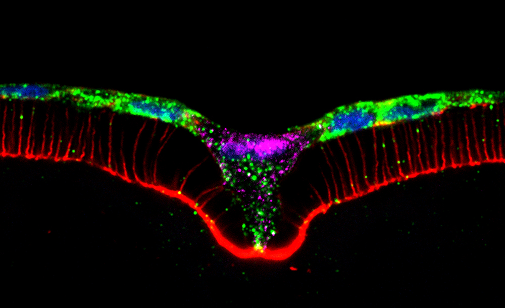
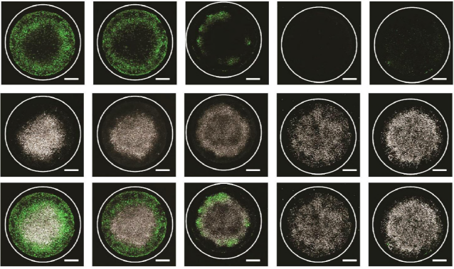
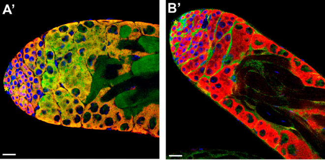
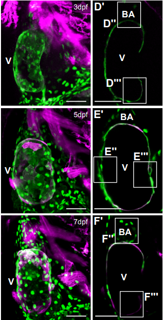
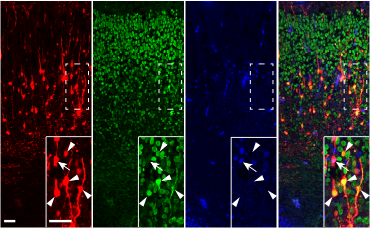
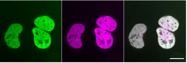
Cross-section of the MC showing Taz (magenta) enrichment in its nucleus (DAPI, blue). Rhodamine-Phalloidin (red) is used as a membrane marker and E-cad (green) is used to mark the contact point between the oocyte and the MC.
Hippo signalling, in both canonical and non-canonical flavours, has been implicated in numerous developmental processes. Have you got any idea how the pathway is directing MC development?
CD Currently, I would give equal importance to both the pathways. Canonical Hippo signalling, by a classical definition, is a kinase cascade, so we need to first find out the phosphorylation status of Taz in the MC in comparison with other cells in the epithelium. This would at least help us to favour one over another.
VL This question remains fully open and, in many cases, it has been shown that canonical and non-canonical Hippo pathways are interconnected. We are just starting to look at whether components of the canonical or non-canonical Hippo pathway are present in the FC layer, and in the MC in particular, so it is too early to rank one pathway over the other.
When doing the research, did you have any particular result or eureka moment that has stuck with you?
CD Yes. At the time I was still working in Freiburg: I started crossing the Taz mutants, but after three to four crossings, I got only unfertilised eggs. This was a kind of eureka moment for me – it was so unusual that hundreds of eggs were left unfertilised.
And what about the flipside: any moments of frustration or despair?
CD During the initial period of my PhD, the mutants I had generated using reverse genetic approaches did not show any detectable phenotype. Therefore, the candidate-based approach did sometimes leave me frustrated!
So what next for you after this paper?
CD I am currently applying for postdoc positions. I wish to continue in the developmental biology field, with a special emphasis on in vitro systems such as organoids or embryonic stem cells.
Where will this work take the Lecaudey lab?
VL This work establishes the MC as a new and exciting system to dissect the mechanisms that underlie the specification of a unique cell fate in a field of otherwise homogeneous cells. This is what we are particularly interested in and want to focus on in the near future. The two main obvious questions that come out of this work are: what are the mechanisms downstream of Taz that drive the differentiation of the MC and what is the nature of the signal that transmits positional information from the oocyte into the overlying FC layer, leading to the specification of a unique cell within? As mentioned above, this signal could be biochemical, biomechanical or both.
Finally, let’s move outside the lab – what do you like to do in your spare time in Frankfurt?
CD Frankfurt being a very international city, I often go out and try cuisines from different countries.
VL I spend as much time as I can with my husband and our two children. We live just outside Frankfurt in a very nice hilly area called ‘Taunus’. We like to hike and bike there. It does not really matter what we do, we just enjoy the time together.
Welcome to our monthly trawl for developmental biology (and related) preprints.
This month’s haul includes a potful of plant development, new ways to mend broken hearts, an Alexa in the lab, and three preprints from Development’s recently appointed Editor Cassandra Extavour.
The preprints were hosted on bioRxiv, PeerJ, andarXiv. Let us know if we missed anything, and use these links to get to the section you want:
In vitro characterization of the human segmentation clock
Margarete Diaz-Cuadros, Daniel Wagner, Christoph Budjan, Alexis Hubaud, Jonathan Touboul, Arthur Michaut, Ziad Al Tanoury, Kumiko Yoshioka-Kobayashi, Yusuke Niino, Ryoichiro Kageyama, Atsushi Miyawaki, Olivier Pourquie
Zebrafish neural tubes from Collins, et al.’s preprint
Germline deletion reveals a non-essential role of the atypical MAPK6/ERK3
Natalia Ronkina, Karin Schuster-Gossler, Florian Hansmann, Heike Kunze-Schumacher, Inga Sandrock, Tatiana Yakovleva, Juri Lafera, Wolfgang Baumgärtner, Andreas Krueger, Immo Prinz, Achim Gossler, Alexey Kotlyarov, Matthias Gaestel
DEAH-box helicase 37 (DHX37) defects are a novel molecular etiology of 46,XY gonadal dysgenesis spectrum
Thatiana Evelin da Silva, Nathalia Lisboa Gomes, Antonio M Lerario, Catherine E Keegan, Mirian Yumie Y Nishi, Filomena M Carvalho, Eric Vilain, Hayk Barseghyan, Alejandro Martinez-Aguayo, Maria Verónica Forclaz, Regina Papazian, Luciani Renata Carvalho, Elaine Frade Costa, Berenice B Mendonca, Sorahia Domenice
Contribution of Retrotransposition to Developmental Disorders
Eugene J Gardner, Elena Prigmore, Giuseppe Gallone, Patrick J Short, Alejandro Sifrim, Tarjinder Singh, Kate E Chandler, Emma Clement, Katherine L Lachlan, Katrina Prescott, Elisabeth Rosser, David R FitzPatrick, Helen V Firth, Matthew E Hurles, Deciphering Developmental Disorders study
PDGFRα signaling in cardiac fibroblasts modulates quiescence, metabolism and self-renewal, and promotes anatomical and functional repair
Naisnana S Asili, Munira Xaymardan, Elvira Forte, Ashley J Waardenberg, James Cornwell, Vaibhao Janbandhu, Scott Kesteven, Vashe Chandrakanthan, Helena Malinowska, Henrik Reinhard, Sile F Yang, Hilda A Pickett, Peter Schofield, Daniel Christ, Ishtiaq Ahmed, James Chong, Corey Heffernan, Joan Li, Mary Simonian, Romaric Bouveret, Surabhi Srivastava, Rakesh K Mishra, Jyotsna Dhawan, Robert Nordon, Peter Macdonald, Robert M Graham, Michael Feneley, Richard P Harvey
Deriving Cardiomyocytes from Human Amniocytes
Colin T. Maguire, Ryan Sunderland, Bradley L Demarest, Bushra Gorsi, Joshua Jackson, ANGELICA LOPEZ-IZQUIERDO, Martin Martin Tristani-Firouzi, H. Joseph Yost, Maureen L Condic
Remote control of alternative splicing in roots through TOR kinase
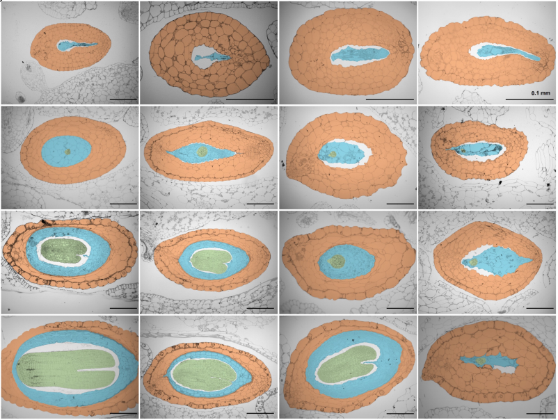
Ezequiel Petrillo, Stefan Riegler, Armin Fuchs, Lucas Servi, Micaela A. Godoy Herz, Maria Guillermina Kubaczka, Peter Venhuizen, Alois Schweighofer, Alberto R Kornblihtt, Craig Simpson, John W.S. Brown, Christian Meyer, Maria Kalyna, Andrea Barta
Functional dissection of the ARGONAUTE7 promoter
J Steen Hoyer, Jose L Pruneda-Paz, Ghislain Breton, Mariah A Hassert, Emily E Holcomb, Halley Fowler, Kaylyn M Bauer, Jacob Mreen, Steve A Kay, James C Carrington
A novel model plant to study the light control of seed germination
Zsuzsanna Merai, Kai Graeber, Per Wilhelmsson, Kristian K. Ullrich, Waheed Arshad, Christopher Grosche, Danuse Tarkowska, Veronika Tureckova, Miroslav Strnad, Stefan A. Rensing, Gerhard Leubner-Metzger, Ortrun Mittelsten Scheid
Regulatory changes in pterin and carotenoid genes underlie balanced color polymorphisms in the wall lizard
Pedro Andrade, Catarina Pinho, Guillem Perez i de Lanuza, Sandra Afonso, Jindrich Brejcha, Carl-Johan Rubin, Ola Wallerman, Paulo Pereira, Stephen J. Sabatino, Adriana Bellati, Daniele Pellitteri-Rosa, Zuzana Bosakova, Miguel A. Carretero, Nathalie Feiner, Petr Marsik, Francisco Pauperio, Daniele Salvi, Lucile Soler, Geoffrey M. While, Tobias Uller, Enrique Font, Leif Andersson, Miguel Carneiro
CRISPR/Cas12a-assisted PCR tagging of mammalian genes
Julia Fueller, Matthias Meurer, Konrad Herbst, Krisztina Gubicza, Bahtiyar Kurtulmus, Julia D. Knopf, Daniel Kirrmaier, Benjamin Buchmuller, Gislene Pereira, Marius K. Lemberg, Michael Knop
Unbiased detection of CRISPR off-targets in vivo using DISCOVER-Seq
Beeke Wienert, Stacia K Wyman, Christopher D Richardson, Charles D Yeh, Pinar Akcakaya, Michelle J Porritt, Michaela Morlock, Jonathan T Vu, Katelynn R Kazane, Hannah L Watry, Luke M Judge, Bruce R Conklin, Marcello Maresca, Jacob E Corn
One scientific story has dominated the news this week: the first report of CRISPR-edited human babies being born. In an associated Node post, we’ve collected the most useful links we could find surrounding the story, and here we reached out to members of the community for their perspectives.
Some responses are hopefully still coming in so look out for updates, and we’d also love to hear your thoughts – just use the comment box below.
MRC London Institute of Medical Sciences, Imperial College London
There is already a prevailing feeling that this work may be false. Certainly it is very difficult to know exactly what has gone on, with the limited information available thus far. I would agree with many other commentators that it is simply too soon to attempt human genome editing in embryos. For one thing there are obvious concerns about off-target effects and overall safety. I am also not convinced of the argument for attempting to make this particular modification. This doesn’t mean such edits are necessarily wrong, but I am not convinced a compelling case has been made especially given the risks.
The story has provoked an interesting debate regarding what type of genome and/or germline modification might be justified. While I agree that in many cases pre-implantation genetic diagnosis (PGD) would be a better and safer option than genome editing, I think many commentators have oversimplified the issue. There are situations in which PGD would be of no use, and genome editing is the only option, and this is not simply restricted to cases in which both parents are homozygous for a recessive disease allele. So to attempt to argue the issue away in this way seems nonsensical. My own group is doing some pre-clinical work to test if a genome editing approach might be curative in just such a scenario. Of course, if such approaches do appear promising, moving forwards to in vivo application will require very careful and meticulous pre-clinical studies to demonstrate safety, as well as proper ethical debate, public scrutiny and legislation/regulation.
The most worrying aspect of this story is that these critical ethical and safety debates have been skipped. I do hope this does not set the field back, or prevent considerate and nuanced debate going forwards. It would be sad if irresponsible use of the technology in the coming years prevents its judicious application in the future, including potentially curative therapies for patients with no other options.
Case Western Reserve University School of Medicine
The germline editing of human embryos is not new – there have already been a handful of scientific papers published on this type of research. What is new about the He case is that He transferred the edited embryos into women’s wombs, with or without full informed consent – that point is unclear. But aside from concerns about informed consent, the most significant ethical line that he crossed involved his many attempts at uterine transfer. Had He confined his editing work to in vitro activities only, he would have made a very small splash in the scientific community. Current ethical recommendations and guidelines, including the 2016 Guidelines of the International Society for Stem Cell Research, all state that it would be unethical to transfer germline edited human embryos into the womb, although in vitro work alone is permissible. Guidelines such as these should cast a wider net of potential actors.
It is not enough for scientific self-regulation to involve just the usual suspects involved in events like the 1st and 2nd International Summit on Human Genome Editing, where most of the participants are basic scientists and ethics and policy experts. Conspicuously absent from international and national discussions are the fertility clinic physicians and other assisted reproduction medical professionals who would eventually be the ones to provide reproductive gene editing procedures as an option for affected couples seeking to have healthy children. These medical professionals from the world of assisted reproductive technologies need to be involved in the discourse around reproductive germline editing. They need to be brought into the discussion surrounding what it means for scientists (and licensed physicians) to self-regulate on the issue of germline engineering. Fertility clinic doctors need to have a seat at the table.
Hospital for Sick Children Toronto, University of Toronto
This announcement from the group in China led by JianKui He is a very unfortunate and unwise development. The consensus from almost every working group internationally, including the National Academies Working Group, of which I was a part, has been that we need to move cautiously on possible germline editing in terms of safety and efficacy and that, even when these barriers are met, this approach would only be used for preventing serious genetic disease, where there is no other option, and where there has been full oversight, ethical approval and societal consensus. None of these applies to this report; ethical review is under question; his own university has disavowed him; the editing, if true, would count as an enhancement and not necessary for the child to be HIV-free; the long-term consequences in terms of susceptibility to other viral diseases could be damaging. The Chinese academies and academics are united in condemning this work and continued work towards international guidelines and regulation is clearly needed.
Paulo Navarro-Costa
The Gulbenkian Institute and at the Institute of Environmental Health in Portugal
As a reproductive biologist I was tremendously relieved by the nearly universal backlash against this purported achievement. The importance of preclinical safety assessment is paramount, particularly when it comes to procedures with a direct impact on our germ cells and resulting embryos. At the moment we still don’t know just how safe human genome editing really is. Another point this controversy makes abundantly clear is the need to ensure a consistent ethical framework across borders. Science and technology are a global enterprise and should be regulated likewise, especially when it comes to the use of human gametes and embryos for research purposes. I’m concerned with the fact that our currently heterogenous regulatory landscape leaves too much room for unethical and exploitative research.
The University of Texas MD Anderson Cancer Center, Houston
Tuesday evening here in Houston, I watched Dr. Jiankui He’s talk live through a video link to the 2nd International Summit on Human Genome Editing in Hong Kong. Dr. He presented a large amount of data about the research that led to the generation of the first humans produced with edited genomes, twin girls. Dr. He said a paper describing the results had been submitted for peer review. The gene that was edited using CRISPR technologies was CCR5. CCR5 encodes a receptor required for HIV infection. There is a relatively common loss-of-function allele called D32 in certain human populations such that there are individuals homozygous for this allele that are apparently normal yet resistant to HIV infection. Dr. He reported his group had generated one infant girl homozygous for CCR5 edited alleles and a twin girl that was heterozygous. He reported that the girls were normal and healthy. He also said that there would be an 18-year follow up on the children.
If the results hold true, then a so-called line has been crossed. In the current situation, normal (wild-type) zygotes were edited to make them resistant to a viral infection. However, in situations to cure a genetic disease, in nearly all cases that I can imagine, there will be carrier embryos and probably wild-type embryos. In these situations, preimplantation genetic diagnosis could identify embryos without the genetic disease for transfer into the womb. Thus, even though human genome editing to generate babies is now apparently possible, I’m not sure how it would be applied for clinical therapies.
Carnegie Institution for Science Department of Embryology
As for the medical reason claimed by He Jiankui on this clinical trial (protecting the babies from HIV), I do not believe it is justified. It appears to me that he is doing an extremely risky, but completely unnecessary, experiment directly on two innocent HUMAN babies. I am therefore totally horrified for what He has done. Meanwhile, I do feel this is an individual case. Although deeply depressing, it is slightly gratifying to see that the whole Chinese Biology Community is unprecedentedly unified to condemn such an irresponsible, unethical, and illegal behavior. Next, I think multiple levels of investigations are needed to first validate the whole case. And we should give the two innocent girls the privacy and a normal life–or at least as close to be normal as possible. As a global community, we should take this case as a hard lesson to find a better and efficient way on implementing the standards and guidelines.
One scientific story has dominated the news this week: the first report of CRISPR-edited human babies being born. The story’s scientific and ethical aspects stirred up heated debate, as did its means of delivery: rather than a published paper, the story broke with reports of clinical trial documents and then a YouTube video from lead researcher He Jiankui (from the Southern University of Science and Technology in Shenzhen), all on the eve of a conference he was due to speak at (and whose organisers were seemingly unaware what we was going to speak about).
In an associated Node post, we asked developmental and reproductive biologists to give their reaction to the story (and we’d love to hear yours too), but here we’ve collated a bunch of hopefully helpful links, and some recent Development commentaries on the issues surrounding gene editing in humans.
The story breaks
On 25 November, Antonio Regaldo in MIT Technology Review reported details of the study’s clinical trial data
The He Lab YouTube channel released this video on the same day (the channel also has four associated videos about the work)
“A surgery that could save a child from a lethal genetic disease like cystic fibrosis or from a life-threatening infection like HIV doesn’t just give that little boy or girl an equal chance at a healthy life. We heal a whole family”
Statement from He’s employer, the Southern University of Science and Technology, stating that the university knew nothing about He’s work and plans to set up an independent committee to investigate (in Chinese – translates page reads quite clearly; 26/11)
Gaetan Burigo gave a helpful thread particularly regarding the science presented by He in the summit (28/11)
OK there are a lot to unravel in this He Jankui talk and panel discussion at the #GeneEditSummit on #CRISPR in human embryos leading to the birth of the twins Lulu and Nana. -> Let's analyze the science, the ethics and controversies -> thread pic.twitter.com/3qOCC6IhvJ
— Dr Gaetan Burgio @gaetan_burgio@mstdn.science (@GaetanBurgio) November 28, 2018
Peter Mills, Assistant Director of the Nuffield Council on Bioethics, gives his thoughts (28/11)
Here at Development we’ve been thinking about issues of human gene editing for some time, and have commissioned content specifically exploring scientific and ethical aspects. We recently published two Spotlight articles on the theme (published in 2017 and 2018 respectively, before the current story broke).
Stem cells are already being used in combating previously untreatable diseases. Nevertheless, stem cells are not delivering their full potential because the production of specific cell types from stem cells cannot yet be managed. Researchers have now discovered the signals that determine the fate of immature cells in the pancreas. The research shows that they are very mobile and that their destiny is strongly influenced by their immediate environment. This breakthrough will facilitate the manufacturing of pancreatic islet cells for combating type 1 diabetes.
We are rapidly approaching the era for safe mass production of specialized neuronal cell types and insulin-producing beta cells. It will then be possible to test whether transplanting such cells will enable paralysed people to walk again or people with type 1 diabetes to restart their own production of insulin. Until now, the engineering of the specialized cells from pluripotent stem cells has largely been based on empirical knowledge of what works. Results published in the prominent journal Nature by a Danish-led research project represent a major leap forward.
“We have now been able to map the signal that determines whether pancreatic progenitor cells will become endocrine, such as insulin-producing beta cells or duct cells. The cells are analogous to pinballs, whose ultimate score is based on the sum of pin encounters. They are constantly moving around within the developing pancreas, leading to frequent environmental changes. We show that the exposure to specific extracellular matrix components determines the ultimate destiny of the cells,” explains Henrik Semb, Professor and Executive Director, Novo Nordisk Foundation Center for Stem Cell Biology, DanStem, University of Copenhagen.
The matrix determines the destiny
Progenitor cells are similar to stem cells since they can both self-renew and differentiate into mature cell types. However, their self-renewal capacity is generally limited compared with that of stem cells. The dynamic behaviour of progenitors during organ formation makes them difficult to study. By seeding individual human stem cell–derived progenitors on micropatterned glass slides, the researchers could study how each progenitor, without the influence of neighbouring cells, reacts to its surroundings.
“This enabled us to discover something very surprising. Our investigation revealed that interactions with different extracellular matrix components change the mechanical force state within the progenitor. These forces result from interactions between the extracellular matrix, which is outside the cell, and the actin cytoskeleton, which is within the cell.”
Pancreatic endocrine cells include all hormone-producing cells, such as insulin-producing beta cells and glucagon-producing alpha cells, within the islet of Langerhans, whereas the duct cells are epithelial cells that line the ducts of the pancreas.
“The experiments show that exposure to the extracellular matrix laminin instructs the progenitor cells towards an endocrine fate by reducing mechanical forces within the cells. Whereas exposure to fibronectin results in a duct fate because of increased mechanical forces.”
Mechanism facilitates exploitation
To exploit their discovery, the researchers had to understand the signalling pathway. They showed that components in the extracellular matrix trigger a signal into the cell via an integrin receptor, resulting in changes in mechanical forces transmitted through the actin cytoskeleton. The yes-associated protein (YAP) then senses these forces to turn on and off specific genes.
“This cascade determines the ultimate fate of the progenitor cell. Perhaps the most astonishing achievement is that our data answer an enigma that has puzzled the field for decades. How some progenitors mature into duct cells, whereas others become endocrine cells via Notch signals.”
The researcher show that the seemingly stochastic regulation of Notch function is in fact mediated by the progenitor’s encounters with extracellular matrix interactions via the force-sensing gene regulator protein YAP. They were even able to validate the physiological relevance in vivo during pancreas development.
“We can now replace significant numbers of empirically derived substances, whose mode of action in current state-of-the-art differentiation protocols is largely unknown, with small molecule inhibitors that target specific components of the newly identified mechanosignalling pathway.”
With this new strategy, insulin-producing beta cells can now be more cost-effectively and robustly produced from human pluripotent stem cells for future treatments against diabetes.
“Our discovery breaks new ground because it explains how multipotent progenitor cells mature into different cell types during organ formation. It also gives us the tools to recreate the processes in the laboratory, to more precisely engineer cells that are lost or damaged in severe diseases, such as type 1 diabetes and neurodegenerative diseases, for future cell replacement therapies.”
”Mechanosignaling via integrins directs pancreatic progenitor fate decisions” has been published in Nature. Henrik Semb, Professor and Executive Director, Novo Nordisk Foundation Center for Stem Cell Biology, DanStem, University of Copenhagen, and head of Institute of Translational Stem Cell Research at Helmholtz Zentrum München is last author. Drs. Anant Mamidi, Assistant Professor, DanStem and Christy Prawiro DanStem share first authorship, and the work is the result of a collaboration with Professor Palle Serup’s group, DanStem.. The Novo Nordisk Foundation has awarded grants of almost DKK 700 million (€92 million) to the Center for research between 2010 and 2018.
Here at The Company of Biologists we’ve been debating the Bank of England’s decision to put a scientist on their new £50 note (the highest denomination note in England). The scientist must be deceased (only the Queen can grace notes while still alive) and ‘have shaped thought, innovation, leadership or values in the UK’.
Each of our five journals was asked to come up with their nominations for the face of the fifty. Here’s who they picked and why they picked them:
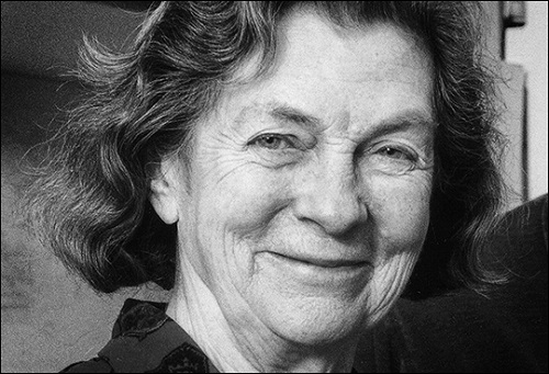
“McLaren was a towering figure in developmental and reproductive biology. She did foundational work in IVF, experimental chimeras and germ cell differentiation, contributed to regulatory policies on human embryo research, and championed pubic engagement”
“She studied in Cambridge, and although a chemist, made a crucial, and often unrecognised, contribution to the discovery of the double helix structure of DNA”
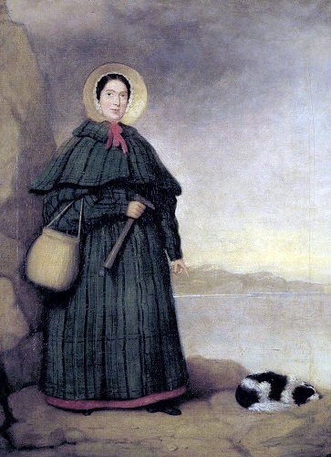
“English fossil collector/palaeontologist. Considered an expert in her field, contributing to important changes in scientific thinking about prehistoric life, at a time when women were mostly excluded from the scientific community”
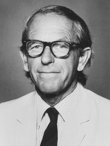
“Modern biology wouldn’t be what it is without him. Double Nobel winner known for sequencing DNA & pioneering work on the structure of proteins. Declined the offer of a knighthood, as did not wish to be addressed as Sir”
The Company of Biologists Twitter feed has a poll where you can pick your favourite out of the four:
What do you think of Development’s choice of Anne McLaren? Which other developmental biologist do you think could be honoured? Let us know in the comments
Regular meetings of scientists such as annual society conferences can create opportunities for scientists to engage the public without extensive effort, making connections between scientists and public audiences. Under the umbrella of a specific topic, events can be created to engage local communities with international researchers and foster forums for discussion of specific areas of research.
With this in mind, we created a space and a time for public engagement and a citizen’s approach to developmental biology in the recent Joint meeting of the Portuguese, Spanish and French Societies for Developmental Biology at Oporto, Portugal (http://devbiomeetingporto2018.pt/).
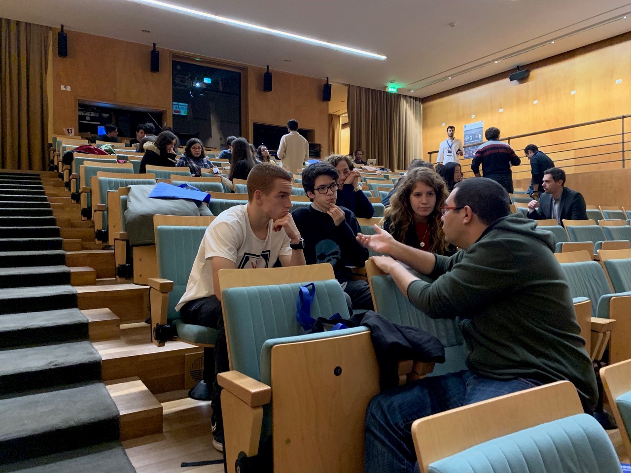
We invited local citizens through social media, the meeting webpage and local secondary school networks. And at the start of the meeting, which took please at the Almeida Garrett Library in Oporto, we organized an open science event for local Oporto high school students (mainly 16-17 year olds), their teachers and other members of the public.
It began with an informal conversation about what is developmental biology and why do we study it. This was done as a dialogue, with a backup of a few slides showing how embryos develop, some historical background and modern applications of the study of developmental biology. For this first part, we used some of the materials available at the BSDB as well as the Droso4schools and HHMI websites.
This was followed by an organized speed-dating with scientists with the help of 12 Portuguese researchers working in national institutions as well as abroad. These volunteers were asked in advance what was the main question they were trying to answer with their research, so they could start their informal conversations from this starting point. They were also asked to bring along an object related to their research as a communication “ice-breaker”. The format of the speed dating consisted of groups of three members of the public to one scientist, with seven and a half minute slots of time available. After this time, a new scientist would take the place of the previous one and the cycle would start again. We found this informal set up allowed for fluid dialogue between scientists and the invited citizens. In addition, the speed-dating format allowed for each person to have the opportunity to speak with 5 or 6 different researchers, all in about 1h. When asked for their opinion about the event, one of the teachers told us:
“As far as the activity with the scientists is concerned, the students liked it immensely. They told me that this type of interaction is much more interesting than just a conference.”
Scientific meetings can play a key role in building bridges between scientific research and public audiences. Let’s try to create more of these opportunities in many other scientific conferences.
Participants in the event:
Sofia J. Araújo, Leonor Saúde, Patrick Lemaire, João Amorim, Tomás Azevedo, Gil Carraco, Ana Gali, André Gonçalves, Sofia Moreira, Paulo Navarro-Costa, Pedro Rifes, Lígia Tavares
MSc/PhD and postdoc positions available in the Zaidel-Bar Cellular and Tissue Morphogenesis Lab.
We study the regulation of the cytoskeleton from single proteins to the entire organism and system levels, using multiple approaches (including bioinformatics, genetics, biochemistry and live imaging) to understand how cells and tissues change shape, move, sense, and generate forces (for more info: celladhesionlab.com).
For the last two years, our interview series ‘The people behind the papers‘ has showcased the faces of developmental biology, and we’re excited to announce that the series will now also be printed in Development.
The Company of Biologists’ journals – Development, Journal of Cell Science, Journal of Experimental Biology andDisease Models & Mechanisms – offer Travelling Fellowships of up to £2,500 to graduate students and post-doctoral researchers wishing to make collaborative visits to other laboratories. These are designed to offset the cost of travel and other expenses. There is no restriction on nationality.
They really are an amazing opportunity for ECRs to learn new things, meet new people and travel to new places.
The current round of Travelling Fellowships closes on 30 November (for travel >14 Jan 2019)

 (No Ratings Yet)


 There is already a prevailing feeling that this work may be false. Certainly it is very difficult to know exactly what has gone on, with the limited information available thus far. I would agree with many other commentators that it is simply too soon to attempt human genome editing in embryos. For one thing there are obvious concerns about off-target effects and overall safety. I am also not convinced of the argument for attempting to make this particular modification. This doesn’t mean such edits are necessarily wrong, but I am not convinced a compelling case has been made especially given the risks.
 This announcement from the group in China led by JianKui He is a very unfortunate and unwise development. The consensus from almost every working group internationally, including the National Academies Working Group, of which I was a part, has been that we need to move cautiously on possible germline editing in terms of safety and efficacy and that, even when these barriers are met, this approach would only be used for preventing serious genetic disease, where there is no other option, and where there has been full oversight, ethical approval and societal consensus. None of these applies to this report; ethical review is under question; his own university has disavowed him; the editing, if true, would count as an enhancement and not necessary for the child to be HIV-free; the long-term consequences in terms of susceptibility to other viral diseases could be damaging. The Chinese academies and academics are united in condemning this work and continued work towards international guidelines and regulation is clearly needed.
 As a reproductive biologist I was tremendously relieved by the nearly universal backlash against this purported achievement. The importance of preclinical safety assessment is paramount, particularly when it comes to procedures with a direct impact on our germ cells and resulting embryos. At the moment we still don’t know just how safe human genome editing really is. Another point this controversy makes abundantly clear is the need to ensure a consistent ethical framework across borders. Science and technology are a global enterprise and should be regulated likewise, especially when it comes to the use of human gametes and embryos for research purposes. I’m concerned with the fact that our currently heterogenous regulatory landscape leaves too much room for unethical and exploitative research.
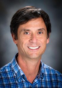 Tuesday evening here in Houston, I watched Dr. Jiankui He’s talk live through a video link to the 2nd International Summit on Human Genome Editing in Hong Kong. Dr. He presented a large amount of data about the research that led to the generation of the first humans produced with edited genomes, twin girls. Dr. He said a paper describing the results had been submitted for peer review. The gene that was edited using CRISPR technologies was CCR5. CCR5 encodes a receptor required for HIV infection. There is a relatively common loss-of-function allele called D32 in certain human populations such that there are individuals homozygous for this allele that are apparently normal yet resistant to HIV infection. Dr. He reported his group had generated one infant girl homozygous for CCR5 edited alleles and a twin girl that was heterozygous. He reported that the girls were normal and healthy. He also said that there would be an 18-year follow up on the children.
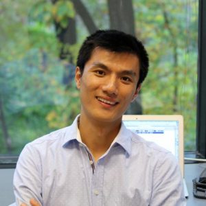 As for the medical reason claimed by He Jiankui on this clinical trial (protecting the babies from HIV), I do not believe it is justified. It appears to me that he is doing an extremely risky, but completely unnecessary, experiment directly on two innocent HUMAN babies. I am therefore totally horrified for what He has done. Meanwhile, I do feel this is an individual case. Although deeply depressing, it is slightly gratifying to see that the whole Chinese Biology Community is unprecedentedly unified to condemn such an irresponsible, unethical, and illegal behavior. Next, I think multiple levels of investigations are needed to first validate the whole case. And we should give the two innocent girls the privacy and a normal life–or at least as close to be normal as possible. As a global community, we should take this case as a hard lesson to find a better and efficient way on implementing the standards and guidelines.
 (6 votes)


