Participants in “Salamander Models in Cross-Disciplinary Research” Vienna, July 2018) Back row: Jeremiah Smith, Jesus Chimal-Monroy, Renee Dickie, Dunja Knapp, Sergej Nowoshilow, Vladimir Soukup, Ryan Kerney, Toshinori Hayashi. Middle Row: Andras Simon, Hans-Georg Simon, Stephane Roy, Jifeng Fei, Moshe Khurgel, Gürkan Ozturk, Kiyokazu Agata, Katia Del Rio-Tsonis, Tatiana Sandoval Guzmán,Ken-Ichi Suzuki (behind Tatiana). Front Row: James Monaghan, Maximina Yun, Alfredo Cruz, Karen Echeverri, Randal Voss, Elly Tanaka, Jessica Whited, Catherine McCusker, James Godwin.
Vienna, Austria
July 2018
The use of salamanders in regeneration and developmental research has a long history filled with luminaries of the life sciences. Thomas Hunt Morgan, Hans Spemann and Hilde Mangold, Ross Granville Harrison, Inez Whipple Wilder, and August Weismann all employed salamanders in their work 1,2. Current experimental research on salamanders largely focuses on studies of regeneration. Their abilities to regenerate limbs, brain, kidney, heart, and tail3 is enviable from the perspective of our limited human regenerative abilities. There is a large and growing toolkit of experimental manipulations, mutant and transgenic lines, and genomic resources that have advanced the use of salamanders to probe critical questions on the evolution of regeneration and its loss. These offer opportunities to discover mechanisms that could lead to new therapies for tissue repair.
The first “Salamander Models in Cross-Disciplinary Biological Research” meeting, organized by Elly Tanaka, Jessica Whited, Karen Echeverri, and Randal Voss, was held this past July (2018) at the Research Institute of Molecular Pathology (IMP) in Vienna, Austria. The meeting was hosted by the intrepid Dr. Tanaka, who is a Senior Scientist at the IMP and a leader in the regenerative biology field. This was a gathering of principal investigators working on various aspects of salamander regeneration, development, genetics, and genomics. The major goals included reviewing recent advances in research tools, along with nuanced tips for employing them, while also establishing a master “to-do” list for the field. Another objective was to lay the groundwork for organizing future salamander meetings intended for the broader community, including postdoctoral and pre-doctoral trainees and potentially representatives from funding agencies.
The Salamanders
Unlike “the” worm or “the” fly, there are multiple salamander models used in molecular studies of development and regeneration. These prominently include the Mexican axolotl (Ambystoma mexicanum), the Iberian ribbed newt (Pleurodeles waltl), the Japanese newt (Cynops pyrrhogaster) and to a lesser extent the North American eastern newt (Notophthalmus viridescens) and several species of lungless salamanders (Plethodontidae). Currently the most commonly used model is the axolotl, which has the longest captive history of any laboratory animal1. This history includes the importation of a founder population to Paris in 1864, some of which contained the mutant “white” phenotype (an edn3 mutant), and deliberate introgression of an A. tigrinum locus found in 1962, which confers albinism through a tyrosinase mutation4,5. These salamander species are representatives of three families (Ambystomatidae, Plethodontidae, and Salamandridae – the newts) out of the ten extant salamander families, which likely had extensive limb regenerative abilities at the base of the their clade6.
Genomics
Our sessions started with a review of the impressive new work in salamander genomics. Recent published genomes for both the Iberian ribbed newt (Pleurodeles waltl7) and Mexican axolotl (Ambystoma mexicanum8) provide tremendous new resources for the field. The axolotl genome is roughly ten times the size of the human genome, making it the largest genome to be sequenced and assembled to date (sorry loblolly pine). The publication of both of these genomes promises to help resolve the loci of multiple established mutant lines5, and offers the opportunity to establish future forward and reverse genetic screens that will improve our mechanistic understanding of regenerative processes. The community identified additional work needed to make consistent annotations, resolving 5’ ends of genes, and approaching a chromosome scale resolution of contiguous sequence. The latter was recently made available through a bioRxiv preprint from the Smith and Voss labs at University of Kentucky. The lack of whole-genome sequences in salamanders has been a major impediment to the field, and though the assemblies will still require extensive refinements, having these resources should prove to be enormously beneficial to labs currently working with these species as well as those interested in starting salamander research.
Tansgenics and Genome Editing
Genome editing approaches are now available for the axolotl9,10, the Iberian newt11,7 and the Japanese fire-bellied newt Cynops pyrrhogaster12 using TALEN and CRISPR-based approaches. The most recent protocols developed by Ji-Feng Fei13 (now at the South China Normal University) for knock-out and knock-in approaches were reviewed. Current best practice techniques focused on improving knock-in strategies without relying on homology-directed repair. These include targeting introns for knock-in’s, screening injected embryo knockouts for efficient guide RNA’s prior to knock-ins, and non-homologous end joining approaches with “ORF Baits.”
The advent of CRISPR and TALEN approaches to genome editing in Pleurodeles was reviewed by Ken-Ichi Suzuki from Hiroshima University. The Suzuki lab is currently working to identify a ROSA-like locus for constitutive expression of knock-in constructs that would also provide a “safe harbor” for exogenous DNA. The intent of this approach is to develop a site that would both be minimally disruptive to normal cellular physiology while experiencing minimal interference from histone modifications in different cell lineages.
A wide range of transgenic and CRISPR edited lines are becoming available, especially in the axolotl. These include constitutive RFP and GFP reporter lines (available from the University of Kentucky’s Ambystoma Stock Center – AGSC), a pax7-mcherry muscle satellite cell marker, a neuronal marker, and a brainbow axolotl14. Discussions focused on prioritizing existing stocks and facilitating their dissemination through the AGSC (primarily in North America), Medipol University, Max Planck Dresden, and MPI (in Europe). The need to pursue financial resources to enable more extensive repository functions for salamanders, including cryopreservation to biobank lines, was also addressed. A long-term goal of the community is to secure resource funding for these types of valuable operations, which will be necessary to advance discoveries in regenerative biology through this growing research community.
Temporal control of transgene expression
Many genes implicated in regenerative processes have pleiotrophic roles in early development. These make knock out experiments in studying adult regeneration difficult as they can be embryonic lethal. Therefore both temporal and spatial control of gene expression is critical for furthering regeneration research.
There are several creative approaches available to address this potential stumbling block. These include inducible cre-lox systems15, constitutive cas-9 expression in genomic “safe harbors” with drug inducible guide RNA’s, and viral delivery of foreign transgenes into regenerative blastemas161718. The latter approach has been championed by both Jessica Whited’s group at Harvard University and the Tanaka lab at MPI. These pseudotyped retroviruses have tremendous potential for further labeling and functional studies of the limb blastema, without raising transgenic embryos or modifying the expression of pleiotrophic genes outside the limb.
Open discussions evaluating approaches and avenues for future research allowed individual labs to share their own research objectives with a collective eye toward developing critical methodologies to advance the community.
Resources needed for the field
The real value of this PI-focused meeting was to allow researchers to define limiting resources they found most pressing for the field. Several additional needs were identified in addition to continued improvements to genome annotations and the temporal control of gene expression. These included detailed histological atlases of regeneration, master lists of validated antibodies, and more stable cell lines for in vitro experiments. One of the most obvious resources needed was the continued communication between labs with subsequent salamander research conferences that should continue to strengthen this growing research community.
Coordination and prioritization of a model system
Several research communities have benefitted from the intentional development and promotion of particular model organisms19. One-stop repositories of information such as Flybase, Wormbase, ZFinBase, Xenbase, and Sal-Site have all expanded the research capabilities of participating labs. While many of these labs focus on Developmental Biology, the reach of these model systems includes studies of neuroscience, evolutionary biology, physiology, and ecology. Emulating this deliberate approach will provide vital cohesion and an undoubted boon to investigators studying salamander regeneration and development. While salamanders are a remarkable “model” for regenerative research they are also remarkable organisms for their unique evolutionary histories, ecological roles, life history variation, and conservation biology. Development of tools and resources for the molecular biologists and biochemists working on salamanders will undoubtedly have unintentional spillover benefits into a wider range of research fields.
Outlook
Salamander models continue to be fertile ground for amazing discoveries on regenerative biology, cell differentiation, and development. This conference generated a tremendous amount of motivation for its participants. We have planned a broader 2019 meeting in Massachusetts, which will be a showcase for these recent developments and an iterative checkup on this rapidly growing experimental field.
References
Reiß, C., Olsson, L. & Hoßfeld, U. The history of the oldest self-sustaining laboratory animal: 150 years of axolotl research. Journal of Experimental Zoology Part B: Molecular and Developmental Evolution324, 393–404 (2015).
Wilder, I. W. The Morphology of Amphibian Metamorphosis. (Smith College, 1925).
In Salamanders in Regeneration Research Methods and Protocols (eds. Kumar, A & Andras, S) (Humana Press, 2015).
Humphrey, R. R. Albino axolotls from an albino tiger salamander through hybridization. J. Hered.58, 95–101 (1967).
Woodcock, M. R. et al. Identification of mutant genes and introgressed tiger salamander DNA in the laboratory axolotl, Ambystoma mexicanum. Scientific Reports7, (2017).
Fröbisch, N. B., Bickelmann, C. & Witzmann, F. Early evolution of limb regeneration in tetrapods: evidence from a 300-million-year-old amphibian. Proc. Biol. Sci.281, 20141550 (2014).
Elewa, A. et al. Reading and editing the Pleurodeles waltl genome reveals novel features of tetrapod regeneration. Nat Commun8, 2286 (2017).
Nowoshilow, S. et al. The axolotl genome and the evolution of key tissue formation regulators. Nature554, 50–55 (2018).
Fei, J.-F. et al. CRISPR-mediated genomic deletion of Sox2 in the axolotl shows a requirement in spinal cord neural stem cell amplification during tail regeneration. Stem cell reports3, 444–459 (2014).
Flowers, G. P., Timberlake, A. T., McLean, K. C., Monaghan, J. R. & Crews, C. M. Highly efficient targeted mutagenesis in axolotl using Cas9 RNA-guided nuclease. Development (Cambridge, England)141, 2165–2171 (2014).
Hayashi, T. & Takeuchi, T. Gene manipulation for regenerative studies using the Iberian ribbed newt, Pleurodeles waltl.Methods Mol. Biol.1290, 297–305 (2015).
Nakajima, K., Nakajima, T. & Yaoita, Y. Generation of albino Cynops pyrrhogaster by genomic editing of the tyrosinase Gene. Zool. Sci.33, 290–294 (2016).
Fei, J.-F. et al. Efficient gene knockin in axolotl and its use to test the role of satellite cells in limb regeneration. Proc. Natl. Acad. Sci. U.S.A.114, 12501–12506 (2017).
Currie, J. D. et al. Live imaging of axolotl digit regeneration reveals spatiotemporal choreography of diverse connective tissue progenitor pools. Dev. Cell39, 411–423 (2016).
Khattak, S. et al. Optimized axolotl (Ambystoma mexicanum) husbandry, breeding, metamorphosis, transgenesis and tamoxifen-mediated recombination. Nat Protoc9, 529–540 (2014).
Kuo, T.-H. & Whited, J. L. Pseudotyped retroviruses for infecting axolotl. Methods Mol. Biol.1290, 127–140 (2015).
Khattak, S. et al. Foamy virus for efficient gene transfer in regeneration studies. BMC Dev. Biol.13, 17 (2013).
Oliveira, C. R. et al. Pseudotyped baculovirus is an effective gene expression tool for studying molecular function during axolotl limb regeneration. Dev. Biol.433, 262–275 (2018).
Abzhanov, A. et al. Are we there yet? Tracking the development of new model systems. Trends Genet.24, 353–360 (2008).
Position Description
The Boston College Biology Department seeks outstanding candidates for a tenure-track faculty position in the area of Cell and Developmental Biology. Applicants are sought at the Assistant Professor level; however, exceptionally strong candidates will be considered at the Associate Professor level. The university provides competitive startup funds and research space with the expectation that the successful candidate will establish, or bring to the university, a vigorous, funded research program. Special consideration will be given to candidates whose research program synergizes with current faculty interests. We especially encourage applicants whose research utilizes embryonic model systems, those who study developmental neurobiology, and/or those who work at the interface of biophysics and cell biology. The successful candidate will join an active and expanding department with current strengths in cytoskeletal regulation, developmental biology, cancer biology, as well as microbiology, infectious disease, and computational biology.
The university is situated on a beautiful campus dating back to the beginning of the twentieth century and is closely located to downtown Boston and Cambridge. The Biology Department has strong ongoing collaborative efforts with other departments including Chemistry and Physics, surrounding institutes including BU, Harvard, MIT, Northeastern University and Tufts University, and has state of the art (core-) facilities including next-gen sequencing, cleanrooms for nanofabrication, FACS, microscopy (including super-resolution microscopy), microfluidics, robotics, NMR and mass-spec. Moreover, the university has recently announced its decision to establish an interdisciplinary Institute for Integrated Science and Society, and the building is scheduled for construction beginning Spring 2019.
How to Apply
Applicants should submit a cover letter, curriculum vitae, a statement of research plans, a statement of teaching interests, and arrange for three letters of reference. All application materials should be submitted via Interfolio at http://apply.interfolio.com/53713. Review of applications will begin on October 1st and will continue until the position is filled.
The University of Virginia invites applications for a tenure-track Assistant Professor position in the Department of Biology. We seek applicants who have a research vision that addresses new or longstanding fundamental questions in cell biology. Located within the College of Arts and Sciences, the Department of Biology provides an interdisciplinary and collaborative environment for basic research and teaching that spans multiple levels of biological organization, enhanced by close collaborations with colleagues at our Schools of Medicine and Engineering & Applied Sciences. A successful candidate is expected to establish a vigorous, independent, and externally funded research program as well as provide instruction and scientific training at the undergraduate and graduate levels. Applicants with a respect for diversity and a passion for making a positive impact on the world in a collaborative environment are strongly encouraged to apply. The position will begin on August 25, 2019.
Applicants must have a Ph.D. or other doctoral degree and post-doctoral research experience. A successful applicant will also have research accomplishments and plans of outstanding quality and significance as well as a commitment to excellence in teaching and mentoring. Commitment to participate in and further develop a diverse, collegial, interdisciplinary, and collaborative environment is strongly preferred.
To apply, jobs.virginia.edu/applicants/Central?quickFind=85482. Complete a candidate profile online, attach a cover letter that succinctly highlights your most significant research accomplishments, experiences, and qualifications; a curriculum vitae; a research statement that describes your vision for your research program at the university (≤ 3 pages); a statement of teaching goals; and the contact information of three references. The deadline for receipt of applications is October 31, 2018.
For questions regarding the position, please contact search chair Keith Kozminski, Associate Professor of Biology, at biocellsearch@virginia.edu; for questions about the application process, please contact Savanna Galambos, Faculty Search Advisor, at: skh7b@virginia.edu
The University of Virginia is an equal opportunity and affirmative action employer. Women, minorities, veterans and persons with disabilities are encouraged to apply.
The Department of Biological Sciences at the University of South Carolina invites applications for a tenure-track position as an Assistant Professor with a research program in neurobiology. The successful candidate will be expected to establish an independent, extramurally funded research program focusing on cell-cell communication in neural development and/or disease. We are especially interested in applicants studying how interactions among neurons or between neurons and other cell types such as astrocytes, oligodendrocytes, Schwann cells, microglia, macrophages or endothelial cells contribute to nervous system development, disease, or response to injury in any experimental or model system. The successful candidate will have a Ph.D. or an M.D., and will have completed post-doctoral training in a relevant area of neuroscience or biomedical sciences. He or she will interact with research groups in Biological Sciences, including the Center for Childhood Neurotherapeutics that includes neuroscientists focusing on mechanisms of neural development and neural repair. Additionally, the University has a highly interactive neuroscience research community that encourages and precipitates collaborations. The candidate will be responsible for teaching at undergraduate and graduate levels in courses appropriate to his/her expertise.
To ensure full consideration, applications should be received by November 16, 2018. All applicants must fill out an online application at the USC employment website at: http://uscjobs.sc.edu/postings/39584. Qualified candidates should include with their application a curriculum vita, research statement (3 pages) and teaching philosophy (1 page). The names, email addresses and phone numbers of at least three references should also be provided. Additional information on the position and the Department of Biological Sciences can be found at http://www.biol.sc.edu/. For questions or further information, please contact Dr. Fabienne Poulain (fpoulain@mailbox.sc.edu).
The University of South Carolina is an affirmative action, equal opportunity employer. Minorities and women are encouraged to apply. The University of South Carolina does not discriminate in educational or employment opportunities on the basis of race, color, religion, national origin, sex, sexual orientation, gender, age, disability, veteran status or genetics.
BBSRC funded postdoc position in the laboratory of Natalia Sánchez-Soriano (https://sanchezlab.wordpress.com), to study the cell biology of neuronal ageing and the underlying mechanisms.
Click to see a version with text
On this project you will study the harmful changes that neurons undergo at the subcellular level during ageing, and unravel the cascade of events that cause them. The focus will be on intracellular degradation systems and the upstream regulatory pathways.
Ideally, applicants should be trained in neuro- and/or in vivo cell biology, and imaging, and have some experience with Drosophila.
The post is available from 1/12/2018 until 30/11/2021.
To apply, please visit: https://recruit.liverpool.ac.uk
Job Ref: 009911, closing Date: 17 September 2018
Welcome to our monthly summary of developmental biology (and related) preLights.
preLighters are early-career researchers who select and highlight preprints which they feel are interesting for the life-science community. While writing highlight posts is mostly an individual effort, plenty of interactions between the preLights team members take place on our Slack channel. This is where last month several preLighters decided to respond to a controversial World View article in Nature about the danger of preprints, and they shared their well-argued (and highly read) commentary here on the Node. Apart from that, the month of August was not short of exciting developmental biology (and related) preLights, we hope you enjoy this selection!
Gene expression and cell fate
There was a good mix of preLights dealing with gene expression in various models, such as fish, flies and living neuronal tissue. Idoia Quintana-Urzainqui discussed a preprint showing that in zebrafish embryos, this first wave of transcription is restricted to a special nuclear compartment. Later on in development, transcription factors, which are often tissue-specific, regulate cell fate. Amanda Haage highlighted how the progression from pluripotent blastula cells to neural crest cells is regulated by a transition from SoxB1 to SoxE transcription factors in zebrafish. Moving to flies, Clarice Hong’s and Natalie Dye’s preLight both dealt with transcription factor function. Clarice discussed how binding site orientation and spacing determines the activity and specificity of Hox transcription factors, while Natalie reported on how the transcription factor Doublesex is involved in the decision making of male vs. female gonad stem cell niches. Finally, Theresa Rayon illustrated how cells make transitions during neurogenesis. The study she preLighted imaged the Hes5 transcription factor in real-time and showed that its fluctuating expression in neuronal progenitors turns oscillatory as cells enter differentiation.
Neurodevelopment and morphogenesis
Neurogenesis during development of the cortex was the focus of Boyan Bonev’s preLight. By using a new labelling approach, the study uncovered the temporal order in the production of diverse neuronal subtypes and found that many of them emerge simultaneously. Ashrifia Adomako-Ankomah highlighted the spectacular morphogenetic events occurring during development of the eye. The research identified an important role for the protein nidogen, secreted by the surrounding neurocrest cells, in shaping the optic cup in zebrafish. Nidogen also featured in the preLight of Nargess Khalilgharibi, and it turned out that this protein is not essential for Drosophila morphogenesis, but still has tissue-specific functions. Lastly, Andreas van Impel reviewed how the local translation of a transcript at the leading edge of migrating endothelial cells regulates blood vessel formation.
Figure taken from preprint by Bryan et al., highlighted by Ashrifia Adomako-Antomah
Tools & Technologies
Undoubtedly, new methodologies are key in paving the way for discoveries in developmental biology. Hannah Brunsdon covered a technique with enormous potential, in which the authors combined CRISPR lineage tracing with single-cell RNA sequencing to decode early mammalian development. Tools to regulate gene expression are highly desired not only for basic research, but also for potential future gene therapies. Tim Fessenden highlighted a clever technique to control the level of transgene expression by integrating synthetic miRNA target sites into the transgene. Tuning gene expression by changing the regulatory 3D interactions was the focus of Ivan Candido-Ferreira’s highlight, which showcased the use of CRISPR technology in combination with optogenetics. While optogenetics is broadly used in many experimental systems, it seems tricky to get it to work in some organisms, such as the C. elegans embryo. Angika Basant discussed an approach in her preLight that showed how such a limitation (in this case germline silencing) could be overcome. Lastly, Samantha Seah preLighted the development of a microfluidic device that allows imaging of sea anemone larvae, which are models for coral symbiosis. Make sure to read the authors’ comments who tell us about how the project was created.
Figure taken from preprint by Chan et al., highlighted by Hannah Brunsdon
Do also visit the preLights website to discover further interesting preprint highlights, such as Gautam Dey’s post on a statistical tool that provides an alternative to the p-value , or Nicola Stevenson’s highlight on the horizontal transfer of RNAs in honeybees!
Last week, Development and our sister journal Journal of Cell Science signed an open letter coordinated by ASAPbio, signalling our intention to publish peer review reports alongside published papers. I’m really delighted to be making this commitment and wanted to take the opportunity to say a few words about our thinking behind this decision.
So why publish peer review reports and why start doing it now? The commentary in Nature that came out to accompany the open letter does, in my opinion, a great job of explaining the both the benefits and the potential pitfalls involved in publishing peer review reports and associated correspondence. Above all, what we gain is transparency: both in terms of providing the reader additional information about the published paper and in opening up the journal’s decision-making process. Referee reports and author point-by-point responses give valuable insight into why a paper is seen by referees as important for the field, what the caveats with the work might be, and how a paper has evolved through the peer review process. I am proud of the job that Development’s Academic Editors do in helping to select papers to be published in the journal, and I’m happy to showcase the work they do in a more transparent manner.
We still have many details to work out in terms of exactly what information we will be making public, and how we will be doing it, but one thing we are clear on is that referees should still have the right to remain anonymous – both to the authors and the reader. We know from talking to the community that many referees would feel uncomfortable signing their name on a report. While open identities are a nice idea in theory, there is a strong risk that forcing referees to sign their names might compromise the quality and rigour of peer review – would you to be happy to openly criticise a paper written by someone you think might review your next manuscript, or sit on your next grant panel? If referees want to sign their name, they are more than welcome to do so, but we do not want to make this essential.
With the protection of anonymity, though, we hope that referees will continue to do the excellent job they do in assessing papers for Development. I was part of the team at The EMBO Journal when they initiated their policy of transparent peer review, and we were concerned that referees would refuse to review papers, or would provide only ‘bland’ reports. Neither of these concerns came to pass, and the positive experience I had with transparent peer review there convinced me that it would be a good thing to implement across journals more broadly.
Development has been considering publishing peer review reports for some years. A few of our editors have been strong proponents of the policy for a long time; others have been more cautious – primarily for the reasons detailed above and in the Nature commentary. A few years ago, we conducted a community survey asking about priorities in peer review innovations, and this told us that there were other things our readers cared about more – such as introducing cross-referee commenting (which we implemented a couple of years ago). Now, however, we feel that the time is right to start planning for publishing referee reports – and this is something that our incoming Editor-in-Chief James Briscoe is keen to implement, with the full support of all our editors. Early reactions to our announcement on social media suggest that this move will be welcomed by the community. We will be working with Journal of Cell Science to implement transparent peer review; the other Company of Biologists journals will be reviewing how things go at Development and Journal of Cell Science and consulting with their communities before deciding on their own plans.
We hope to introduce this policy in early 2019. For now, though, we welcome any feedback you may have, and look forward to sharing further details as our plans progress.
The Developmental Biology Unit seeks to understand the general principles and mechanisms underlying the development of multicellular organisms. Researchers in the unit combine the power of genetic model organisms with quantitative imaging and -omics technologies, synthetic biology, reduced (in vitro) systems and theoretical modelling, to create a cross-cutting approach to modern developmental biology.
Research in the Developmental Biology Unit is firmly embedded within the overall EMBL environment, with extensive in-house collaborations, access to outstanding graduate students and postdoctoral fellows, and support from cutting-edge facilities, including genomics, transgenesis, metabolomics, mass-spectrometry, and microscopy. EMBL brings together the most talented scientists, empowering them to explore bold new areas of biological inquiry and carry out interdisciplinary research.
We are seeking to recruit outstanding group leaders who aim to establish novel approaches to investigate multicellular development at all scales, from the cellular and tissue, to the whole organism level.
For more information and for the application please go to the following links:
Axon guidance relies on the reception and integration of molecular cues from the environment by growth cones, and defective pathfinding results in misplaced projection patterns in the mature nervous system. A new paper in Development investigates this process in the Drosophila neuromucular system, as well as the consequences of axonal miswiring to locomotion. We caught up with lead author Jaqueline Kinold and her supervisor Hermann Aberle, Professor in the Department of Functional Cell Morphology at Heinrich Heine University, Düsseldorf.
Hermann and Jaqueline
Hermann, can you give us your scientific biography and the questions your lab is trying to answer?
HA In retrospect, I find it quite interesting that my work was all the time somehow associated with cell adhesion and cellular junctions. I started off as a PhD student in Rolf Kemler‘s lab in Freiburg, working on vertebrate Cadherin-Catenin complexes, which connect neighbouring epithelial cells at adherens junctions. As a postdoctoral fellow, I switched topics and changed to Drosophila neuromuscular junctions (NMJs) in Corey Goodman‘s lab at UC Berkeley, where I was involved in a large-scale mutagenesis screen searching for genes that affected the morphology of these terminals. After cloning and functional characterization of the wishful thinking gene, I moved to Christiane Nüsslein-Volhard‘s lab at the MPI for Developmental Biology in Tübingen. There, we cloned several other genes that came out of the screen, including tolloid-related, mical, ankyrin-2 and neuroligin-1. During this time, and inspired by work of Darren Gilmour, we also developed tools and techniques to image migrating motor axons in living Drosophila embryos. Receiving a grant from the German Research Foundation and a generous invitation to join Christian Klämbt‘s department, we moved to the University of Münster, where we not only had fantastic imaging opportunities but also focussed more and more on the function of the axon guidance molecule sidestep (side), which also came out of the screen. In 2010, I received a call from the Heinrich Heine University in Düsseldorf, where we developed the idea to search for behavioural consequences of wiring defects.
Jaqueline, how did you come to join the lab, and what drives your research?
JK I studied biology at the University of Kassel and did my diploma thesis on the topic of spermatogenesis in Drosophila under the supervision of Mireille Schäfer. Afterwards, I wanted to continue my research with Drosophila, but I could imagine a change of topic and laboratory for my PhD very well. Hermann’s job advertisement and also his project description during my interview fascinated me very much. On the one hand, the correct wiring of the muscles is a multifaceted process in which many different components interlock and, on the other hand, the different methods and starting points spoke in favour of joining Hermann’s laboratory.
Control and side mutant larvae expressing the postsynaptic marker ShGFP and dsRed in motor acons and salivary glands. From Figure 1 in the paper.
When did your lab first get interested in the link between neuronal wiring and locomotion in flies?
HA Since the phenotype of side mutants is really strong, I was once asked after a talk if this has any consequences for viability or eclosion. This question stuck in my mind and I started to realize that most papers in the field dealt with the guidance process itself, in embryos, and hardly anybody was exploring postembryonic stages, when the neuromuscular system is actually functional and constantly contracting. Together with the Drosophila community moving more and more into circuit analysis and behavioural studies, I asked myself which behaviour could be affected by our mutations. It took a while until I realized that locomotion and movements are the underlying basis for most, if not all, behaviours. However, it took a while until we had all the necessary technical equipment. In fact, everything truly started as the high-speed video camera arrived.
Can you give us the key results of the paper in a paragraph?
HA We examined the final innervation pattern on all muscles in side mutant third instar larvae. From this survey, we derived three major conclusions. First, innervation defects were permanent, i.e. even if potential rescue mechanisms exist, they failed to restore NMJs at non-innervated muscles. Second, loss of side affected all peripheral motor pathways and thus all body wall regions. Third, innervation errors were non-stereotypical and appeared in each hemisegment differentially. Since Side functions as a substrate-bound attractant, ventral bypass phenotypes or lack of NMJs at distal-most muscles, could be explained by insufficient attraction, which either inhibits defasciculation or slows axonal growth, respectively.
Overexpression of Side (E’) and Side-Cherry (F’) in muscle progenirots leads to premature stalling of motor axons, from Figure 3 in the paper
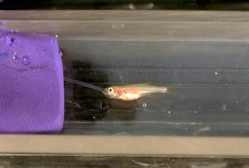
JK Also, overexpression of Side attracted motor axons at the wrong time to the wrong place, resulted in innervation defects. This was particularly evident when we expressed Side in muscle precursors. Attraction was that strong that individual axons travelling in the ISN were stretched into opposite directions leading to split pathways and complete lack of NMJs on dorsal-most muscles. This caused also amazing locomotion phenotypes. During crawling, larvae detached from the substrate and excessively lifted their head and tail segments into the air, despite lack of dorsal innervation. Nobody was expecting such a phenotype. Crawling seems to be much more complex than anticipated.
A dorsal view of third instar larvae showing some of the variability in the side phenotype, from Figure 2 in the paper.
side mutants show considerably variable phenotypes, even in adjacent hemisegments. What do you think this tells us about how axon guidance works in flies?
HA At the beginning of the screen, in Corey Goodman’s lab, we discussed a lot what kind of phenotypes we might possibly find. One such idea was that we might find genes that affect only a single NMJ on a specific muscle fibre, because we imagined a key-and-lock mechanism for wiring 30 muscle fibres, where unique ligands on motor axons connect to cognate receptors on muscles. Inactivating such a receptor by point mutations should in theory prevent the formation of the associated NMJ. Unfortunately, we did not find this phenotypic class, nor did anybody else. We therefore think that motor axon guidance in Drosophila is not hard-wired by high-affinity ligand-receptor complexes but rather functions via co-operative actions of several guidance molecules. Newer finding in Pablo Labrador‘s laboratory also push this older idea originally developed in the Keshishian and Goodman labs. Guidance decisions therefore seem to be made at several points and sum up along the entire path. If several decisions go wrong in a row, phenotypes become noticeable.
Do the locomotion defects you found in larvae have anything to tell us about movement disorders in mammals?
HA Oh yes, I think this is one of the interesting parts of our story. Similar to Drosophila, there are only a few mammalian studies that correlate axon guidance errors with muscle innervation patterns. But these studies made several interesting observations. First, in mice, if a motor nerve branch fails to develop or is severely stalled, entire muscle fields are not innervated after birth causing muscle atrophy. In limbs, this can lead to paralysis and aberrant locomotion patterns. Second, the phenotypes are not necessarily symmetrical and occasionally affect only one side of the body, i.e. very similar to the unilateral defects observed in side mutants. Third, feet and ankles of affected limbs are occasionally twisted inward in newborn mice, a deformity highly reminiscent to congenital clubfoot in humans. The aetiology of clubfoot is still not fully understood, but since it frequently develops unilaterally, one speculative possibility is that it could develop due to innervation defects.
When doing the research, did you have any particular result or eureka moment that has stuck with you?
JK There was not one key moment that has stuck with me, but several small highlights, especially at the laser-scanning microscope. I am over and over again enthusiastic about how “beautiful” the axonal pattern in the embryo or larva is. Further, I had such special moments when larvae showed particularly strong or unusual phenotypes during crawling, which looks sometimes quite funny.
Drosophila larva overexpressing Side in muscles and showing an extreme crawling phenotype.
And what about the flipside: any moments of frustration or despair?
JK Yes, there were some of these frustrating moments, especially handling living larvae for videography. During my research, I have realized that Drosophila larvae have sometimes their own mind, as they decided not to crawl at all or not to crawl in a straight line for the high-speed videography, but rather to crawl away from the nicely prepared agar block.
What next for you after this paper?
JK Since this paper is only a partial project of my PhD thesis, I plan for the next months to press ahead with another project for publication – hopefully we can submit the manuscript successfully at the end of this year.
And where will this work take the Aberle lab?
HA One direction we are heading is how locomotion in animals with hydrostatic skeletons works after all. Crawling behaviour is much more sophisticated and fine-tuned than it appears at first glance. We would like to understand how specific muscle groups contribute to particular movements and how this is antagonized by the liquid-filled body cavity. Which muscle groups, for example, induce rolling escape behaviours during attacks of predatory wasps or coordinate jumping in some dipteran larvae. Ultimately we would like to find mutations or conditions which activate or inactivate particular movements, in order to better understand the underlying circuitry and machinery. Another line of research will certainly be the functional analysis of the entire Sidestep family. There are 7 Side paralogs in the Drosophila genome and virtually nothing is currently known about their functions.
The Düsseldorf skyline. Image credit: Ingo Valentin, from Wikipedia.
Finally, let’s move outside the lab – what do you like to do in your spare time in Düsseldorf?
JK As I am a very water-loving person, I like to spend my spare time along the Rhine river – whether for a walk or to make myself comfortable on a blanket on its shores and let my soul dangle watching the ships passing by. I also like to take advantage of the city’s concert events, as two of my favourite bands, “Die Toten Hosen” and “Broilers”, come from Düsseldorf and regularly give concerts in the city. Furthermore, the whole laboratory and I like to celebrate the Düsseldorf Carnival, which takes place every spring.
HA Düsseldorf has a quite lively art scene, and since I enjoyed landscape photography very much during my undergraduate years, I am particularly drawn to the “Düsseldorf School of Photography”, founded by Bernd and Hilla Becher. Whenever there are exhibitions by Andreas Gursky, Axel Hütte, Thomas Ruff, to name a few, I try to not miss the vernissage. I also love to shop for coffee-table books in local stores.
Welcome to our monthly trawl for developmental biology (and other related/just plain cool) preprints.
This month we found a tonne of papers dealing with various aspects of inheritance in worms, a flush of fly mechanics, and more single cell sequencing than you could shake a stick at. And as summer draws to a close, it’s raining cats and dogs (and wolves) in our ‘Why not…’ section.
The preprints were hosted on bioRxiv, PeerJ, andarXiv. Let us know if we missed anything, and use these links to get to the section you want:
Conserved cell types with divergent features between human and mouse cortex
Rebecca D Hodge, Trygve E Bakken, Jeremy A Miller, Kimberly A Smith, Eliza R Barkan, Lucas T Graybuck, Jennie L Close, Brian Long, Osnat Penn, Zizhen Yao, Jeroen Eggermont, Thomas Hollt, Boaz P Levi, Soraya I Shehata, Brian Aevermann, Allison Beller, Darren Bertagnolli, Krissy Brouner, Tamara Casper, Charles Cobbs, Rachel Dalley, Nick Dee, Song-Lin Ding, Richard G Ellenbogen, Olivia Fong, Emma Garren, Jeff Goldy, Ryder P Gwinn, Daniel Hirschstein, C Dirk Keene, Mohamed Keshk, Andrew L Ko, Kanan Lathia, Ahmed Mahfouz, Zoe Maltzer, Medea McGraw, Thuc Nghi Nguyen, Julie Nyhus, Jeffrey G Ojemann, Aaron Oldre, Sheana Parry, Shannon Reynolds, Christine Rimorin, Nadiya V Shapovalova, Saroja Somasundaram, Aaron Szafer, Elliot R Thomsen, Michael Tieu, Richard H Scheuermann, Rafael Yuste, Susan M Sunkin, Boudewijn Lelieveldt, David Feng, Lydia Ng, Amy Bernard, Michael Hawrylycz, John Phillips, Bosiljka Tasic, Hongkui Zeng, Allan R Jones, Christof Koch, Ed S Lein
t-sne plot of an E8.5 mouse embryo, from Chan, et al.’s preprint
Molecular recording of mammalian embryogenesis
Michelle Chan, Zachary D Smith, Stefanie Grosswendt, Helene Kretzmer, Thomas Norman, Britt Adamson, Marco Jost, Jeffrey J Quinn, Dian Yang, Alexander Meissner, Jonathan S Weissman
Planar cell polarity pathway and development of the human visual cortex
Jean Shin, Shaojie Ma, Edith Hofer, Yash Patel, Gennady Roshchupkin, Andre M Sousa, Xueqiu Jian, Rebecca Gottesmann, Thomas H Mosley, Myriam Fornage, Yasaman Saba, Lukas Pirpamer, Reinhold Schmidt, Helena Schmidt, Bernard Mazoyer, Amaia Carrion-Castillo, Joshua Bis, Shuo Li, Qiong Yang, Michelle Luciano, Sherif Karama, Lindsay Lewis, Mark Bastin, Matthew A Harris, Ian Deary, Joanna M Wardlaw, Markus Scholz, Markus Loeffler, Veronica Witte, Frauke Beyer, Arno Villringer, Hieab HHH Adams, M Arfan Ikrum, William S Kremen, Nathan A Gillespie, Nenad Sestan, Zdenka Pausova, Sudha Seshadri, Tomas Paus
Bovine ovary sections in Plewes, et al.’s preprint
Mapping cell types in the foetal human neocortex, from Polioudakis, et al.’s preprint
A single cell transcriptomic analysis of human neocortical development
Damon Polioudakis, Luis de la Torre-Ubieta, Justin Langerman, Andrew G Elkins, Jason L Stein, Celine K Vuong, Carli K Opland, Daning Lu, William Connell, Elizabeth K Ruzzo, Jennifer K Lowe, Tarik Hadzic, Flora I Hinz, Shan Sabri, William E Lowry, Kathrin Plath, Daniel H Geschwind
Epigenetic factors coordinate intestinal development
Julia Ganz, Ellie Melancon, Catherine Wilson, Angel Amores, Peter Batzel, Marie Strader, Ingo Braasch, Parham Diba, Julie A Kuhlman, John H Postlethwait, Judith S Eisen
Analysis of novel domain-specific mutations in the zebrafish ndr2/cyclops gene generated using CRISPR-Cas9 RNPs
Ashley N Turner, Reagan S Andersen, Ivy E Bookout, Lauren N Brashear, James C Davis, David M Gahan, John P Gotham, Baraa A Hijaz, Ashish S Kaushik, Jordan B McGill, Victoria L Miller, Zachariah P Moseley, Cerissa L Nowell, Riddhi K Patel, Mia C Rodgers, Yazen A Shihab, Austin P Walker, Sarah R Glover, Samantha D Foster, Anil Kumar Challa
Functional dissection of the ARGONAUTE7 promoter
J Steen Hoyer, Jose L Pruneda-Paz, Ghislain Breton, Mariah A Hassert, Emily E Holcomb, Halley Fowler, Kaylyn M Bauer, Jacob Mreen, Steve A Kay, James C Carrington
Gene Correction for SCID-X1 in Long-Term Hematopoietic Stem Cells
Mara Pavel-Dinu, Volker Wiebking, Beruh T Dejene, Waracharee Srifa, Sruthi Mantri, Carmencita Nicolas, Ciaran Lee, Gang Bao, Eric J Kildebeck, Niraj Punjya, Camille Sindhu, Matthew A Inlay, Nivi Saxena, Suk See DeRavin, Harry Malech, Maria Grazia Roncarolo, Kenneth I Weinberg, Matthew Porteus
Neural crest stem cells from Stebbins, et al.’s preprint
Signalling pathways drive heterogeneity of ground state pluripotency
Kirsten R McEwen, Sarah Linnett, Harry G Leitch, Prashant Srivastava, Lara Al-Zouabi, Tien-Chi Huang, Maxime Rotival, Alex Sardini, Thalia E Chan, Sarah Filippi, Michael Stumpf, Enrico Petretto, Petra Hajkova
Need for high-resolution Genetic Analysis in iPSC: Results and Lessons from the ForIPS Consortium
Bernt Popp, Mandy Krumbiegel, Janina Grosch, Annika Sommer, Steffen Uebe, Zacharias Kohl, Sonja Ploetz, Michaela Farrell, Udo Trautmann, Cornelia Kraus, Arif B Ekici, Reza Asadollahi, Martin Regensburger, Katharina Guenther, Anita Rauch, Frank Edenhofer, Juergen Winkler, Beate Winner, Andre Reis
A human cell model of cardiac pathophysiological valvulogenesis
Tui Neri, Emylie Hiriart, Piet Van Vliet, Emilie Faure, Russel Norris, Batoul Farhat, Julie Lefrancois, Thomas Moore-Morris, Stephane Zaffran, Randolph Faustino, Alexander Zambon, Jean-Pierre Devisgnes, David Salgado, Yukiko Sugi, Robert Levine, Jose Luis de la Pompa, Andre Terzic, Sylvia Evans, Roger Markwald, michel Puceat
Caenorhabditis uteleia under SEM, from Stevens, et al.’s preprint
Comparative genomics of ten new Caenorhabditis species
Lewis Stevens, Marie-Anne Félix, Toni Beltran, Christian Braendle, Carlos Caurcel, Sarah Fausett, David HA Fitch, Lise Frézal, Taniya Kaur, Karin C Kiontke, Matt D Newton, Luke M Noble, Aurélien Richaud, Matthew V Rockman, Walter Sudhaus, Mark Blaxter
The Genomic Basis of Arthropod Diversity
Gregg W. C. Thomas, Elias Dohmen, Daniel S. T. Hughes, Shwetha C. Murali, Monica Poelchau, Karl Glastad, Clare A. Anstead, Nadia A. Ayoub, Phillip Batterham, Michelle Bellair, Gretta J Binford, Hsu Chao, Yolanda H Chen, Christopher Childers, Huyen Dinh, HarshaVardhan Doddapaneni, Jian J Duan, Shannon Dugan, Lauren A Esposito, Markus Friedrich, Jessica Garb, Robin B. B Gasser, Michael A. D. Goodisman, Dawn E Gundersen-Rindal, Yi Han, Alfred M Handler, Masatsugu Hatakeyama, Lars Hering, Wayne B Hunter, Panagiotis Ioannidis, Joy C Jayaseelan, Divya Kalra, Abderrahman Khila, Pasi K Korhonen, Carol Eunmi Lee, Sandra L Lee, Yiyuan Li, Amelia R.I. Lindsey, Georg Mayer, Alistair P McGregor, Duane D. McKenna, Bernhard Misof, Mala Munidasa, Monica Munoz-Torres, Donna M Muzny, Oliver Niehuis, Nkechinyere Osuji-Lacy, Subba R. Palli, Kristen A. Panfilio, Matthias Pechmann, Trent Perry, Ralph S. Peters, Helen C Poynton, Nikola-Michael Prpic, Jiaxin Qu, Dorith Rotenberg, Coby Schal, Sean D Schoville, Erin D Scully, Evette Skinner, Daniel B Sloan, Richard Stouthamer, Michael R Strand, Nikolaus U Szucsich, Asela Wijeratne, Neil D Young, Eduardo E Zattara, Joshua B Benoit, Evgeny M Zdobnov, Michael E Pfrender, Kevin J Hackett, John H Werren, Kim C Worley, Richard A Gibbs, Ariel D Chipman, Robert M Waterhouse, Erich Bornberg-Bauer, Matthew W Hahn, Stephen Richards
Spatial Organization of Rho GTPase signaling by RhoGEF/RhoGAP proteins
Paul Markus Mueller, Juliane Rademacher, Richard D Bagshaw, Keziban Merve Alp, Girolamo Giudice, Louise E Heinrich, Carolin Barth, Rebecca L Eccles, Marta Sanchez-Castro, Lennart Brandenburg, Geraldine Mbamalu, Monika Tucholska, Lisa Spatt, Celina Wortmann, Maciej T Czajkowski, Robert William Welke, Sunqu Zhang, Vivian Nguyen, Trendelina Rrustemi, Philipp Trnka, Kiara Freitag, Brett Larsen, Oliver Popp, Philipp Mertins, Chris Bakal, Anne-Claude Gingras, Olivier Pertz, Frederick P Roth, Karen Colwill, Tony Pawson, Evangelia Petsalaki, Oliver Rocks
The Signaling Pathways Project: an integrated ‘omics knowledgebase for mammalian cellular signaling pathways
Scott Ochsner, David Abraham, Kirt Martin, Wei Ding, Apollo McOwiti, Zichen Wang, Kaitlyn Andreano, Ross Hamilton, Yue Chen, Angelica Hamilton, Marin Gantner, Michael Dehart, Shijing Qu, Susan Hilsenbeck, Lauren Becnel, Dave Bridges, Avi Maayan, Janice Huss, Fabio Stossi, Charles Foulds, Anastasia Kralli, Donald McDonnell, Neil McKenna