A Postdoctoral Research Associate position is available in the Conduit lab to study the role of the nuclear envelope in centrosome assembly. Our lab studies how microtubule formation is regulated in cells, including how microtubule organising centres (MTOCs) are assembled. The post-holder will use Drosophila melanogaster to follow up on our recent discover that the nuclear envelope may help regulate centrosome assembly. They will use CRISPR/HDR to generate a series of new fly strains, and then use advanced live-cell fluorescence imaging, including super-resolution imaging, and biochemistry to establish how the nuclear envelope helps regulate centrosome assembly.
The appointment will be for a period of up to three years starting 2nd October 2017, or as soon as possible thereafter.
Candidates are expected to have (or soon have) a PhD in cell/developmental biology and have experience in fluorescent microscopy. Prior experience in Drosophila, centrosome biology, molecular biology and/or basic biochemistry would be an advantage, but not essential.
Applications should include a C.V. and a brief statement of your scientific background and why you would like to join the lab.
Fully funded postdoctoral positions are presently available in the Conlon Lab whose studies focus on identifying the molecular networks that are essential for early heart development and how alterations in these networks lead to congenital heart disease. For these studies, we use a highly integrated approach that incorporates developmental, genetic, proteomic, biochemical and molecular based studies in mouse, Xenopus and stem cells.
Recent advances and projects of interest in the Conlon lab include studies that define the cellular and molecular events that lead to cardiac septation, those that explore cardiac interaction networks as determinants of transcriptional specificity, the mechanism and function of cardiac transcriptional repression networks and, the regulatory networks of cardiac morphogenesis.
Candidates should have recently obtained or be about to obtain a Ph.D. or M.D. in a field of biological science and should have a strong publication record. Outstanding and highly motivated candidates should apply by email to Dr. Frank L. Conlon and include a CV/resume, three references and description of your specific interest in our research programs.
Daily life changes when you set foot in Woods Hole. There is a beauty in your surroundings and energy in the air that invigorates you. The days are long (8am to 2 or 3 am most days!) and we have a full schedule but we are all so excited to be here – to learn – to question – to move the field forward.
Storm rolls in over Eel Pond
A typical day in the course starts with morning lecture at 9am. The lecture is broken down into two parts, the first part is general information about the model system we are working with and the second is about the current research project in the speaker’s lab. After lecture we head to the ‘Sweat Box’ where the students ask the speaker questions. This includes grilling them about their research, it was said this session is like a qualifying exam for the professors! We can also use this time to ask them about their career trajectory and for career advice. We then break for lunch; two students take the speaker to lunch where they get to talk more in depth about science and life.
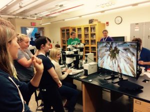
Students looking at a Sea Urchin
After lunch we head to the lab, we get a ‘cookbook’ for each organism; this includes information on how to take care of them and many protocols. In this ‘cookbook’ there are instructions on how to fertilize the egg, manipulate the embryo and the organism as well as a list of reagents and tools that are available to us. We then brainstorm ideas in groups and have full access to the teaching assistants, professors and course directors to plan and execute our experiments.
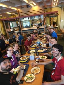
Student eat a meal at Swope
We break in the early evening to take a run, shoot some hoops or practice softball for the annual softball match (watch out physio) and eat dinner. Two students also get to take the speaker out to dinner at a local Woods Hole restaurant during this time. Then it’s back to the lab to continue our experiments into the wee hours of the morning!
How do we survive this schedule for six weeks? We drink a lot of coffee!
Follow the course here on the blog and on twitter #embryo2017
A postdoctoral position is available to study the extrinsic modulation of intestinal proliferation in the research group of Golnar Kolahgar at the Department of Physiology, Development and Neuroscience at the University of Cambridge. The candidate is expected to hold a PhD in cellular, molecular or developmental biology.
Our goal is to identify the components of the extracellular space that contribute to maintaining and remodelling the adult intestine in response to various physiological conditions. We use the genetically tractable Drosophila gut as a paradigm to investigate cell fate decisions in vivo (e.g. Kolahgar et al, Dev Cell, 2015; Suijkerbuijk et al, Curr. Biol, 2016). The aim of this project is to explore how integrin signalling promotes intestinal stem cell proliferation and contributes to gut plasticity, using a combination of Drosophila genetics, lineage tracing and clonal analysis, confocal imaging and whole genome sequencing, thus experience with one or several of these techniques is required.
The post is funded for an initial period of 3 years.
Development of the placental vasculature – known as the labyrinth – is critical for foetal development. Today’s paper comes from the most recent issue of Development and addresses the signalling events involved in placental vascular maturation. We caught up with lead author Lijun Chi and her PI Paul Delgado-Olguin of the Hospital for Sick Children and University of Toronto.
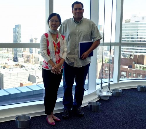
Lijun and Paul
Can you give us your scientific biography and the main questions your lab is trying to answer?
PD-O As a Ph.D. student at the Universidad Nacional Autónoma de México (UNAM) under supervision of Dr. Ramón Coral Vázquez and Dr. Félix Recillas Targa, I investigated the transcriptional regulation of genes expressed in skeletal muscle and whose mutations cause muscular dystrophies. My reading during my Ph.D. studies led me to numerous landmark papers from Eric Olson’s lab on skeletal muscle gene regulation. I soon discovered Dr. Olson’s and Dr. Deepak Srivastava’s seminal work on the transcriptional pathways controlling heart development, a process that I always found fascinating. Working at Dr. Recilla’s lab, I became very interested in the function of chromatin modifiers in gene control, which led me to mine the scientific literature to learn about the function of chromatin structure regulators in cardiac gene control and development. This made me realize how little was known on the subject at the time, and brought me across a pioneering paper from Dr. Benoit Bruneau’s lab describing an essential function of Baf60c, a subunit the SWI/SNF chromatin remodelling complex, in heart development. This helped me decide what I wanted to do as a postdoc.
I obtained my Ph.D. degree in June 2005, and by August I was in Dr. Bruneau’s laboratory ready to start my postdoctoral research. In Dr. Bruneau’s lab I investigated the function of the histone methyltransferase Ezh2 in the heart. This enzyme tri-methylates the lysine 27 of histone H3 to repress gene expression, and its global deletion causes lethality early in mouse development. Surprisingly, we found that deletion of Ezh2 in cardiac progenitor cells, despite altering embryonic gene expression, did not alter heart development, and mutant mice had normally structured hearts. However, adult mutants developed heart disease. This raised the possibility that epigenetic alterations in differentiating cardiovascular progenitor cells early in development might program adult heart disease susceptibility. To address this possibility, my lab at The Hospital for Sick Children (SickKids) has been studying the function of several histone modifiers in cardiac and vascular development, and in regulating adult cardiovascular system function since 2012. Because the placental vasculature is required for proper embryo development, and its malfunction can affect heart development and program adult disease in the offspring, my lab is also interested in uncovering the mechanisms controlling its development. My lab will continue to focus on uncovering the mechanisms controlling cardiovascular development and programming postnatal disease during embryogenesis.
PD-O Toronto is a great place to do science. It is home for numerous world-class scientists, research institutes, numerous hospitals with very strong research programs, and the University of Toronto, which has an outstanding research curriculum. These attributes make Toronto and ideal place to develop multidisciplinary research of the highest quality. For example, my lab investigates fundamental biological processes, and being at SickKids and in Toronto’s rich scientific environment allowed establishment of key collaboration with clinician scientists in neighbouring institutions, which has facilitated me to explore the translation potential of my lab’s research.
Lijun, how did you come to join the Delgado-Olguin lab?
I obtained my PhD. at the University of Oulu, Finland under the supervision of Professor Seppo Vainio. My PhD thesis, published in 2007, describes the role of Sprouty2 in development of the urogenital system. I then pursued a postdoctoral fellowship in Dr. Norman Rosenblum’s laboratory at SickKids in Toronto, Canada. During my fellowship I investigated the function of the cilia protein Kif3a, whose deficiency causes polycystic kidney in human and mouse models. After finishing my fellowship in 2011, I wanted to learn about cardiovascular development, and I knew Paul was just about to Join SickKids. Working at the Delgado-Olguin lab has given me the opportunity to work in exciting projects to understand the basis of cardiovascular development and disease.
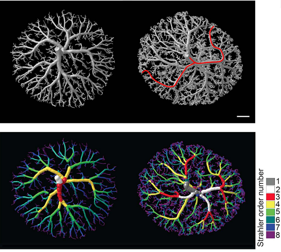
Surface renderings of placentae, from Fig. 2, Chi et al. 2017
What was known about signalling pathways controlling placental vascular maturation before you started this work?
PD-O Because of its relevance in embryogenesis and in postnatal health, I was surprised to find out how little we knew about pathways controlling placental vascular maturation before we started our work. Perhaps the most informative report on the subject is from Knox & Barker (2008), who performed global gene expression analyses on the embryonic portion of mice placental vasculature, known as the labyrinth, at consecutive days of development. This analysis revealed a sharp molecular transition defining the developmental and the maturation phases of the placenta. In this transition, over 700 genes change their expression from embryonic day 12.0 to E13.5, and functions associated with these genes provided a general idea of some of the processes occurring in each phase. For instance, genes that are expressed in the developmental phase are involved in growth, metabolic processes, DNA and RNA processing, and cell cycle regulation. While genes active in the maturation phase are involved in pregnancy and reproduction. However, these studies were done on whole labyrinth, and thus tell us little on the pathways controlling this transition in specific cell types. To the best of my knowledge, our work is the first one to address the regulation of the transition from development to maturation in placental vascular maturation, and to define pathways active in endothelial cells regulating this process.
Can you give us key results of the paper in a paragraph?
PD-O We found that the histone methyl transferase G9a activates the Notch pathway in endothelial cells to promote maturation of the placental vasculature. We inactivated the histone methyltransferase G9a in endothelial progenitors and their derivatives, and found that mutant embryos died with placental defects. Closer examination revealed that the gross morphology of the placentae from mutant embryos appeared normal at E12.5, but had a smaller vascularized area at E13.5. Because the transition from the developmental to the maturation phase of the placenta occurs precisely between these stages, this raised a possible function for G9a as a regulator placental vascular maturation. Analysis of cell proliferation revealed that growth of the labyrinth is coordinated with decreased growth of the spongiotrophoblast during the development to maturation transition, and that this balance is lost in G9a mutant placentae. To uncover regulatory pathways we performed global gene expression analysis, which revealed that effectors of the Notch pathway were downregulated in G9a mutant placental endothelial cells. We then introduced a transgene to activate the Notch pathway in G9a mutants, and we found that the placental morphology was rescued! This opened the possibility that a G9a-Notch axis might be disrupted in placental diseases with vascular maturation defects. Indeed, we found that G9a, and Notch regulators were downregulated in human placentae from pregnancies affected with intra uterine growth restriction.
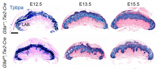
Tpbpa in situs, from Fig. 4, Chi et al. 2017
How might your work inform efforts to diagnose or even treat placental defects during pregnancy?
PD-O These are very exciting possibilities. Intra uterine growth abnormalities are diagnosed only when the foetus is smaller than expected for the gestational age or when the placenta is already malfunctioning. The ability to identify preclinical placental insufficiency is limited because we know very little about the regulation of placental vascular development, and on the events that precede placental malfunction. We found that imbalanced growth of the labyrinth vs the spongiotrophoblast precedes the appearance of gross morphological abnormalities in G9a mutant placentae. Based on these results, we think that being able to define the growth ratio of these placental cell types, and detect imbalances might offer a way of identifying foetuses at risk of defective intrauterine growth. In terms of prevention or treatment, our findings open the possibility that activating the Notch pathway might be investigated as a means to promote placental vascular maturation. Our mouse model, combined with availability of pharmacologic compounds that activate the Notch pathway, will allow us to further investigate these possibilities.
When doing the research, did you have any particular result or eureka moment that has stuck with you?
LC In the initial stage of the research, we were mainly focused on identifying cardiac defects in G9a mutants. However, I noticed that mutants had placentae with reduced vascularization. I decided to analyze embryos at consecutive developmental stages and I found that the vascular defect was obvious only at E13.5 and onwards. When I realized that the transition from the developmental to the maturation phase occurs precisely from E12.5 to E13.5, I hypothesized that G9a might regulate this transition. Given that the regulation of placental vascular maturation is very poorly understood, we decided to investigate further and test the hypothesis.
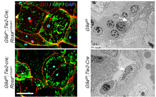
Endothelial cells by fluorescence and TEM, from Fig. 3, Chi et al., 2017
What about the flipside: any moments of frustration or despair?
LC As often happens with genome wide gene expression data, it was difficult to identify potentially relevant targets downstream of G9a from our RNAseq results. Fortunately, we found published reports demonstrating the involvement of the Notch signalling pathway in vascular maturation in the retina. We also found a report showing downregulation of some Notch regulators in placentae from pregnancies affected by intrauterine growth restriction. These reports encouraged me to confirm downregulation of Notch effectors in G9a mutant placental endothelial cells, and later on to test the effect of activating the Notch pathway in G9a mutant endothelial cells. These experiments were nerve wracking, because if the vascular phenotype were not to be corrected at least partially, I would have had to keep trying to identify functionally relevant G9a targets.
What are your career plans following this work?
LC With the completion of this current study, I am planning to test whether pharmacologically activating the Notch pathway in the G9a mutant placental vasculature promotes the maturation process and ameliorates or prevents placental defects. This might open the door to new experiments to try to promote vascular maturation in other models of intrauterine growth restriction.
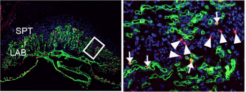
The labyrinth endothelium in mutant placentae, from Fig. 4, Chi et al. 2017
And what next for the Delgado-Olguin lab?
PD-O We will delve deeper into the mechanisms by which G9a controls placental vascular maturation and the effects of activating Notch signalling in the placental vasculature. There are outstanding questions from our work. Particularly intriguing is how G9a activates the Notch pathway, as it is predominantly known as a transcriptional repressor, while its function as a transcriptional activator is less understood. Dissecting the mechanisms of action of G9a in placental endothelium will likely reveal additional regulatory pathways and potential approaches to promote vascular maturation. More broadly, our lab will continue to investigate the mechanisms by which postnatal cardiovascular disease is programmed during embryogenesis.
Finally, what do you two like to do in Toronto when you are not in the lab?
PD-O I enjoy hiking the many trails and parks in the city with my family and dogs, visiting museums, and fishing. Also, being an avid foodie, living in a city where there is food from all over the world available close by is a great bonus!
LC During my spare time, I am a passionate reader who enjoys a diversity of novels. I am also a skilful cook who loves exploring different recipes and trying out new styles. To stay physically well rounded, swimming is one of my weekly activities.
24 students along with faculty, teaching assistants and course assistants arrived over the weekend to embark on a life changing summer experience. Since my days in undergraduate research in cell biology, I always heard fascinating stories about the science and life at Woods Hole. I knew it was an experience that I wanted to take part in during my training. The MBL at Woods Hole is a mecca of research, creativity, ingenuity and curiosity it gives everyone who sets foot on its campus the opportunity to learn new concepts and techniques. This course and the community here allow you to test your cool and creative hypotheses and push you out of your comfort zone. It’s an experience that empowers scientific adventure and will leave you wanting to come back year after year.
Students introduce each other at the first gathering of the 2017 Embryology Course
In the first days here as a student in one of the longest standing classes at the MBL I have been welcomed into an amazing group of scientists. My fellow students and I had the opportunity to share our research with our peers and some of the course faculty during an informal poster session over pizza, listened to amazing lectures on echinoderm development, and started to learn how to make the tools and run the microscopes that will enable us to conduct our experiments.
I can’t wait to see what we learn and discover this summer!
Follow the course here on the blog and on twitter #embryo2017.
One of the research topics in Michel Milinkovitch’s laboratory (https://www.lanevol.org) at the University of Geneva (Switzerland) is to understand how squamates (lizards and snakes) generate such a tremendous variety of colours and colour patterns.
Colours
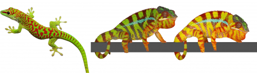
The colour of a lizard’s patch of skin is generally the result of the combination among structural and pigmentary elements found in various types of chromatophores(1-5). Pigmentary colours are produced by brown/black melanins in melanophores, as well as by yellow and red pteridines/carotenoids in xanthophores and erythrophores, respectively. On the other hand, structural colours are produced by light interference in iridophore cells containing layers of guanine nanocrystals(5): the wavelengths specifically reflected by these periodic structures is a function of the mean distance between successive layers of guanine nanocrystals — the longer the distance, the longer the wavelengths that are reflected. For example, many species of reptiles and amphibians are green despite that their skin does not contain any green pigment! They produce their chlorophyll-matching colour in a more sophisticated way(4): a layer of iridophores selectively reflects most of the incoming green and blue wavelengths but a layer of yellow pigments absorbs blue. As all other wavelengths of the visible range (yellows, oranges, and reds) go through the skin (and are absorbed by deeper tissues), the only colour that bounces back from the skin is pure bright green(4), as in day geckos (Fig. 1a).
The Milinkovitch lab also discovered that chameleons change colour by manipulating structural colours rather than by dispersion/aggregation of pigment-containing organelles within chromatophores. Indeed, combining microscopy, videography, RGB photometry and photonic band-gap modelling, they showed that chameleons shift colour through active tuning of a 3D lattice of guanine nanocrystals within a superficial layer of dermal iridophores(5). In other words, chameleons manipulate light interference by changing the distance among their nanocrystals of guanine. Take an adult male panther chameleon. In its cryptic state, it is green for the same reason as the Phelsuma lizards: its dermal iridophores reflect green and blue, and the latter is being absorbed by a yellow pigment. But if another mature male enters its territory, the two animals increase the distance between the nanocrystals within their iridophores … such that they both turn yellow or orange or red to become as visible as possible and impress each other (Fig. 1b and link to YouTube videos). This usually suffices for one of the two males to give up…otherwise they will start a physical fight.
These studies provided some new answers but also opened many new questions. How do iridophores generate and spatially organise nanocrystals? What is the cellular mechanism involved in the tuning of the distance among nanocrystals in iridophores of chameleons when they change colour? Milinkovitch is now teaming up with biochemists/cell biologists such as Marcos Gonzalez-Gaitan for investigating these questions.
Fig. 1 (a- left) A day gecko (Phelsuma grandis); (b-right): A male panther chameleon (Furcifer pardalis) changing colour.
Colour change in chameleons YouTube playlist.
Colour Patterns
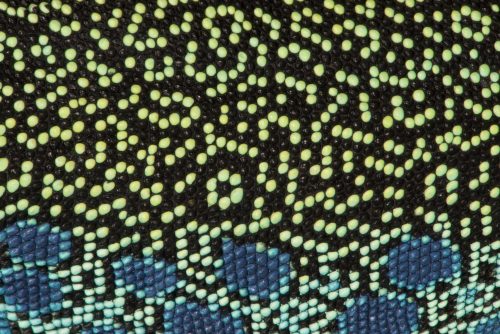
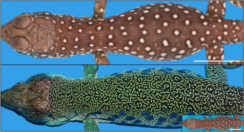
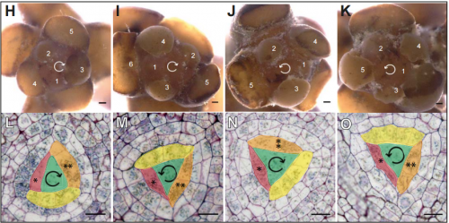
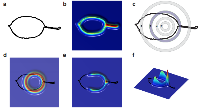
Animals display colours, but they often additionally exhibit colour patterns, i.e., symmetry-breaking regularities (stripes, spots, tessellations, meanders, and labyrinths) that result from short-range and long-range interactions among chromatophores(6-11). At the macroscopic scale, these dynamical processes obey reaction-diffusion (RD) equations discovered by the mathematician Alan Turing(12-14). Strikingly, the formation of skin colour in the ocellated lizard (Timon lepidus) seems to conflict with this RD framework as skin scales, rather than individual chromatophore cells, establish the pattern. Indeed, the brown juvenile lizard gradually transforms its skin colour as it ages to reach an intricate adult labyrinthine pattern where each scale is either green or black (Fig. 2).
Figure 2: The colour pattern changes drastically in about three years from (top) the juvenile to (bottom) the adult. The inset shows a juvenile on the same scale as the adult. Scale bar = 11 mm.
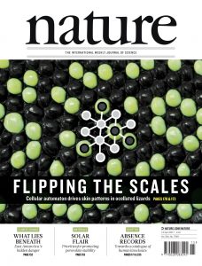
But why would the pattern form at the level of scales, rather than at the level of biological cells? This is the questions that the Milinkovitch team solved recently, as reported recently in the journal Nature(15).
To tackle this question, two PhD students, Liana Manukyan (computer scientist) and Sophie Montandon (developmental biologist), followed individual lizards during three to four years of their development from hatchlings crawling out of the egg to fully mature animals. For multiple time points, they reconstructed the geometry and colour of the network of scales on three animals by using R2OBBIE-3D (YouTube playlist), a very high resolution robotic system(16) developed previously in the Milinkovitch laboratory by a physicist PhD student: Antonio Martins.
R2OBBIE-3D YouTube playlist
R2OBBIE-3D allows to reconstruct the 3D geometry and colour texture of objects up to 1.5 meters with a resolution of … 15 microns ! The Swiss team then set up a software pipeline that allowed them to automatically detect scales on each animal at each time point and match these networks. This was not a trivial task because the size of the animal, the positions of its body parts, and its skin pattern all change from scan to scan. Fortunately, the number of scales is invariant for a given individual throughout its life. This analysis indicated that the brown juvenile scales change to green or black then, surprisingly, continue flipping colour (between green and black) during the life of the animal (Fig. 3).
Figure 3: Close-ups of the same individual as in Fig 2 illustrating the pattern time evolution. Orange arrows show two examples of colour switching between two time points.
This very strange observation prompted Milinkovitch to suggest that the skin scale network forms a so-called ‘Cellular Automaton’. This esoteric computing system was invented in 1948 by the mathematician John von Neumann. Cellular automata are lattices of elements in which each element changes its state (here, its colour, green or black) depending on the states of neighbouring elements. The elements are called cells but are not meant to represent biological cells; in the case of the lizards, they correspond to individual skin scales. These abstract automata were extensively used to investigate computing systems and to model natural phenomena, but the Geneva team discovered what seems to be the first case of a genuine 2D automaton appearing in a living organism. Analyses of the four years of colour change allowed to confirm Milinkovitch’s hypothesis: the scales were indeed flipping colour depending of the colours of their neighbour scales. Computer simulations implementing the discovered mathematical rule generated colour patterns that could not be distinguished from the patterns of real lizards.
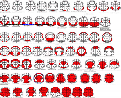
How could the interactions among pigment cells, described by Turing equations, generate a von Neumann automaton exactly superposed to the skin scales? The skin of a lizard is not flat: it is very thin between scales and much thicker at the center of them. Given that Turing’s mechanisms involves movements of cells, or the diffusion of signals produced by cells, Milinkovitch understood that this variation of skin thickness could impact on the Turing’s mechanism. Liana Manukyan, but also Anamarija Fofonjka, a third PhD student in Milinkovitch’s team, then performed computer simulations and saw a cellular automaton behaviour emerge, demonstrating that the development of Cellular Automata as computational systems is not just an abstract concept developed by John von Neumann, but also corresponds to a natural process generated by biological evolution.
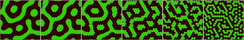
However, the automaton behaviour was imperfect as the mathematics behind Turing’s mechanism and von Neumann automaton are very different. Milinkovitch called in Stanislav Smirnov, Professor of mathematics at the University of Geneva. Stanislav was awarded in 2010 the Fields Medal (the equivalent of the Nobel Price in Mathematics). Before long, Smirnov derived a so-called discretisation of Turing’s equations that would constitute a formal link with von Neumann’s automaton. Anamarija Fofonjka implemented Smirnov new equations in computer simulations, obtaining a system that had become un-differentiable from a von Neumann automaton (Fig. 4). The highly multidisciplinary team of researchers had closed the loop in this amazing journey, from biology to physics to mathematics.
Figure 4. A CA behaviour rapidly emerges from the continuous RD process when diffusion coefficients are reduced by a factor 1-P > 0.8 in the inter-scale regions (from left to right, 1-P=0.2, 0.6, 0.8, 0.9, 0.95, 0.99).
Hiroshi Hamada is the Director of the RIKEN Center for Developmental Biology (CDB) in Kobe, Japan. His lab focusses on the establishment of left-right asymmetry in the mouse embryo, and the role of cilia in the symmetry-breaking event. Hiroshi’s work has been recognised by various awards, including the Keio Medical Science Prize in 2014, and election as an EMBO Associate Member in 2016. We met with Hiroshi on a recent visit to the CDB, to talk about his career and current interests, and the prospects for developmental biology in Japan.
How did you become interested in science generally and developmental biology in particular?
I was interested in science from when I was really young. I don’t know why, but I used to like to read science books and in particular I was interested in basic medical science, not in clinical medicine. So I thought I wanted to be a basic research scientist. But my interests in developmental biology are relatively recent. When I started in research, I wanted to contribute to medicine, and because cancer was the most serious disease at that point (as well as now), that was what I wanted to study. But, to start, I thought I needed to get into molecular biology (which didn’t really exist as a field at the time), so as a graduate student I studied the structure of RNA – I was a biochemist – and then I learned a lot of cloning techniques when they came along. These were very useful when I moved to the NIH for my postdoc to work on cell transformation, to look at oncogenic mutations.
You first established your own lab in Newfoundland, Canada. How did that come about?
At that time, I wanted to be more independent, so I looked for a job in the USA rather than moving back to Japan. I had a relatively decent publication record and I thought I would have a good chance of choosing where I wanted to go. But I interviewed at several places in the USA and Canada, and Newfoundland was the only place I got an offer.
And how was it there?
It was a wonderful place – the people were very kind and the university was very decent. Most of the people there were more senior and were focussing mainly on education, so they said that because I had a grant I could focus on the research and didn’t have to take on other responsibilities. So I was very happy there.
But then you did eventually go back to Japan?
Yes, but that was not my plan. I really enjoyed Newfoundland, but I had two young kids and Newfoundland wasn’t the best place to be for their education. So I was planning to move to another university in Canada, where I had an offer. Then my supervisor from Japan called me – he was planning to retire in 5 years, and his associate professor had been promoted and moved elsewhere, so he needed someone to take over from him. I decided to return to Japan, first to Tokyo and then later to Osaka.
How did you find the differences in the science culture between North America and Japan?
When I first moved back, the facilities in Japan weren’t as good – even compared with Newfoundland. Now, of course, things are much better here, but at the time all the equipment and so on was quite old. And of course there were huge differences in the culture… I felt almost half-Western by then, so I found it very different when I moved back. Fortunately, my professor ensured that I could do my own research and be independent and kindly assigned the best graduate students to me.
So when did you actually become a developmental biologist?
When I was in Canada, I started working on embryonal carcinoma (EC) cells, and I was interested in differentiation – these cells differentiated very nicely. One of the best EC lines, P19 cells, was established by Michael McBurney at University of Ottawa, and he showed me how to induce the differentiation. So I took that system to Newfoundland, and I tried to answer several questions, such as what determined the undifferentiated state and what triggered differentiation into neural cells.
We tried to identify the genes specifically expressed in undifferentiated cells, and the transcription factors that recognised the undifferentiated cell-specific enhancers. This second approach was very successful – we used enhancer trapping to identify an enhancer active in undifferentiated cells. And then when I came back to Japan, we identified the specific transcription factor that bound this enhancer – which turned out to be Oct4, though we originally called it Oct3. We and two other groups all reported the same transcription factor around the same time; two of us called it Oct3, and Hans Schöler called it Oct4, which was the name that eventually stuck.
You then discovered Lefty, also in a tissue culture screen, and this led you into working on axis establishment and left-right (LR) asymmetry, which has been your focus for the past two decades. What do you think are the big open questions in the field?
I think we have been able to find out some of the important principles underlying LR axis determination, but there are still many important unanswered questions. For example, we know that fluid flow is crucial – the rotation of motile cilia in the node generates directional flow – but we don’t really know how embryos sense this flow. The first molecular asymmetry we see is the degradation of a particular mRNA on one side of the node, but it’s not clear how this is induced. Of course, we know that immotile cilia are involved in flow-sensing, but how do they do this? Calcium signalling seems to be involved, but the pathway still isn’t clear and this is an active area of research for us.
Another question is what determines the direction in which the cilia rotate, which in turn sets up left versus right. We know there must be inputs from the anterior-posterior and dorsal-ventral patterning systems, and also that there must be some chirality in the system to set up the asymmetry. But there is a lot still to figure out for us to understand why the cilia always rotate in the clockwise direction. Also, it seems like the node cilia are the only ones in the body that rotate as opposed to beat, but why is that?
Your research encompasses a broad range of scales – from the molecular architecture of cilia to the developmental consequences of disrupting LR determination in mice. How do you stay abreast of the latest techniques required to investigate the problem at such diverse levels?
For myself, I think that the question always comes first. I don’t think about what I can do with a particular technique, I think about the question and then try to work out what kind of techniques are necessary to address it. And then of course I have to find the people who can use that technique – either through collaboration or people in my lab. Fortunately, I’ve had good people come to my lab, including people with engineering backgrounds, who’ve been very good at analysing the flow and the mechanics. I didn’t try to recruit them, but they somehow found me, and that’s been great.
I understand you’ve also become interested in mathematical and computational approaches to investigating axis establishment. How do you think such approaches can contribute?
Initially, I wasn’t really sure what mathematical modelling or computational science can do for real biology. But I was exposed to the work of my friend Shigeru Kondo, who is interested in Turing patterns and reaction-diffusion models in pigmentation. Then, when I was studying the very dynamic patterns of gene expression during LR patterning, we did lots of experiments but they didn’t really get at the principle of what’s going on. We thought that reaction-diffusion might be involved, so we decided to try and model it. And this has really helped, and I realised what modelling can do for you. When it works, it really clarifies things in a way that lab experiments couldn’t. Personally, I’m not good at mathematics and physics, but I collaborate with people who understand this, and I find it very useful.
You took over as Director of the CDB (probably the most prestigious developmental biology institute in Japan) a couple of years ago, in the wake of the problems with and fallout from the STAP cell papers. How is the institute moving forwards from that difficult period and what do you hope to achieve as director?
Masatoshi Takeichi was stepping down as Director, and partly because I was on the advisory council of the CDB and was familiar with the institute, they asked me if I would take over. And I have a great respect for the CDB and the people, including Takeichi-sensei, and how they run the institute, which has always been very fair. For example, they always make sure to select the best people for positions rather than choosing their friends, which is of course very important. And everything they do is like this. So I knew that this was a very important institute and we couldn’t see it run into difficulty or disappear – we had to support it, and I hope I have done that as Director.
I see my job mainly as to rejuvenate things – several people have moved on and we have recruited and keep recruiting some very good new people, so we are looking to the future. One thing we want to do is to extend the scope of the CDB a bit. Traditionally, we have mainly focussed on embryonic development, but we now see development as something that continues through life. And so we want to recruit people working on maintenance and homeostasis – adult stem cells – as well as regeneration and ageing.
Traditionally, we have mainly focussed on embryonic development, but we now see development as something that continues through life
More broadly, Japanese science has a long and prestigious history in developmental biology, and is now at the forefront of stem cell research, which of course depends on insights from developmental biology. How do you see the balance between basic and translational research in Japan, particularly in terms of funding?
Fortunately RIKEN headquarters appreciate the importance of developmental biology and they are committed to supporting the field. But we cannot go on with just basic developmental biology – we have to have a balance between basic and more translational science, and this is sensible. Fortunately for us, Masayo Takahashi, who has pioneered transplantation of induced pluripotent stem cell-derived cells into human patients, is based here at the CDB. She likes to be here because she knows that the basic science is important and that the reason she can do translational work is because of the basic foundations. There has to be interaction between basic and translational science, and that can be a little bit difficult, but we have to make sure we bring these people together and I am trying to promote this.
More broadly, I think that there is a larger proportion of grant money in Japan going to stem cell and other translational research, and basic scientists are having a hard time, but I think that’s true everywhere. However, it is essential to maintain the diversity in research, because, as history tells us, big breakthroughs do not always come from top-down projects but from unexpected directions.
What advice would you give to young scientists starting out their career in developmental biology?
Try to find an interesting question, and one that you think is important; then, stick with it, be patient and don’t compromise to easier questions. It might take a long time, but I think you have to do it. I guess the current situation makes it difficult for young people – who have to publish – so maybe I’m saying something unrealistic. But what I used to do was to have side projects that were maybe easier questions, but always returned to the main theme.
Try to find an interesting question, and one that you think is important; then, stick with it, be patient and don’t compromise to easier questions
Away from the lab, what might Development readers be surprised to find out about you?
I very much enjoyed my time in Newfoundland – it was a great place with lots of small fishery towns and wonderful wildlife, and I would love to go back. They have a big Irish population, and I discovered Irish music there, which I really like. The first time I visited, when I went for the job interview, I went to my host’s house for dinner. And I could hear some Irish music playing in the background. As a result, I am probably one of the few Japanese people who love traditional Irish music.
Our latest monthly trawl for developmental biology (and other cool) preprints. See last year’s introductory post for background, and let us know if we missed anything
This month featured a host of preprints on plants, stem cells, connectomics and modelling. Plus the first preprint from Nobel laureate and recent Development interviewee Eric Wieschaus, an introduction to the concept of the Human Cell Atlas, and a plea from Science Editor-in-Chief Marcia McNutt and colleagues for transparency in author contributions. The preprints were hosted on bioRxiv, F1000Research, PeerJ and arXiv.
Organization Of The Drosophila Larval Visual Circuit. Ivan Larderet, Pauline Fritsch, Nanae Gendre, Larisa Maier, Rick D. Fetter, Casey Schneider-Mizell, James Truman, Marta Zlatic, Albert Cardona, Simon Sprecher
Kenyon cells from a first instar fly larvae in Eichler, et al.’s preprint
The Complete Connectome Of A Learning And Memory Center In An Insect Brain. Katharina Eichler, Ashok Litwin-Kumar, Feng Li, Youngser Park, Ingrid Andrade, Casey M. Schneider-Mizell, Timo Saumweber, Annina Huser, Claire Eschbach, Bertram Gerber, Richard D. Fetter, James W. Truman, Carey E. Priebe, L. F. Abbott, Andreas S. Thum, Marta Zlatic, Albert Cardona
A Complete Electron Microscopy Volume Of The Brain Of Adult Drosophila melanogaster. Zhihao Zheng, J. Scott Lauritzen, Eric Perlman, Camenzind G. Robinson, Matthew Nichols, Daniel Milkie, Omar Torrens, John Price, Corey B. Fisher, Nadiya Sharifi, Steven A. Calle-Schuler, Lucia Kmecova, Iqbal J. Ali, Bill Karsh, Eric T. Trautman, John Bogovic, Philipp Hanslovsky, Gregory S. X. E. Jefferis, Michael Kazhdan, Khaled Khairy, Stephan Saalfeld, Richard D. Fetter, Davi D. Bock
Whole-Brain Serial-Section Electron Microscopy In Larval Zebrafish. David Grant Colburn Hildebrand, Marcelo Cicconet, Russel Miguel Torres, Woohyuk Choi, Tran Minh Quan, Jungmin Moon, Arthur Willis Wetzel, Andrew Scott Champion, Brett Jesse Graham, Owen Randlett, George Scott Plummer, Ruben Portugues, Isaac Henry Bianco, Stephan Saalfeld, Alex David Baden, Kunal Lillaney, Randal Burns, Joshua Tzvi Vogelstein, Alexander Franz Schier, Wei-Chung Allen Lee, Won-Ki Jeong, Jeff William Lichtman, Florian Engert
Highly Efficient Maternal-Fetal Zika Virus Transmission in Pregnant Rhesus Macaques. Sydney M Nguyen, Kathleen M Antony, Dawn M Dudley, Sarah Kohn, Heather Simmons, Bryce Wolfe, M Shahriar Salamat, Leandro BC Teixeira, Gregory J Wiepz, Troy H Thoong, Matthew T Aliota, Andrea M Weiler, Gabrielle L Barry, Kim L Weisgrau, Logan J Vosler, Mariel S Mohns, Meghan E Breitbach, Laurel M Stewart, Mustafa N Rasheed, Christina M Newman, Michael E Graham, Oliver E Wieben, Patrick A Turski, Kevin M Johnson, Jennifer Post, Jennifer M Hayes, Nancy Schultz-Darken, Michele L Schotzko, Josh A Eudailey, Sallie R Permar, Eva G Rakasz, Emma L Mohr, Saverio Capuano III, Alice F Tarantal, Jorge E Osorio, Shelby L O’Connor, Thomas C Friedrich, David H O’Connor, Thaddeus G Golos
Olfactory Receptors Are Required For Social Behavior And Neural Plasticity In Ants, As Evidenced By CRISPR-Mediated Gene Knockout. Hua Yan, Comzit Opachaloemphan, Giacomo Mancini, Huan Yang, Matthew Gallitto, Jakub Mlejnek, Kevin Haight, Majid Ghaninia, Lucy Huo, Alexandra Leibholz, Jesse Slone, Xiaofan Zhou, Maria Traficante, Clint A. Penick, Kelly Dolezal, Kaustubh Gokhale, Kelsey Stevens, Ingrid Fetter-Pruneda, Roberto Bonasio, Laurence J. Zwiebel, Shelley Berger, Juergen Liebig, Danny Reinberg, Claude Desplan
Development of a high-density, ~2M SNP genotyping array and 670k SNP imputation array for the domestic horse. Robert J Schaefer, Mikkel Schubert, Ernest Bailey, Danika L. Bannasch, Eric Barrey, Gila Kahila Bar-Gal, Gottfried Brem, Samantha A. Brooks, Ottmar Distl, Ruedi Fries, Carrie J. Finno, Vinzenz Gerber, Bianca Haase, Vidhya Jagannathan, Ted Kalbfleisch, Tosso Leeb, Gabriella Lindgren, Maria Susana Lopes, Nuria Mach, Artur da Câmara Machado, James N. MacLeod, Annette McCoy, Julia Metzger, Cecilia Penedo, Sagi Polani, Stefan Rieder, Imke Tammen, Jens Tetens, Georg Thaller, Andrea Verini-Supplizi, Claire M. Wade, Barbara Wallner, Ludovic Orlando, James R. Mickelson, Molly E. McCue
The Human Cell Atlas. Aviv Regev, Sarah Teichmann, Eric S. Lander, Ido Amit, Christophe Benoist, Ewan Birney, Bernd Bodenmiller, Peter Campbell, Piero Carninci, Menna Clatworthy, Hans Clevers, Bart Deplancke, Ian Dunham, James Eberwine, Roland Eils, Wolfgang Enard, Andrew Farmer, Lars Fugger, Berthold Gottgens, Nir Hacohen, Muzlifah Haniffa, Martin Hemberg, Seung K. Kim, Paul Klenerman, Arnold Kriegstein, Ed Lein, Sten Linnarsson, Joakim Lundeberg, Partha Majumder, John Marioni, Miriam Merad, Musa Mhlanga, Martijn Nawijn, Mihai Netea, Garry Nolan, Dana Pe’er, Anthony Philipakis, Chris P. Ponting, Stephen R. Quake, Wolf Reik, Orit Rozenblatt-Rosen, Joshua R. Sanes, Rahul Satija, Ton Shumacher, Alex K. Shalek, Ehud Shapiro, Padmanee Sharma, Jay Shin, Oliver Stegle, Michael Stratton, Michael J. T. Stubbington, Alexander van Oudenaarden, Allon Wagner, Fiona M. Watt, Jonathan S. Weissman, Barbara Wold, Ramnik J. Xavier, Nir Yosef
TIRF-M images and associated kymographs from Tan, et al.’s preprint
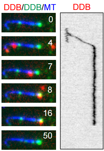
Loss Of PTEN Promotes Formation Of Signaling-Specific Clathrin-Coated Pits. Luciana K. Rosselli-Murai, Joel A. Yates, Sei Yoshida, Julia T. Bourg, Kenneth K. Y. Ho, Megan White, Julia Prisby, Xinyu Tan, Megan Altemus, Liwei Bao, Zhi-Fen Wu, Sarah L. Veatch, Joel A. Swanson, Sofia D. Merajver, Allen P. Liu
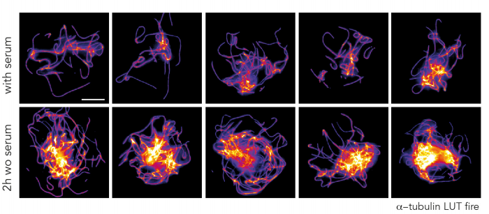
Drosophila embryos and aspect ratios, from Atia, et al.’s preprint
Universal geometric constraints during epithelial jamming. Lior Atia, Dapeng Bi, Yasha Sharma, Jennifer A. Mitchel, Bomi Gweon, Stephan Koehler, Stephen J. DeCamp, Bo Lan, Rebecca Hirsch, Adrian F. Pegoraro, Kyu Ha Lee, Jacqueline Starr, David A. Weitz, Adam C. Martin, Jin-Ah Park, James P. Butler, Jeffrey J. Fredberg
Aberrant Cortical Activity In Multiple GCaMP6-Expressing Transgenic Mouse Lines. Nicholas A. Steinmetz, Christina Buetfering, Jerome Lecoq, Christian R. Lee, Andrew J. Peters,Elina A. K. Jacobs, Philip Coen, Douglas R. Ollerenshaw, Matthew T. Valley, Saskia E. J. de Vries, Marina Garrett, Jun Zhuang, Peter A. Groblewski, Sahar Manavi, Jesse Miles, Casey White, Eric Lee, Fiona Griffin, Joshua D. Larkin, Kate Roll, Sissy Cross, Thuyanh V. Nguyen, Rachael Larsen, Julie Pendergraft, Tanya Daigle, Bosiljka Tasic, Carol L. Thompson, Jack Waters, Shawn Olsen, David J. Margolis,Hongkui Zeng, Michael Hausser, Matteo Carandini, Kenneth D. Harris
SCENIC: Single-Cell Regulatory Network Inference And Clustering. Sara Aibar, Carmen Bravo González-Blas, Thomas Moerman, Jasper Wouters, Vân Anh Huynh-Thu, Hana Imrichová, Zeynep Kalender Atak, Gert Hulselmans, Michael Dewaele, Florian Rambow, Pierre Geurts, Jan Aerts, Jean-Christophe Marine, Joost van den Oord, Stein Aerts
Accuracy, Reproducibility And Bias Of Next Generation Sequencing For Quantitative Small RNA Profiling: A Multiple Protocol Study Across Multiple Laboratories. Maria D. Giraldez, Ryan M. Spengler, Alton Etheridge, Paula Maria Godoy, Andrea J. Barczak, Srimeenakshi Srinivasan, Peter L. De Hoff, Kahraman Tanriverdi, Amanda Courtright, Shulin Lu, Joseph Khoory, Renee Rubio, David Baxter, Tom A. P. Driedonks, Hank P. J. Buermans, Esther N. M. Nolte-‘t Hoen, Hui Jiang, Kai Wang, Ionita Ghiran, Yaoyu Wang, Kendall Van Keuren-Jensen, Jane E. Freedman, Prescott G. Woodruff, Louise C. Laurent, David J. Erle, David J. Galas, Muneesh Tewari
Opportunities And Obstacles For Deep Learning In Biology And Medicine. Travers Ching, Daniel S. Himmelstein, Brett K. Beaulieu-Jones, Alexandr A. Kalinin, Brian T. Do, Gregory P. Way, Enrico Ferrero, Paul-Michael Agapow, Wei Xie, Gail L. Rosen, Benjamin J. Lengerich, Johnny Israeli, Jack Lanchantin,Stephen Woloszynek, Anne E. Carpenter, Avanti Shrikumar, Jinbo Xu, Evan M. Cofer, David J. Harris, Dave DeCaprio, Yanjun Qi, Anshul Kundaje,Yifan Peng, Laura K. Wiley, Marwin H. S. Segler, Anthony Gitter, Casey S. Greene
A persistent lack of International representation on editorial boards in biology. Johanna Espin, Sebastian Palmas-Perez, Farah Carrasco-Rueda, Kristina Riemer, Pablo Allen, Nathan Berkebile, Kirsten Hecht, Renita Kay Kastner-Wilcox, Mauricio Nunez-Regueiro, Candice Prince, Maria Constanza Rios-Marin, Erica P Ross, Bhagatveer Singha, Tia Tyler, Judit Ungvari Martin, Mariana Villegas, Tara Cataldo, Emilio Bruna
Lee lab has been studying on cell specification process using human pluripotent stem cells, particularly peripheral nervous system and skeletal muscle cells.
We are looking for highly motivated postdoc(s) who have experience on optogenetic studies. Recently, by using CRY2-PHR or LOV2 domains, we started to explore how light illumination can spatially and temporally modulate human stem cell fates, which can lead us to have unprecedented precise control as well as to interrogate/measure necessary/sufficient input amounts to elicit a certain biological event.
Previous experience on human pluripotent stem cells is not necessarily required (but basic cell culture experience will be great) and we value more on the ‘non-stem cell’ expertise, such as molecular cloning. Please send your application (CV and three reference contact info) to Gabsang Lee (leelabjob@gmail.com).
 (No Ratings Yet)
 (1 votes)
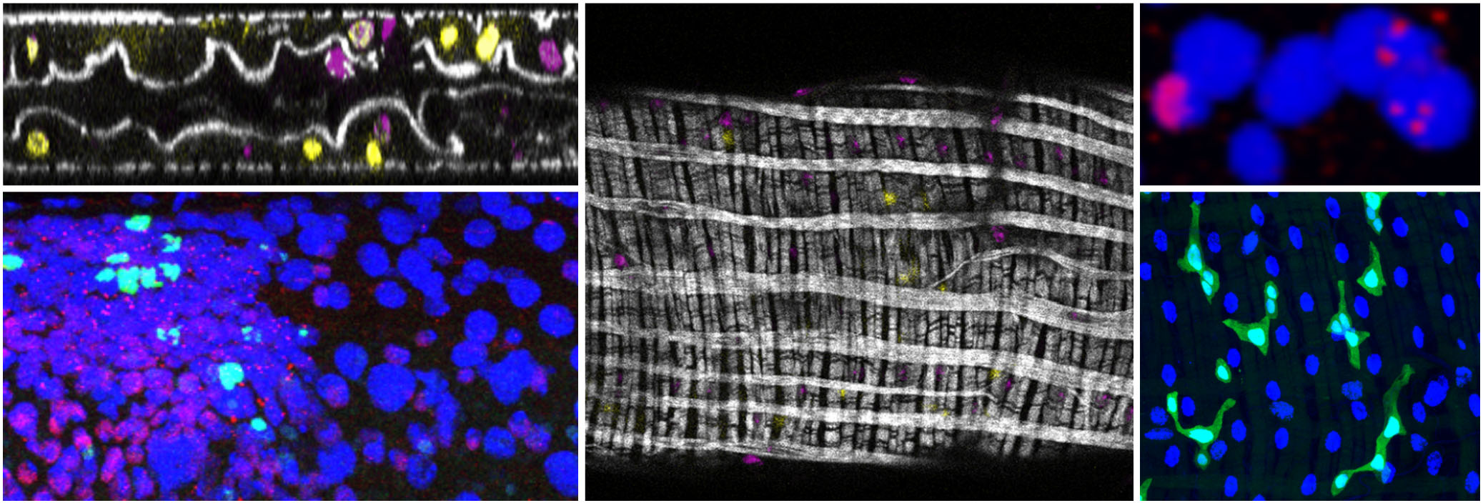

 o tackle this question, two PhD students, Liana Manukyan (computer scientist) and Sophie Montandon (developmental biologist), followed individual lizards during three to four years of their development from hatchlings crawling out of the egg to fully mature animals. For multiple time points, they reconstructed the geometry and colour of the network of scales on three animals by using R2OBBIE-3D (YouTube playlist), a very high resolution robotic system(
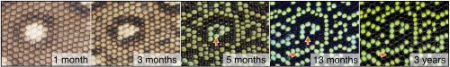
