Two postdoctoral positions are available in the School of Life Sciences at the University of Sussex supervised by Professor Claudio Alonso (http://www.sussex.ac.uk/lifesci/alonsolab/) within the broad field of Molecular and Developmental Neuroscience. The aim of the project is to investigate the genetic factors underlying the control of movement with a focus on the roles played by small RNAs in the process. The work will combine state-of-the-art molecular, biochemical, genetic, imaging and behavioural approaches to determine the roles of RNA regulation on neural development and behaviour in Drosophila. The work builds on a recent discovery made in the Alonso Lab that microRNAs can affect behaviour and complex movements in Drosophila (Picao-Osorio et al. 2015 Science 350:815-20).
The posts are funded by the Wellcome Trust and will contribute to an ambitious research programme funded by a Wellcome Trust Investigator Award made to Prof. Claudio Alonso. The project will be fostered by the scientific excellence of Sussex Neuroscience ranked within the Top-10 UK academic units within Neuroscience and Biological Sciences in the REF2014 (http://www.sussex.ac.uk/sussexneuroscience/).
One of the Postdocs will be an RNA Biologist (http://www.sussex.ac.uk/aboutus/jobs/1554) and the other a Neurobiologist (http://www.sussex.ac.uk/aboutus/jobs/1552). Successful candidates will be outstanding, committed and highly motivated postdocs seeking to develop an original an independent project within the broad area of Molecular and Developmental Neuroscience. Applicants should have PhD in Biology, Biochemistry, Neuroscience or other relevant disciplines.
The University of Sussex is located 10-min away from the lively and cosmopolitan seaside city of Brighton on the UK South Coast, 60-min away from central London, 30-min away from London Gatwick Airport and with full access to the beautiful country side of the Sussex South Downs.
Applications are invited for a 4-year Medical Research Council Industry CASE PhD studentship, which will be jointly supervised by Dr. Laurentiat the University of Cambridge and Dr. Francis at GlaxoSmithKline (GSK), to commence in October 2017.
The Laurenti laboratory combines state-of-the-art experimental and computational methods to study the unique biological and molecular properties of human haematopoietic stem cells (HSCs). GSK is a world leading research-based pharmaceutical company. In May 2015, the first autologous ex vivo gene therapy product, developed by the Cell and Gene Therapy (CGT) platform, was recently approved by the European Medicines Agency. CGT supports numerous cell and gene therapy projects from early phase to commerical launch, and the development of innovative technologies to enable improvements to cell and gene therapy manufacture.
The principal research aim of this project is to determine to what extent the gene therapy protocol affects the biology of HSCs. The project will combine single cell transcriptomics, lentiviral transduction technology, flow cytometry and single cell functional assays in vitro and in vivo. Adult HSCs and progenitor cells will be subjected to the gene therapy protocol and changes in their fate choices and transcriptome will be determined by single cell functional assays and single cell RNA-seq. This information will provide insights into how changes in the molecular circuitry of HSC alter their function under stress conditions, and will be used to guide process improvements to increase HSC functionality after transduction.
The primary research will be carried out mostly in Dr Laurenti’s laboratory but the student will spend a minimum of 6 months at GSK during the time of the fellowship.
We encourage applications from students with mathematical and/or bioinformatics skills.
Eligibility
Applicants should hold or be about to achieve a First or Upper-Second (2.1) class degree in a relevant subject. Students with a Bachelor-level degree are encouraged to apply.
Submit your application documents which should include your Application Form, CV, and your Degree transcripts, in pdf format to: sci-phd@stemcells.cam.ac.uk
Please ask your referees to submit references directly to the SCI Graduate Administrator by the application deadline:sci-phd@stemcells.cam.ac.uk, using “MRC iCASE 4-Year PhD studentship (Laurenti)” in the subject header. It is your responsibility for ensuring that both references are received by the closing date.
Application Deadline: Tuesday 14th February 2017 and shortlisted candidates will be interviewed between 27th-28th February 2017.
Informal Academic Enquiries to: Dr Elisa Laurenti el422@cam.ac.uk
Application Process Enquiries to: Graduate Administrator sci-phd@stemcells.cam.ac.uk.
For further details about our group and the institute, please visit:
Department/Location: Wellcome Trust – Medical Research Council Cambridge Stem Cell Institute, University of Cambridge
Salary: £29,301-£38,183
Reference: PS11020
Closing date: 18 January 2017
Fixed-term: The funds for this post are available until 31 March 2018 in the first instance.
The Pluripotent Stem Cell Platform (PSCP) is a hub in the UK Regenerative Medicine Platform, a joint research council programme to tackle the critical challenges in developing new regenerative treatments (www.ukrmp.org.uk). PSCP is a multi-disciplinary collaboration focussed on the quality controlled manufacturing and differentiation of human pluripotent stem cells suitable for clinical applications (http://www.ukrmp.org.uk/hubs/cell-behaviour-differentiation-and-manufacturing/).
The research is centred on optimising the generation of genetically modified human embryonic and induced pluripotent stem cells with reduced immunogenicity for development of cell based therapies, in particular megakaryocytes and platelets for transfusion.
Candidates should have a PhD with experience in the culture and analysis of pluripotent stem cells and/or haematological cell differentiation processes.
Applications are encouraged from candidates with experience of work in this area and an appreciation of cell production for clinical use and trials.
Technical support is available and access to a range of flow cytometry, imaging and qPCR instrumentation.
To apply online for this vacancy and to view further information about the role, please visit: http://www.jobs.cam.ac.uk/job/12444. This will take you to the role on the University’s Job Opportunities pages. There you will need to click on the ‘Apply online’ button and register an account with the University’s Web Recruitment System (if you have not already) and log in before completing the online application form.
The closing date for all applications is the Wednesday 18 January 2017.
Please upload your Curriculum Vitae (CV) and a covering letter in the Upload section of the online application to supplement your application. If you upload any additional documents which have not been requested, we will not be able to consider these as part of your application.
With over 200 votes counted, the cover of Development’s Special Issue on Plant Development has won the voter’s favourite cover for 2016! The fruit bat came second, and in joint third the fly nervous system and the fly legs. A fitting variety of model organisms for a great year in developmental biology!
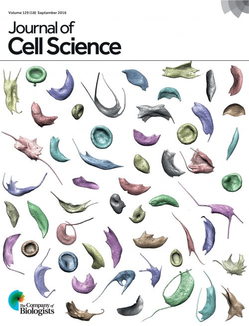
In our in-house competition, the fly nervous system won the Development team’s vote (showing we’re not too far from public opinion!) , but sadly lost out to these beautiful blood cells from our sister journal in the Company of Biologists, The Journal of Cell Science.
The cover images for the 24 issues ofDevelopment in 2016 showcase the breadth and beauty of developmental biology today. Model systems from plants to bats were imaged in various modalities – confocal and electron microscopy, microCT, good old fashioned skeletal preps and darkfield – and we also featured some in silico modelling.
Which one is your favourite? You can vote below the gallery (click to expand), and tell us why in the comments section.
Three-dimensional rendering of a confocal image stack showing a single alveolar type 1 cell with expansive cellular extensions (green, GFP, genetically labelled with HopxCreER/+; RosamTmG/+) intertwined with the vasculature (blue, ICAM2) in the mouse lung. From Yang et al., p. 54.
Embryonic day 8 chicken hindgut was cultured for 72 hours on a fibronectin-coated surface in the presence of supplemental glial-derived neurotrophic factor (Gdnf). Immunostaining with a neural crest cell marker (Hnk1; red) and a neuronal marker (Tuj1; green) demonstrates robust migration of the enteric neural crest cells. From Nagy et al. p. 264.
Ovules of Arabidopsis bel1 cna phb phv mutants. The ovulate axis of extant angiosperms does not branch, whereas some fossil gymnosperms show branching ovules. The branched ovulate axis of this mutant might thus bridge the gap in ovule body plan. From Yamada et al., p. 422.
Differing Six2/SIX2 transcriptional networks in mouse and human kidney. E15.5 mouse kidney next to a 15.5 week human fetal kidney with Six2/SIX2 (cyan) marking the nephron progenitors and cytokeratin (red) highlighting the collecting duct system. Nuclei are in blue. From O’Brien et al., p. 595.
Stage 19 short-tailed fruit bat (Carollia perspicillata). On the left side is an image of the fixed embryo before staining; the right side shows the embryo after Alcian Blue staining for cartilage. This image, taken by Idoia Quintana-Urzainqui, Paola Bertucci, Peter Warth and Chi-Kuo Hu at the 2014 Woods Hole MBL Embryology course, was chosen by readers of the Node.
Actin filaments (red) are enriched at the borders of enveloping layer cells surrounding the blastoderm (nuclei, blue), in the yolk cell cortex, and in a band adjacent to the blastoderm, where they function in cell movements of epiboly and maintain integrity of the blastoderm and yolk cell, a process that is disturbed in zebrafish split top embryos. From Langdon et al., p. 1016.
A segmented brain MRI scan for a heterozygous Tubb5 knockout mouse. Depicted are the olfactory bulbs (yellow), cortex (cyan), putamen (dark blue), lateral ventricles (red), hippocampus (green), colliculi (maroon) and cerebellum (pink). These mice have microcephaly reminiscent of patients with TUBB5 mutations. From Breuss et al., p. 1126.
A skeletal preparation of an embryonic bamboo shark (ventral view) showing the gill arch branchial rays and pectoral fins. The branchial rays of cartilaginous fishes and the paired fins/limbs of jawed vertebrates are patterned by a common Shh-dependent signalling mechanism. From Gillis and Hall, p. 1313.
Stage 17 Drosophila melanogaster embryo (ventral view) showing Elav (green; neuronal nuclei), Spalt (yellow; subset of neuron and muscle nuclei), BP102 (red; CNS axons), Eve (magenta; subset of CNS nuclei, and ring of nuclei around anal pad), HRP (grey; neuronal cell bodies and axons) and DAPI (blue; nuclei) staining. The image was taken by Connie Rich (University of Cambridge, UK) at the 2014 Woods Hole MBL embryology course and was chosen by readers of the Node
Cardiac atrium of a 6-week-old zebrafish labelled by priZm multicolour fate-mapping, with colour recombination generated by an atrial cardiomyocyte-specific inducible Cre recombinase. Coloured patches are multicellular muscle clones derived from individual atrial cardiomyocytes present at 3 days post-fertilization, illuminating the morphological changes and proliferation dynamics that shape the maturing chamber. From Foglia et al., p. 1688.
Müller glia-derived progenitor cells in acutely damaged chick retina. The retinal section was labelled with antibodies to Sox9 (red), neurofilament (green), phospho-histone H3 (blue) and CD45 (magenta). From Zelinka et al., p. 1859
3D volume-rendered heart of a 75 hpf Tg(kdrl:EGFP) zebrafish larva. Between the atrium and ventricle (right and left side of image, respectively) is the atrioventricular canal, where the emerging valve leaflet is recognisable as a folded structure. Such 3D analyses provide fundamental insights into the cellular rearrangements underlying cardiac valve formation in zebrafish. From Pestel et al., p. 2217
The mosaic of Rhodopsin 5 (blue)- and Rhodopsin 6 (red)-expressing R8 photoreceptors in the retina of Drosophila melanogaster. The insulator protein BEAF-32 is required for Hippo pathway activity to correctly specify R8 subtypes. From Jukam et al., p. 2389
A frontal section of a mouse heart at embryonic day 17.5 showing normal anatomy, which can be disrupted by short-term exposure to hypoxia during gestation. The image was generated using optical projection tomography. From Shi et al., p. 2561.
Mouse E16.5 dorsal tongue filiform papillary epithelia are highly patterned as rosettes, ordered between regions of intercalating lamina propria. Epithelium is demarcated by membranously expressed E-cadherin (Rhodamine Red-X); nuclei are stained with DAPI (blue). The mitotic spindle orientation factor LGN has multiple distinct functions in regulating oral epithelial development by controlling cell division orientation. From Byrd et al., p. 2803.
The hyaloid blood vessel network (red) with associated macrophages (green) isolated from a mouse eye. This, along with the retinal vessel network, was used to provide new insights into the role of apoptosis during angiogenesis and vessel regression. From Watson et al. p. 2973
Posterior (left) and lateral (right) views of the squid Doryteuthis pealeii at hatching scanned using microCT. Segmented reconstructions of brain regions correspond to a fate map generated during early embryogenesis. Depicted are the pedal (purple), buccal (cyan), paliovisceral (green) and cerebral (pink) ganglia, the optic lobes (dark blue), the retina (red) and the lens/iris (yellow). From Koenig et al., p. 3168
Computational modelling predicts an asymmetric Aux1 pattern in the root during halotropism (right, stylised picture). The asymmetric Aux1-YFP pattern observed in an Arabidopsis thaliana root during halotropism (left) confirms the model predictions. See Research article by van den Berg et al., p. 3350.
Leg phenotypes obtained after a progressive reduction in the dose of Sp family genes in Drosophila. Clockwise from top left: wild type, btd mutant, Sp1 mutant, Sp1 mutant with one mutant copy of btd and Sp1, btd double deletion mutant. From Córdoba et al., p. 3623.
Multiple stages of Tg(fli1a:egfp)y1 zebrafish embryos immunostained for pERK (red) and EGFP (green). In endothelial cells, ERK is activated in distinct signalling contexts to promote angiogenesis and lymphatic morphogenesis during development. From Shin et al., p 3796.
Spectral karyotyping of a metaphase cerebellar granule neuron progenitor from a P3 mouse with conditional deletion of Atr demonstrates complex chromosomal rearrangements, including fusions and translocations, revealing that ATR plays a crucial role in maintaining genomic integrity during brain development. Each chromosome is labelled in a different colour. From Lang et al., p. 4038.
Sea star gastrula larva showing neural precursor soxc-expressing cells (green) scattered throughout the ectoderm, dividing to produce lhx2/9-expressing daughters (pink) in the anterior ectoderm. DAPI staining shows nuclei (blue). Conserved anteroposterior patterning domains control the progression of neurogenesis in this deuterostome. See From Cheatle Jarvela et al., p. 4214.
Scanning electron microscopy of E10.5 control (left) and Six3neo/− (right) mouse embryos. The telencephalic vesicles (pseudocolored in red), and the medial (pseudocolored in yellow) and lateral (pseudocolored in green) nasal prominences are well separated in control embryos; however, only one small telencephalic vesicle and no medial nasal prominences are present in Six3neo/− embryos. From Geng et al., p. 4462.
3D reconstructions of mid-gastrula (left) and mid-neurula (right) Ciona embryos electroporated with transgenes driving H2B:Cherry (red) in the epidermis and YFP-CAAX (green) in the neural plate. Nodal and FGF control the cellular behaviours underlying morphogenesis of the neural tube during Ciona development. From Navarette and Levine, p. 4665.
The Alberto Monroy Fellowship is awarded to an Italian citizen to support their attendance in the Embryology Course next summer at the Marine Biological Laboratory in Woods Hole, Massachusetts.
The amount of the fellowship is 5,500.00 euros (about $5,750.00). The full text of the announcement of the award competition for 2017 can be found at http://www.giomolab.it/AAM.
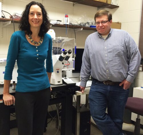
The neural crest is a remarkable multipotent cell population that has become a model system for how epithelial cells become mesenchymal and migrate during development. In today’s post, we feature a paper from the recent issue of The Journal of Cell Biologythat investigates the role that proteolysis of cell-cell adhesion receptors plays in this process. We caught up with first author and postdoc Andrew Schiffmacher, and his advisor Lisa Taneyhill, Associate Professor in the Department of Animal and Avian Sciences at the University of Maryland.
Lisa and Andy
So Lisa, can you tell us your scientific biography and what questions your lab is interested in?
LT Well, I did not start out as a developmental biologist, but am sure glad I ended up as one! On the contrary, I was trained as a cancer biologist working with cultured cells in Dr. Arnie Levine’s lab at Princeton, from where I received my Ph.D. But what I lacked at the time was experience in a live animal model. My interest in developmental biology truly blossomed while a postdoc in the lab of Dr. Marianne Bronner at Caltech. Here Marianne’s enthusiasm for development, and the chick embryo as a model, was truly infectious. I fell in love with developmental biology and soon realized that I could couple my interest in cancer, and in particular metastasis, with development by looking at how neural crest cells form and migrate.
In 2007, I started my own lab at the University of Maryland, where we use both the chick embryo and cell culture to ask questions related to embryo development. In my lab, we aim to elucidate how cells interact and communicate to form new tissues, and specifically, the fine balance that exists between dismantling and assembling cellular junctions to generate migratory cells and mediate intercellular interactions, respectively. We are investigating these processes by examining the cranial neural crest cell epithelial-to-mesenchymal transition, or EMT, and the formation of the cranial ganglia, which occurs through interactions between two different migratory cell types, cranial neural crest and placode cell-derived sensory neurons.
“We aim to elucidate how cells interact and communicate to form new tissues, and specifically, the fine balance that exists between dismantling and assembling cellular junctions to generate migratory cells and mediate intercellular interactions”
And Andy, how did you come to join the Taneyhill lab?
I was a doctoral student in Dr. Carol Keefer’s lab investigating gene networks that are responsible for maintaining pluripotency and directing lineage segregation in early mammalian embryos. Due to limited embryo resources, most of my thesis work entailed the use of cell lines as models for this stage of development. While I became highly interested in stem cell biology and the achievements being made (IPS cell technology came out around this time), I was far more interested in studying stemness and developmental potential within the context of embryonic development. Lisa’s new research program revealed to me the most fascinating cell type- the neural crest cell. As a cell biologist, I knew there were so many fundamental questions that could be addressed by studying neural crest development. In addition, Lisa was an obvious choice as a mentor as she was very successful and strong in areas where I knew I needed improvement.
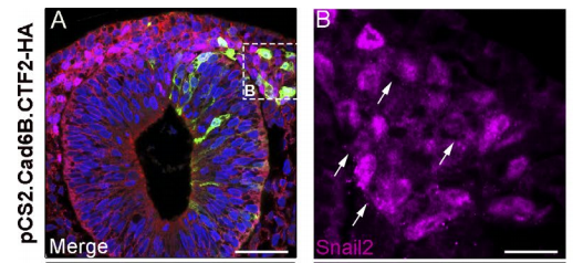
Transverse section through midbrain expressing CTF2-HA, from Fig. 3, Schiffmacher, et al. 2016 JCB.
What are the benefits of chicken embryos as a model to study EMT?
LT The chick is a phenomenal developmental model because of the ability to conduct in ovo/in vivo and ex vivo assays with the neural crest. So one can look at the impact of gene perturbation on EMT in either an embryo section or in a cell culture dish containing neural crest cells dissected out of the embryo. This inherent experimental flexibility lends itself to both fixed and live cell imaging experiments. There is a also wealth of historical literature on chick neural crest development that can help drive our studies.
“The chick is a phenomenal developmental model”
AS Chick cranial neural crest EMT occurs en masse at a very specific time in development, which allows us to perform genetic perturbations at specific times pre-EMT, and then evaluate effects during EMT. The biochemistry experiments I needed to perform to address our questions in vivo required harvesting sufficient amounts of tissue. With the chick, I could electroporate DNA or morpholinos into many embryos at one sitting and collect tissue after a short post-incubation time. The chick model offers the convenience of setting up large, synchronized experiments and does not require an animal facility.
What was known about the roles of Cadherins in EMT prior to your current paper?
LT Dating from my work in Marianne’s lab, we knew that a reduction of at least one cadherin in the chick head, Cad6B, was critical for neural crest cells to undergo EMT and migrate, as perturbation of Cad6B impacts these processes. Initial results revealed that loss of Cad6B transcripts occurred through the activity of the transcription factor Snail2 directly repressing Cad6B transcription during EMT. Work from other postdocs in Marianne’s lab, such as Pablo Strobl-Mazzulla and Crystal Rogers, refined this mechanism and also began uncovering roles for N-cadherin and E-cadherin during EMT, respectively.
But it became very clear that transcriptional repression of cadherins was not the only way to modulate cadherin levels during EMT. My lab showed that Cad6B protein is internalized by both clathrin-mediated endocytosis and macropinocytosis during cranial neural crest EMT, which further reduces membrane Cad6B. Chaya Kalcheim’s lab also published a paper almost 10 years ago now that described how the proteolysis of N-cadherin was important to permit chick trunk neural crest cells to undergo EMT and migrate. And Dom Alfandari, Jubin Kashef, and others have published many studies revealing how Cadherin-11 proteolysis in Xenopus cranial neural crest is key for migration. Andy’s studies on Cad6B proteolysis during EMT follow on from these.
“It became very clear that transcriptional regulation of cadherins was not the only way to modulate their levels during EMT”
ASOur work published in MBOC was definitively inspired by Chaya Kalcheim, Dom Alfandari, and Jubin Kashef’s research and essentially established that cadherins expressed in chick cranial neural crest cells undergoing EMT are also regulated by proteolysis. Our goal was to identify and validate the proteases involved and assess their importance as regulators of EMT. Our next goal was to figure out what the cleaved Cad6B byproducts were doing. Fortunately for us there is an enormous wealth of information on Cadherin biology and proteolysis that helped steer our experiments in the right direction. I like to think our JCB manuscript pays tribute to all of that work.
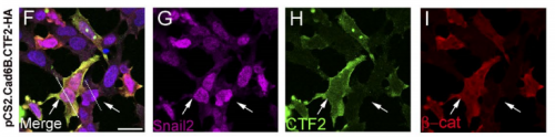
Neural crest explants expressing CTF2-HA, from Fig. 4, Schiffmacher, et al. 2016. JCB
Can you sum up the key results from your paper in a paragraph?
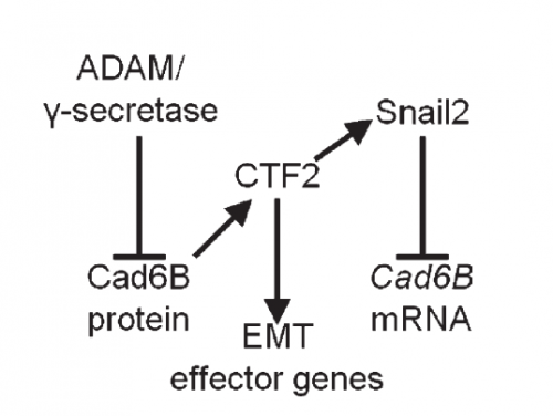
AS We found that prior to and during EMT, Cad6B levels are regulated by ADAM and γ-secretase proteases, and as a result, N-terminal and C-terminal Cad6B fragments are being produced. Following cleavage by γ-secretase, the soluble C-terminal fragment or CTF2 remains associated with β-catenin. This complex protects both proteins from degradation, and allows them to accumulate. This leads to increased nuclear importation, where β-catenin can exert greater regulatory input into modulating EMT effector gene expression, most notably Snail2. Interestingly, we found that the Cad6B CTF2 is not merely along for the ride and separates from β-catenin upon nuclear import, but also co-associates with the chromatin.
Regulatory circuit for neural crest EMT, from Fig. 6, Schiffmacher, et al. 2016. JCB.
It seems like proteases are at the top of your EMT network. What regulates their levels and activities?
LT It’s clear that precise control over these proteases is crucial to limit their activity to defined times during the development of a neural crest cell. Some of this is dictated by the spatiotemporal expression pattern of the proteases themselves, and we certainly see that for the ADAMs and γ-secretase. The other thing to bear in mind is that these proteases will likely process multiple substrates, so their presence in the neural crest is not solely to cleave cadherins. This adds another layer of complexity to trying to understand what modulates their levels and activities. The short answer is that we don’t yet know the upstream pathways regulating these proteases, but this is a question that is currently being addressed by my lab.
And what is going on with the other Cadherins at the same time? Is there any crosstalk between them?
LT We’re intrigued by this possibility, but it is a tricky question to address. Premigratory cranial neural crest cells express multiple cadherin proteins, but at specific developmental times, and it’s quite possible that other cadherins are expressed whose identity has yet to be discovered.
Do you think the proteolysis-transcriptional regulation mechanism you have revealed will be involved in other developmental or pathological events?
AS Most certainly. I would hypothesize it is occurring in any cell where cadherins and metalloproteinases are co-expressed, and especially when cadherin levels need to be under tight regulatory control, including events like EMT and migration. This mechanism also most likely contributes to epithelial carcinoma cell dissemination during metastatic progression in tumors.
When doing the research, was there a particularly exciting result or eureka moment that has stayed with you?
AS For the MBOC article, I was disheartened when we realized that CTF2 overexpression resulted in no dramatic neural crest cell migration phenotype. As part of that experiment, I happened to perform Cad6B co-immunohistochemistry as a premigratory neural crest marker. I’ll never forget sitting in my hotel room in Cancun, Mexico (SDB/ISDB meeting, 2013) staring at the images and then realizing that Cad6B levels were diminished in electroporated neural crest cells! This eureka moment was tied to the next one, where I decided to perform Snail2 quantitative PCR to determine if Snail2 levels were altered in order to explain why CTF2 overexpression downregulated full-length Cad6B. It was relieving to see that Snail2 levels were increased.
“I’ll never forget sitting in my hotel room in Cancun staring at the images and then realizing that Cad6B levels were diminished in electroporated neural crest cells!”
And what about the flipside: any specific moments of frustration or despair?
AS Nearly every Western blot was performed using hundreds of excised midbrain dorsal neural tubes for the collective treatments. One mistake and I would lose weeks/months worth of tissue electroporation/collection. On the bright side, I learned I work well under that kind of pressure.
Finally Andy, what are your plans following this work?
AS I am currently seeking a position as a principal investigator in academia, and would like to continue my research investigating how metalloproteinase-mediated proteolysis generates other substrate fragment-based regulatory inputs into a cell’s gene network during neural crest development.
And Lisa, where next for the Taneyhill lab?
LT:We aim to decipher how CTF2s provide transcriptional input into the neural crest gene regulatory network, and uncover additional targets in this process. We are also investigating potential functions associated with the Cad6B shed ectodomain. Finally, we are examining the molecules required to mediate cell-cell adhesion and interactions as the cranial ganglia assemble. All of these objectives lead to an overarching goal to understand how cells communicate with one another to allow for fundamental, and important, changes during vertebrate development, such as the generation of a migratory neural crest cell or a new tissue from multiple cell types.
Doug Melton is Xander University Professor at Harvard University, co-director of the Harvard Stem Cell Institute and a Howard Hughes Medical Institute Investigator. His lab investigates the development of the pancreas, and uses insights from this process to direct the production of insulin-producing beta cells from stem cells. We met Doug at the 2016 Society for Developmental Biology-International Society of Differentiation (SDB-ISD) joint meeting in Boston, USA, where he gave the Jean Brachet Lecture.
You’re at the SDB-ISD meeting to deliver the ISD Jean Brachet Lecture. What does the award mean to you?
Of course it’s nice to get recognised, particularly so with an award named for Jean Brachet, who in a sense was a molecular biologist, though at the time he would have been called a chemical embryologist. In my own career, I was in a wave of people applying molecular biology and cloning to embryological problems, so it’s nice for me to imagine that I fit into his tradition.
Brachet was responsible for showing the importance of RNA and was right on a path to demonstrate that protein synthesis occurred at ribosomes when World War II intervened. After the war, in the early 1950s, researchers in Brussels couldn’t get hold of radioactive amino acids, but the Americans could, and they won the Nobel Prize. When Brachet read the papers demonstrating protein synthesis from RNA, for which he had all the circumstantial evidence, he said he couldn’t have been happier even if he’d been the author. I just thought: what a gracious thing to say! So it’s especially nice to have been asked to speak in his honour.
What inspired you to become a biologist?
There wasn’t really one event, one moment of epiphany. When I was a little boy I liked frogs and salamanders, and remember being puzzled by how the eggs, which looked so similar, knew how to make a tail or not make a tail, and that sort of piqued my interest.
But more seriously, when I got into college, I read an article in Scientific American by John Gurdon on cloning, and I thought that was the coolest thing, just so, so cool! John is one of the clearest writers of science, and that sort of simple reporting on an important question – what the scientist is interested in and why is it important – is extremely powerful.
So you did your BSc in Illinois, went to Cambridge as a Marshall Scholar and got a BA in the History and Philosophy of Science, and then did a PhD with John Gurdon at the LMB. How did these academic experiences influence your career?
In the late stages of my undergraduate degree, I got interested in philosophy of science and philosophy in general. I was very lucky to get this Marshall Scholarship – I’d never really been outside of Illinois – and Cambridge University in the UK was really good for me. It taught me right away that the philosophy of science was really interesting, but also – and this is maybe the embarrassing part – that I would never do anything original in philosophy but would instead spend my life commenting on really original thinking by others. My ego didn’t want me to spend my life commenting on what others did.
“I showed up at John Gurdon’s door and asked if I could wash dishes or just help out. He didn’t really know what to make of it!”
So then I remembered the article in Scientific American, and I showed up at John’s door and asked if I could wash dishes or just help out. He didn’t really know what to make of it! But he let me play around in the lab while I was finishing up this degree, which was unusual: there weren’t other undergraduates at the MRC LMB at that time. And I’m forever grateful to John for taking a chance on me: I cannot overestimate the influence he had on me, in showing me how to do science and have fun, and how to think about what questions are worth asking. The simplicity with which he’s approached science is breathtaking. He’s a great example for how to do science.
You then returned to the USA and established your own lab at Harvard. What were the main questions you were interested in, and what were the main contributions of the lab?
When I left John’s lab, what really interested me was an older embryology question concerning things in the egg called cytoplasmic determinants. This was around the time that factors such as Bicoid in Drosophila were being discovered, and I worked on a localised mRNA in the frog egg to see if we could find things that help explain Nieuwkoop’s induction work. It was a very exciting time: great people all working in the same area. You’d come to a meeting like this and someone would have pointed their finger to a gene to say ‘that gene’s important’; that was where we were, just trying to find genes responsible for induction.
“It was a very exciting time: great people all working in the same area. You’d come to a meeting like this and someone would have pointed their finger to a gene to say ‘that gene’s important’; that was where we were, just trying to find genes responsible for induction.”
In terms of contribution, I like to think my lab was one player in that general movement. One technology that we developed and turned out to be useful was to make RNA in vitro with SP6. And I think we were one of the first to make the argument that when development occurs, paradoxically the nervous system is the default pathway, and to make endoderm is harder. This really seemed to annoy some neurobiologists, who thought that being a neuron was the highest thing to which you could aspire!
The most important thing to me was the students I got, and being part of this exciting community of excellent scientists. Jim Smith, Eddie de Robertis, Jonathan Slack, Masamoto Asashima, Mark Kirschner; a really large group of people trying to figure out the genes responsible for induction. It really was a lot of fun and just a very exciting time, as I’m sure being in development now is.
I guess now it’s still as exciting, just bigger?
Maybe that’s it. I think developmental biology has expanded, to take on morphology formation in other animals (evo-devo), and into something that I try to practise, applied developmental biology. Rather than asking how nature works, in an almost anti-intellectual way we ask how we can apply this to something, for instance human health.
I love developmental biology because of how lucky one is to be able to work in a field that answers questions about nature, but also can offer something to help people.
In the late 1990s, your lab shifted focus to work on pancreas development and diabetes. What were the big open questions at the time?
I don’t think it’s an exaggeration to say that most people in the diabetes field at the time were working on how to provide insulin, the molecular biology of the insulin gene, how to manipulate the protein’s solubility, and what happens when a patient presents with the disease. For a developmental biologist, that’s not how you would think about it: the obvious question is where do pancreatic cells come from? What genes are involved, and can we remake pancreatic tissue?
This is not some flash of insight: it’s the sort of question an undergraduate might ask. If there were people asking these questions at the time then they were few and far between, and it wasn’t discussed at a meeting like this. So when I went into the lab and told them we were going to change our focus, some people were a little disappointed as it didn’t quite align with what they were doing, but I like to think that the transition was handled well and they could all finish up their projects. Within a year we were working on really basic problems, such as which cells make the pancreas. At that time if you looked in the textbooks, the pancreas was unquestionably a derivative of the neural crest, not the endoderm. The idea came from the fact that three particular genes were expressed in the neural crest, and also in the pancreas! I actually use this as an example when I’m teaching: I’m a big fan of the Boston Celtics, and I’ll wear my Celtics jersey and say: ʻthis is a marker, like a gene, but does it make me a Boston Celtic?’ They remember this, especially as the jersey’s hanging down to my knees!
But back to your question, the lab was really going back to basics: where do the cells come from and what genes are important? The long-term idea being that you might be able to use that information for diabetes.
And then, after that, there was the excitement surrounding stem cells. Before human stem cells became well known – and this is probably a Whiggish form of history – I would say most of the people who worked on stem cells were using them as tools to knock genes out and study gene function. At that time, there wasn’t a huge enterprise in trying to control their differentiation, and that’s the side that we ended up working on.
What does developmental biology bring to the table for understanding and treating diabetes?
In simple form, diabetics either don’t have beta cells or have dysfunctional beta cells, and stem cells can make any cell in the body. Let’s try and connect the dots.
“In simple form, diabetics either don’t have beta cells or have dysfunctional beta cells, and stem cells can make any cell in the body. Let’s try and connect the dots.”
You’ve been working with stem cells to understand diabetes for many years. How has the stem cell field grown and changed since the early days?
This field has grown slowly, but is now burgeoning. Fortunately, we’re past the political concerns and restrictions that characterised the 1990s and early 2000s. People just don’t talk about that sort of thing any more. The restrictions did not stop the research but made it hard, and I feel those days were a real missed opportunity for the field.
Your group has recently shown that you can produce beta cells en masse. Are we now done with the developmental biology and left with the practicalities of getting the cells into the body?
We’re close to it. The developmental aspects for me now come in with the longevity of the cells we can make, moving more towards developmental physiology and function and away from developmental anatomy (in the sense that making the cells, and making the embryo, is an anatomical problem).
The other part of the problem is the immune attack: we don’t really know much about why beta cells are picked out for autoimmunity. As a card-carrying developmental biologist, I would like to reconstruct the system, and watch which cells and genes are involved. This will be quite a challenge. My grant applications regarding this question have been uniformly rejected for two reasons. One is that I’m not an immunologist – that’s true! And two, that my plans are simple-minded and overly ambitious, and I’d say both of those are also true! But I’m a persistent person, and I’m going to work on the question of how beta cells are mistakenly targeted by the immune system.
“We don’t really know much about why beta cells are picked out for autoimmunity. As a card-carrying developmental biologist, I would like to reconstruct the system, and watch which cells and genes are involved”
I don’t mean to imply that we’re done with the first part, but we’re at the goal line. I’d like to get more clinicians involved to understand how the disease develops. There’s this fascinating period called the honeymoon period where you present with the symptoms, then your body suddenly makes enough insulin that you’re not ʻdiabetic’. So in that period your body must be ramping up the amount of insulin made by residual beta cells; it’s an interesting problem.
You were an editor for Development from 1987 to 2001 and are on our Advisory Board today. How has the field and the journal changed since the eighties, and where do you think it’s going?
When Chris Wylie took over the Journal of Experimental Embryology and Morphology (JEEM), changed it to Development in 1987 and recruited me as an editor, we thought a little bit about what we’re today calling applied developmental biology. That is a shift that has started and is worth thinking a bit more about. Not that Development should become a medical journal, but let’s think about how to use the basic ideas of development – induction, lineage, things like that – to think about disease. Developmental biology will also be deeply informative as to the question of whether we call ageing a disease or not.
Let’s think about how to use the basic ideas of development … to think about disease
I’d also like to see Development expand its activities in ʻnon-model organisms’. It’s interesting to reflect on what model organisms have and haven’t been. If you look back in history there were a lot of papers at one point on Dictyostelium, but we don’t seem to have many ʻDicty’ talks here at the SDB. Also, the sea urchin: still important, but not so central. And if you go to a Xenopus meeting today, they angst over whether or not it’s still relevant. The age-old question is: what does it mean to be a model? Philosophically, it’s an interesting one – it depends so much on what we think is important at the time to be able to do or to look at.
Do you have any advice for young researchers today?
Read and think about the history of the field. And go read John Gurdon’s early papers!
What might people be surprised to find out about you?
This might not be surprising, but I’ve tried to give up cars and I ride my bike all the time. I just treated myself to a new road bike and just came back from a cycling trip and hiking in the Dolomites.
Today’s paper comes from the final issue of Development for 2016, and reveals a link between bioelectricity and reactive oxygen species during tail regeneration in Xenopus. We caught up with first author Fernando Ferreira and his advisor Min Zhao, Professor in Dermatology at UC Davis.
Fernando and Min
So Min, can you give me the brief history of your lab, and what questions you are interested in?
MZ The main goal of my lab is to electrically heal wounds and regenerate tissues. Demonstrated over one and half centuries ago, the minute electric signals naturally produced at wounds are very poorly understood and appreciated. Epithelial cells and many other types of cells follow the guidance of the electric signals and migrate and grow directionally. We demonstrated that in epithelial sheets, the guidance effect of physiological electric signals overrides other co-existing guidance cues.
I had medical training in trauma with Zhengguo Wang, one of the founding fathers of trauma surgery in China. I then had research training with Geoffrey Burnstock at University College London, and Colin McCaig and John Forrester at University of Aberdeen. I started my lab at the University of Aberdeen with a Welcome Trust University Award and held professor/personal chair and honorary consultant positions at University of Aberdeen and Aberdeen Royal Infirmary (Scotland), before moving to University of California at Davis to take up a professorship in the Department of Dermatology and Department of Ophthalmology.
Supported by grants from NIH, NSF, California Institute of Regenerative Medicine and other federal and state agencies, my lab is interested in answering the following questions:
How do cells sense and respond to physiological electric fields?
How are the wound electric currents/fields produced and regulated?
Can we exploit the “electrical signalling” to enhance wound healing and induce regeneration?
And Fernando, how did you end up in Min’s lab?
FF During my Master’s degree, my adviser and Min met in an international conference. In the discussion they mentioned me; on arrival, my adviser told me that Min’s lab would be willing to host me and that could be an opportunity that I probably should not let escape. By this time, I already knew and admired much of Min’s work and thought it could be an excellent opportunity to test some old and new ideas falling within the framework of his lab. Thus, naturally, I readily set the goal to work with him.
Meanwhile, however, I applied for other closer positions, but I was oddly satisfied or left with a bitter-sweet feeling after receiving no or negative responses; I guess that this proved that I really wanted to join Min’s lab. Things aren’t straightforward though, and after a considerable despair for not getting a 1 year long Fulbright grant to visit Min’s lab, I took a risky move and entered a PhD programme in the Portuguese university (Minho) without funding. I was working in non-scientific part-time jobs to cover the tuition fees and, simultaneously, applied for a studentship grant sponsored by the Portuguese Science and Technology Foundation (FCT). With the Fulbright denial lingering in my mind, I had low expectations. Fortunately, I got the grant and after a way too long “quarantine” process (~8 months), I finally joined the lab in the USA…
It is pressing and fair to recognise that after joining the University of Minho, I received precious encouragement and help from the PhD programme professors and especially my supervisor Andreia Gomes, who accepted me without second thought, always supported me and gifted me with wise advice. All the happiness and sorrow of this journey, allied with the sheer size of the Atlantic Ocean plus the departure delay, made the farewell from family particularly hard; it is a natural cliche to be grateful for family care and support, and undoubtedly I am.
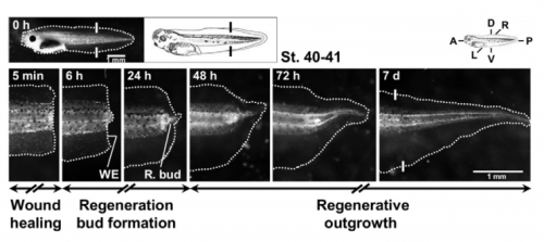
The process of regeneration over time in Xenopus tadpoles, from Figure 1 in the paper
Do you think bioelectricity outside of the nervous system is adequately appreciated in developmental and regenerative biology?
MZ Bioelectricity is not as adequately appreciated in developmental and regenerative biology as I’d wish, which I believe perhaps is due to the following. Research in bioelectricity (not the traditional electrophysiology) was restarted by Lionel Jaffe and his students Richard Borgens, Richard Nuccitelli and Ken Robinson with some wonderful results from the 1960s-1990s. This happened in an era that coincided with the revolutionary discovery of the double helix and the great advances in biology that ensued. Genetic, molecular and biochemical mechanisms are in the lime light of biology, including developmental and regenerative biology. Great advances in technologies and tools in genetics and molecular biology provided developmental and regenerative biologists with powerful tools to understand some of the most fundamental mechanisms. Research technology in bioelectricity, however, has since stayed virtually unchanged. It is also worthwhile to mention that some “charlatan claims” in bioelectricity have tarnished and discredited this field.
“Great advances in technologies and tools in genetics and molecular biology provided developmental and regenerative biologists with powerful tools to understand some of the most fundamental mechanisms. Research technology in bioelectricity, however, has since stayed virtually unchanged”
Bioelectricity is therefore off the radar of most developmental and regenerative biologists. Very few laboratories have continued the efforts in bioelectricity, for example Michael Levin at Tufts University, USA and Colin McCaig at University of Aberdeen, Scotland. Some of their research has provided very impressive results in combination with genetics and molecular biology.
Was anything known about the connection between reactive oxygen species (ROS) and bioelectricity in regeneration before your paper?
MZ & FF Strictly in regeneration, we couldn’t find studies demonstrating a direct connection between redox and bioelectric states. When we started this study we already knew – from decades old evidence – that electric currents were important for a successful regeneration, especially in amphibians; however, no evidence existed about ROS (excluding wound healing in small-scale injuries). Then, a wave of papers came, showing that ROS were also required for regeneration in widespread models, such as Xenopus tadpoles and adult zebrafish. The link between ROS and bioelectricity (membrane potential, transepithelial potential (TEP) and electric currents/fields), remained, however, elusive. In the regeneration field, our study thus appeared in a timely fashion to link the fairly old but re-emerging field of bioelectricity with the emergent field of redox biology.
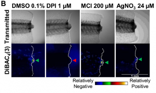
Assaying changes membrane potential following drug treatments, from Figure 2 in the paper.
Could you give us the key results of your paper in a paragraph?
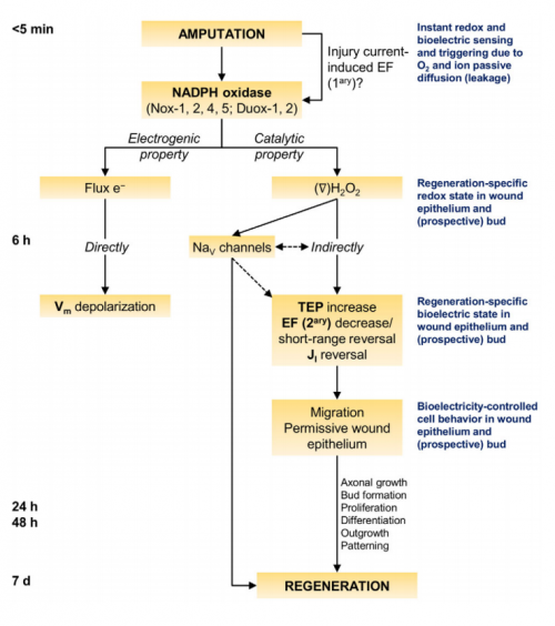
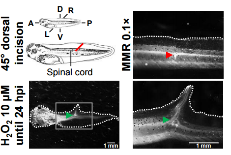
MZ & FF The general take home message is that redox and bioelectric activities interact during regeneration. More specifically, there is a two-way regulation of bioelectric activities by NADPH oxidases: the driven electron flow depolarizes the membrane potential, whereas the produced H2O2 increases the magnitude of TEP (positive inside) and switches the direction of electric current (to inward) in the regeneration bud. The depletion of ROS during the regenerative period mimics the abnormally low TEP and non-reversed electric currents measured during the refractory (non-regenerative) period. The external application of H2O2 for a short period normalizes the bioelectric activities and, by doing so, rescues and induces regeneration. External H2O2 was also inductive enough to form ectopic tails in injuries severing the spinal cord during the regenerative period. Finally and molecularly speaking, H2O2 regulates voltage-gated Na+ channels in order to modulate regeneration.
The model from Figure 7 in the paper
Your paper ends with a model including the proposition that immediately post-amputation, an electrical signal activates the redox signal. How did you come to this hypothesis?
MZ & FF To better understand this hypothesis we need to go back a bit and point out the underlying assumptions. One of the highlights of this study is that ROS are immediately required for regeneration. Those ROS are produced from NADPH oxidases, holoenzymes with complex assembly and regulation; therefore, an ultra-fast signal must activate the enzyme to generate sufficient ROS for the task ahead.
One of the highlights of this study is that ROS are immediately required for regeneration…an ultra-fast signal must activate NADPH oxidases to generate sufficient ROS for the task ahead.
An electric short-circuit is an instantaneous response to amputation, which results in the so-called injury current and subsequent electric field. This immediate and automatic electric field is, according to the hypothesis, what activates the NADPH oxidases. In fact, there is some evidence showing that applied electric fields induce production of ROS in cells in vitro. We are currently designing experiments to test this hypothesis during regeneration. If true, a redox-bioelectric feedback module would exist in regeneration. This is, the injury-induced electric signals activate the upstream redox signals that regulate downstream electric signals. With caution, we think that the now hypothetical feedback module could be used as theoretical evidence, because it could allow a more tight or efficient regulation of regeneration owing to evolution.
Before speculating or designing more experiments to understand how the electric fields would activate the NADPH oxidases, it is more pressing to test whether the hypothesis is true.
How do you think your mechanism might relate to the ‘canonical’ intercellular signalling pathways that are also involved in regeneration?
MZ & FF The integration between ROS and bioelectric activities occurs very early in the regeneration process. Many of the ‘canonical’ signalling pathways, such as Wnt, BMP and Notch, appear to be activated later on. By itself, this may indicate that those pathways are regulated by ROS and/or bioelectricity, i.e., act downstream. In fact, several studies in the regeneration context have shown that ROS or bioelectricity, independently, regulate some signalling pathways (and also cell behaviours), such as Wnt, FGF and Delta.
Given the high penetrance in regeneration, we think that pathways like the ones already mentioned and others might be affected by redox and bioelectric activities; we also think that follow up studies will unveil this, aiming for a higher level of mechanistic integration in regeneration.
Why do you think the H2O2 treatment induced the formation of ectopic tails?
MZ & FF The induction of ectopic tails was a thrilling finding, but it is important to note that the purpose of the assay was to check whether H2O2 induced ectopic tails and not why; the why deserves further research. That said, studies found that fin wounds in both Xenopus tadpoles and adult zebrafish generate ROS, likely H2O2. Unpublished results from us show that blocking the production of ROS impairs healing in Xenopus fin wounds. Therefore, we think that a threshold of H2O2 could define or tune the morphogenetic outcome, meaning that if the threshold is passed we may get an ectopic tail instead of just healing.
“The induction of ectopic tails was a thrilling finding”
Mechanistically speaking, another study found that Wnt signalling induced ectopic tails in the same model as ours. H2O2 could thus regulate Wnt for the same purpose, a pathway that could be mediated by bioelectric activities. Not mutually exclusive, another, maybe more speculative, possibility is an analogy with the accessory limb and blastema formation in axolotls and Xenopus, respectively. To induce them, it is required extra neuronal tissue, usually a deviated nerve. The incision we made in the tail severed the spinal cord, therefore, H2O2 treatment could, maybe via bioelectricity, affect the neuronal tissue so it becomes inductive.
Exogenous hydrogen peroxide induces ectopic tails on severing the spinal cord, from Fig. S15 in the paper.
When doing the research, was there a particularly exciting result or eureka moment that has stayed with you?
FF As many have at one point or another, I had the privilege to experience both life-guiding eureka moments and exciting results. A first eureka-like moment occurred back in my last bachelor year. Anxiously to find what path to follow, I learned about ROS and the caudal regeneration in lizards came to my mind. I had a minor thought experiment: I grabbed a common lizard making it autotomize the tail; with the inner tissues now exposed to the atmosphere, I imagined the oxygen entering the amputation plane down its chemical gradient and then pictured its transformation into ROS; as the levels rose, ROS alarmed local cells that something went wrong and triggered regeneration without any delay. In my innocence at the time, I let this single moment guide me through science ever since; there is no regret!
A second eureka-like moment occurred just before my arrival in the USA. When analysing a review paper, I read that the NADPH oxidases are electrogenic; this triggered a late night chain reaction that led me to the core of this manuscript. As NADPH oxidases work, they transfer electrons through the plasma membrane, so I thought that this was the origin of the membrane depolarization previously shown in regeneration. Then, I thought that since ROS are produced, these would, in turn, affect other facets of the bioelectric phenomena, namely electric currents.
“When analysing a review paper, I read that the NADPH oxidases are electrogenic; this triggered a late night chain reaction that led me to the core of this manuscript”
During the research, the exciting moments were when I was performing the critical experiments testing the redox-bioelectric crosstalk during regeneration. I blocked the production of ROS and imaged the membrane potential and measured the electric currents; during experiments, before getting the positive evidence, I was lightly sweating and my hands and belly were shivering. I know that these “symptoms” are analogous of a romantic encounter, guess that made them even more exciting!
Other unforgettable exciting moment was when, in the very first attempt, I saw a well-defined ectopic tail induced by H2O2. I was so impressed by it that I was childish enough to call other lab members to see an undisclosed “hopeful monster” in the microscope.
And what about the flipside: any particular moments of frustration and despair?
FF Other than now and then when, by procedural vicissitudes – for example, compromised batches of tadpoles (fungal infection, deficient animals, etc.), or all electrodes ending up breaking when touched, or readings were too noisy or strange because the earth wire was somehow disconnected – or for no obvious reason, experiments didn’t work out, I didn’t have any moments of frustration or despair worth noting. If you are surprised, so am I; not having major setbacks by the end of this manuscript surprised me and sometimes even “scared” me, since frustrations are a common theme in the research process and I really don’t want to think that the hypothesis and experimental design put forth were bullet proof. So, for modesty’s sake let’s just call it ‘beginners luck’; probably, the use of well-established techniques and methods helped. Just to highlight the surprise, we are about to submit a new study, where I had my share of despair moments, which I guess covers their absence in this paper, or at least I joke in that way! In fact, I was fortunate enough to get some serendipitous findings which we might end up following.
And finally, Min: where do you think this work will take you next?
MZ We followed an interdisciplinary approach hoping to merge apparently disparate research fields during regeneration. We think that this approach is important and potentially rewarding, and so several lines of research can be pursued with that in mind. We will have at least two exciting possible directions. One is to detail the molecular mechanisms of the redox-bioelectric interplay and to integrate them with ‘canonical’ signalling pathways in the Xenopus and other regeneration models. To help, we are currently establishing a redox and bioelectric sensor facility in the lab. The other is to take advantage of some of the cutting-edge technologies in wound healing and tissue regeneration in mammals. It appears that we are able to manipulate local electric fields and/or redox activities. A combined approach may provide promising therapies for chronic and non-healing wounds.
“I would be tempted to propose a term, “electrobiology”, hoping to suggest that electricity in biology has significant roles”
Evidence is accumulating suggesting bioelectricity as a different layer of mechanism, usually very upstream, together with the fundamental genetics and molecular/cellular processes that orchestrate during development and regeneration. I would be tempted to propose a term, “electrobiology”, hoping to suggest that electricity in biology has significant roles, in contrast to the more than the phenomenological nature of the word “bioelectricity”.
A post-doctoral research associate position in the Department of Zoology, located in Central Cambridge on Downing Street, is available from 1 March 2017 for up to thirty-six months. This is a Leverhulme Trust-funded post, to work with Dr. Andrew Gillis on the embryonic development of gill arch appendages in a cartilaginous fish, the little skate (Leucoraja erinacea).
Cartilaginous fishes possess paired appendages (branchial rays) that project from their gill arches, and over a century ago, Carl Gegenbaur famously proposed that such appendages represent the evolutionary antecedents of paired fins and limbs. We have recently found evidence of developmental parallels between the branchial rays of cartilaginous fishes and the fins/limbs of jawed vertebrates. We now wish to further dissect mechanisms underlying the development of skate branchial rays, in order to test Gegenbaur’s classical hypothesis of gill arch-paired fin serial homology. Duties will include the design and execution of experiments to test for shared embryonic origin, gene regulatory and patterning mechanisms between branchial rays and fins/limbs, and the preparation of results for publication.
The successful applicant should have a Ph.D., completed or completion imminent, in developmental biology, evolutionary biology, comparative anatomy or a related field, with a strong interest in evolutionary-developmental biology. Prior molecular biology and/or bioinformatic training would be beneficial. Skate brood stock is maintained at the Marine Biological Laboratory in Woods Hole, U.S.A., so willingness to travel to the MBL during summer months for experimental work with skate embryos would be beneficial. Enthusiasm, determination and the capacity to work independently are essential.
Further information on this vacancy may be found here.
The closing date for applications is Monday, 23 January 2017. To apply online, please visit http://www.jobs.cam.ac.uk/job/12377/ and click on the ‘Apply’ button. This will route you to the University’s Web Recruitment System, where you will need to register an account (if you have not already) and log in before completing the online application form.
Please quote reference PF10959 on your application and in any correspondence about this vacancy.
The University values diversity and is committed to equality of opportunity.
The University has a responsibility to ensure that all employees are eligible to live and work in the UK.
Biomedical research is experiencing what has been termed a ‘reproducibility crisis’. There is much talk about how we can improve the rigor and robustness of our research to increase its value and predictiveness. Many remedies are being discussed, such as increasing statistical power, reducing bias by improving internal validity, fostering transparency by open data policies, and publication of NULL results, among many others. In general, this debate is about increasing the quality of our research. Errors, mistakes and mishaps negatively impact on quality. In a work environment as complex as experimental biomedical research a substantial number of errors may occur on a daily basis, which can jeopardize the quality of our work, and may waste resources, or even endanger personel. Surprisingly, the issue of errors, and how to avoid them, has not yet received any attention in the current ‘biomedical research waste’ debate.
There is no way for professionals to not make mistakes from time to time. What really makes a difference is how we deal with such mistakes. Often mistakes can even teach you that a certain strategy may not be sufficient to solve a given problem. In any case, we do not want to keep repeating mistakes, so we have to learn how to avoid them. However, while this may work for the person responsible for or witnessing a mistake, this information is lost for the surrounding community if not properly communicated. While most people may consider it as helpful to learn from other’s mistakes, they may not want to admit and communicate their own mistakes in front of others. Potential reasons include just feeling ashamed or concerns that a certain mistake may put the own position at risk. So the question is: How can we facilitate the reporting of errors in an open, non-punitive manner?
Systems to report critical errors and incidents were already in place during World War II in order to improve safety for military pilots. Today, critical incident reporting systems (CIRS) can be found in the energy sector, aviation, or clinical medicine. The basic concept of such CIR systems is that they offer a way to report mistakes and (critical) incidents without the need to reveal the identity of the reporter. While CIRS are mandatory in the context of clinical medicine, structured ways to report errors are virtually unknown in the context of academic preclinical research.
We have therefore developed, tested, and implemented a CIRS for biomedical research. The Department of Experimental Neurology, with approximately 100 students, researchers, and technicians, carries out academic research in preclinical biomedicine. At the moment nine workgroups with different research focus, reaching from spinal cord injury to neuroimmunology work in our department, using techniques like cell culture, microscopy, MRI, animal behavioral studies, molecular biology or biochemistry.
We first encountered the challenge how to handle errors and critical incidents in a structured way in 2012 when we the decided to implement a quality management system to improve the quality and validity of our research. The system we choose as a framework was the ISO 9001:2008 norm, which requested a statement on how we handle critical incidents. During the implementation process we learned a lot about quality management (QM) in general, since QM is very rare in academic basic research. We therefore had to adapt and even invent many features of our QM on the go. The development of CIRS for the laboratory environment is a typical example for this learning by doing approach.
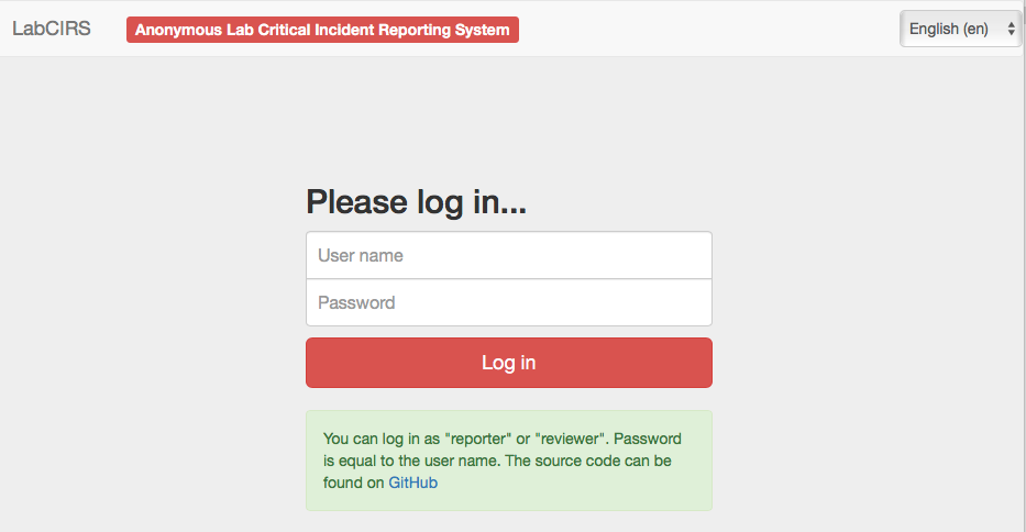
Our first version of an error reporting system was paper based, just a form sheet on our pinboard. While this form already covered all necessary questions, it was almost completely ignored by our staff. Trying to understand the reasons, we found out that the paper version was not convenient and more importantly, not confidential enough. Our colleagues were worried that the reported incident could reveal their identity and may put their position at risk. Acknowledging this obstacle, the idea was born to use an online tool, which does not require user specific information, nor requiring or logging any personal information. Since there was no out of the box system available, which met out needs (anonymous, browser based, structured, but not as complex as a medical CIRS), Sebastian Major, a member of our department designed the LabCIRS from the scratch. The source code and a documentation can be found at github.
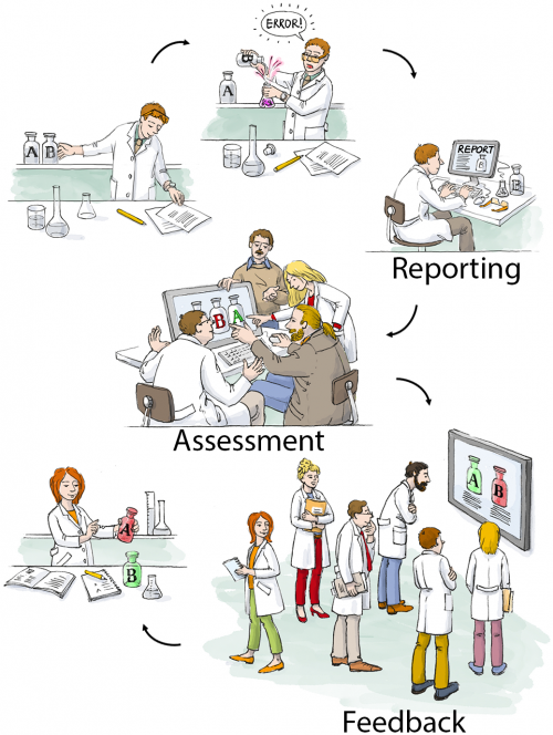
After some fine tuning and beta testing, LabCIRS went online at the end of 2013. This is how it works for the researcher (student, technician, postdoc, etc.): First, one has to log in with a shared login for all department members which does not point to a single user. The system is bilingual (English /German), so depending on their preferences, users can choose the language they feel more comfortable with. After login, the user sees all formerly published incidents and can either search and read through them or report a new incident.
While reporting an incident the reporter can assign a date, describe what happened, and if make suggestions on how to avoid the event in the future. In addition, images can be uploaded, and as a last step, the reporter is asked if the reported incident should be available for all users of the LabCIRS, or reported just to responsible personel.
The reported incident is then checked by a “reviewer” who has privileged access to LabCIRS. This reviewer translates the reported incident and makes sure no personal information is reported. In a next step, the reported incident is internally discussed in our monthly quality meeting with the focus on how the avoid a recurrence of the same incident in the future. Then, if the reporter agreed, it is published inside the LabCIRS, via mail and in addition reported at one of our weekly department meetings.
While it is of course desirable to make all reported incidents available to the public, we found it important to leave this decision to the reporter.
At the very beginning, only about half of the reported entries were cleared by the reporter to be openly published in the department. Over time, however, when it became evident to the reporters that reporting is appreciated and the idea is not to blame anyone, but to learn from mistakes and, if possible, to avoid them in the future, the mindset slowly changed. For more than one year now, all of the reported incidents are cleared for publication. This demonstrates a change in error culture, away from hiding mistakes towards an open discussion and prevention.
Clearly, it is not software which makes people change their minds about quality and error culture, but for us, the LabCIRS was and is a helpful tool helping us in this process.
If you would like to give it try for yourself to check out if this could be useful for you as well, feel free to check it out under http://labcirs.charite.de and download it (or even commit something) at github.
You can find a more detailed report on our LabCIRS in our publication in PLOS Biology.
 (No Ratings Yet)
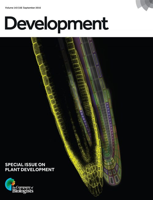


 (1 votes)