[Editorial from Development’s latest Special Issue ‘Uncovering Developmental Diversity’, edited by Cassandra Extavour, Liam Dolan and Karen Sears.]
Scanning through early issues of the Journal of Embryology and Experimental Biology (the previous name for this journal) reveals the diverse range of organisms that were investigated by developmental biologists in the 1950s and 1960s. However, the rise of molecular genetics in subsequent decades led to a narrowing in species choice to a small repertoire of well-characterised model organisms for which there were genetic tools for functional experimentation. In recent years, however, technological advances, including genome and transcriptome sequencing, flexible genome-editing approaches and high-resolution four-dimensional imaging, provide an opportunity to once again study developmental questions in organisms across all kingdoms of life. Given the current global challenges of climate change and biodiversity loss, it is particularly important that we turn our attention to understanding development in an unstable world.
This important topic was the basis of Development’s Journal Meeting, ‘Unconventional and Emerging Experimental Organisms in Cell and Developmental Biology’ in 2023, which you can learn more about in the Meeting Review published here (Lemke et al., 2024). Fuelled by the success of the meeting, we chose to focus this Special Issue, led by Academic Editor Cassandra Extavour, together with Liam Dolan and Karen Sears as Guest Editors, on a related topic: Uncovering Developmental Diversity. We are particularly delighted that multiple attendees from our meeting have contributed both research and review-type articles to this issue.
The 28 research papers in this Special Issue highlight 32 different organisms from across the multicellular tree of life, featuring cnidarians (Garschall et al., 2024), insects (Matsuda et al., 2024; Bai et al., 2024; Beaven et al., 2024; Pallarès-Albanell et al., 2024) and annelids (Bideau et al., 2024), as well as echinoderms (Barone et al., 2024; McDonald et al., 2024; Jackson et al., 2024; Clarke et al., 2024) and chordates (Gigante et al., 2024; Johnson et al., 2024), including vertebrates (Rees et al., 2024; Pérez-Gómez et al., 2024), many of which are various fishes (Leclercq et al., 2024; Li et al., 2024; Woronowicz et al., 2024; Peloggia et al., 2024; Jin et al., 2024). These articles demonstrate the importance of finding the best model to address a developmental question, such as making use of the curved epithelium in the sea star embryo to investigate cell organisation and packing (Barone et al., 2024) or using the regenerative capacity of annelids to learn more about cell plasticity (Bideau et al., 2024). In addition to annelids, a Perspective in this issue highlights five more ‘extraordinary’ model systems for regeneration across scales from single cells to whole organisms (Accorsi et al., 2024).
Not limited to animals, the Special Issue also embraces a wide array of studies uncovering fundamental developmental processes, such as axis formation and organogenesis in photosynthetic organisms, including brown algae (Vigneau et al., 2024; Boscq et al., 2024), liverworts (Attrill and Dolan, 2024; Attrill et al., 2024; Sakai et al., 2024), and vascular plants such as ferns (Woudenberg et al., 2024) and angiosperms (Mody et al., 2024; Spiegelhalder et al., 2024). Photosynthetic organisms feature heavily in the issue’s review-type content, too, with articles describing how brown algae can inform us about the transition to multicellularity (Batista et al., 2024), how the environment and climate change influence development through the lens of stomata (Chua and Lau, 2024) and what we can learn about the evolution of plant development through the fossil record (Hetherington, 2024).
The evo-devo field, in particular, has benefitted from the appreciation of biodiversity and increased taxonomic sampling. Reflecting this, two Reviews discuss fundamental evolutionary concepts, including how phenotypes can be maintained by different underlying genetic architecture through developmental systems drift (McColgan and DiFrisco, 2024), as well as a cautionary tale of how reports on the low-hanging fruit of simple genetic explanations of evolution should not change our perception that evolution is inherently complex (Cooper, 2024).
A broad selection of available organisms also allows the study of rare evolutionary innovations, such as the ability of Nematostella to degrow in response to food availability (Garschall et al., 2024) or of teleost fish to adapt ionocyte differentiation to regulate osmotic levels within aquatic environments (Peloggia et al., 2024). Adaptive plasticity is also the focus of a Review article describing how organisms assess environmental cues across scales and respond via phenotypic changes (Hill et al., 2024). Furthermore, capturing developmental biodiversity furthers our understanding of complex life cycles (McDonald et al., 2024; Peloggia et al., 2024) – a topic motivating a Hypothesis for unravelling cellular rejuvenation (Berger, 2024). Indeed, studying organisms with metamorphic life cycles allows us to learn about the intrinsic developmental process, such as how the rhinoceros beetle remodels its horn (Matsuda et al., 2024) or neuronal cell survival in Ciona (Gigante et al., 2024).
Importantly, research using emerging model systems relies on new tools that facilitate functional experiments. Our Techniques and Resources section features methods for the delivery of proteins and nucleic acids into oocytes in a variety of species (Clarke et al., 2024), as well as approaches for generating stable genetic lines (Jackson et al., 2024) and tools for quantifying diversity (Mody et al., 2024). However, not all species are amenable to being cultured in the lab, and a Perspective describes the importance of fieldwork for developmental biology in unconventional model systems (Brown et al., 2024). In addition, a Spotlight article describes how modern innovations in stem cell technology might be employed for species conservation (Hutchinson et al., 2024), highlighting how understanding biodiversity is the first step to its preservation, an increasingly prevalent topic in the context of climate change.
Overall, we hope that this issue demonstrates both how technological advances have made it possible to understand development and regeneration in previously intractable organisms, as well as the importance of this pursuit. We continue to ensure Development is an appropriate home for your studies in developmental biology, stem cells and regeneration using any organism. We welcome your submissions.
Maria Victoria Serrano, Stephanie Cottier, Lianzijun Wang, Sergio Moreira-Antepara, Anthony Nzessi, Zhiyu Liu, Byron Williams, Myeongwoo Lee, Roger Schneiter, Jun Liu
Aaron M. Savage, Alexandra C. Wagner, Ryan T. Kim, Paul Gilbert, Hani D. Singer, Erica Chen, Elane M. Kim, Noah Lopez, Kelly E. Dooling, Julia C. Paoli, S.Y. Celeste Wu, Sebastian Bohm, Rachna Chilambi, Tim Froitzheim, Steven J. Blair, Connor Powell, Adnan Abouelela, Anna G. Luong, Kara N. Thornton, Benjamin Tajer, Duygu Payzin-Dogru, Jessica L. Whited
Peggy P. Hsu, Ansley S. Conchola, Tristan Frum, Xiangning Dong, Lila Tudrick, Varun Ponnusamy, Michael S. Downey, Manqi Wu, Mengkun Yang, Yusoo Lee, Emma Niestroy, Yu-Hwai Tsai, Angeline Wu, Sha Huang, Ian A. Glass, Sofia D. Merajver, Jason R. Spence
Ling S. Loh, Joseph J. Hanly, Alexander Carter, Martik Chatterjee, Martina Tsimba, Donya N. Shodja, Luca Livraghi, Christopher R. Day, Robert D. Reed, W. Owen McMillan, Gregory A. Wray, Arnaud Martin
Tim Ott, Amelie Brugger, Emmanuelle Szenker-Ravi, Yvonne Kurrle, Olivia Aberle, Matthias Tisler, Martin Blum, Sandra Whalen, Patrice Bouvagnet, Bruno Reversade, Axel Schweickert
Yuchen Liu, Tianli Qin, Xin Weng, Bernice Leung, Karl Kam Hei So, Boshi Wang, Wanying Feng, Alexander Marsolais, Sheena Josselyn, Pingbo Huang, Bernd Fritzsch, Chi-Chung Hui, Mai Har Sham
Qiao Wu, Jian Zhang, Bing Long, Xiao Hu, Bruna Mafra de Faria, Stephen Maxwell Scalf, Kutay Karatepe, Wenxiang Cao, Nikolaos Tsopoulidis, Andres Binkercosen, Masaki Yagi, Aaron Weiner, Mary Kaileh, Enrique M. De La Cruz, Ananda L Roy, Konrad Hochedlinger, Shangqin Guo
Stephen Spurgin, Ange Michelle Nguimtsop, Fatima N. Chaudhry, Sylvia N. Michki, Jocelynda Salvador, M. Luisa Iruela-Arispe, Jarod A. Zepp, Saikat Mukhopadhyay, Ondine Cleaver
Sera Lotte Weevers, Alistair D. Falconer, Moritz Mercker, Hajar Sadeghi, Jaroslav Ferenc, Albrecht Ott, Dietmar B. Oelz, Anna Marciniak-Czochra, Charisios D. Tsiairis
Theopi Rados, Olivia S. Leland, Pedro Escudeiro, John Mallon, Katherine Andre, Ido Caspy, Andriko von Kügelgen, Elad Stolovicki, Sinead Nguyen, Inés Lucía Patop, Thiberio Rangel, Sebastian Kadener, Lars D. Renner, Vera Thiel, Yoav Soen, Tanmay A.M. Bharat, Vikram Alva, Alex Bisson
Nora Ditzer, Ezgi Senoglu, Theresa M. Schütze, Aikaterina Nikolaidi, Annika Kolodziejczyk, Katrin Sameith, Sevina Dietz, Razvan P. Derihaci, Cahit Birdir, Anne Eugster, Mike O. Karl, Andreas Dahl, Pauline Wimberger, Franziska Baenke, Claudia Peitzsch, Mareike Albert
Audrey J. Marsh, Sergei Pirogov, Abby J. Ruffridge, Suresh Sajwan, Tyler J. Gibson, George Hunt, Yadwinder Kaur, Melissa M. Harrison, Mattias Mannervik
Dimitris Botskaris, Ioannis K. Deligiannis, Ioanna Peraki, Haroula Kontaki, Marianna Stagaki, Matthieu D. Lavigne, Celia P. Martinez-Jimenez, Iannis Talianidis
Surbhi Sood, Aktan Alpsoy, Guanming Jiao, Alisha Dhiman, Charles Samuel King, Gabriella Grace Conjelko, Judy E. Hallett, Sagar M Utturkar, Jill E Hutchcroft, Emily C Dykhuizen
Olga M. Sigalova, Mattia Forneris, Frosina Stojanovska, Bingqing Zhao, Rebecca R. Viales, Adam Rabinowitz, Fayrouz Hamal, Benoît Ballester, Judith B Zaugg, Eileen E.M. Furlong
Yuliia Haluza, Joseph A. Zoller, Ake T. Lu, Hannah E. Walters, Martina Lachnit, Robert Lowe, Amin Haghani, Robert T. Brooke, Naomi Park, Maximina H. Yun, Steve Horvath
Rita Manco, Camilla Moliterni, Gauthier Neirynck, Maxime De Rudder, Corinne Picalausa, Leana Ducor, Montserrat Fraga, Frédéric Lemaigre, Christine Sempoux, Alexandra Dili, Isabelle A. Leclercq
Yingnan Lei, Mai Chi Duong, Nuša Krivec, Charlotte Janssens, Marius Regin, Anfien Huyghebaert, Edouard Couvreu de Deckersberg, Karen Sermon, Diana Al Delbany, Claudia Spits
Julian Weihs, Fatima Baldo, Alessandra Cardinali, Gehad Youssef, Katarzyna Ludwik, Harald Stachelscheid, Nils Haep, Peter Tang, Igor Sauer, Pavitra Kumar, Cornelius Engelmann, Susanna Quach, Philip Bufler, Namshik Han, Milad Rezvani
Milad Rezvani, Kyle Lewis, Susanna Quach, Kentaro Iwasawa, Julian Weihs, Hasan Al Reza, Yuqi Cai, Masaki Kimura, RanRan Zhang, Yuka Milton, Praneet Chaturvedi, Konrad Thorner, Ramesh C. Nayak, Jorge Munera, Phillip Kramer, Brian R. Davis, Appakalai N. Balamurugan, Yeni Ait Ahmed, Marcel Finke, Rose Yinghan Behncke, Adrien Guillot, René Hägerling, Julia K. Polansky, Philip Bufler, Jose A Cancelas, James M. Wells, Momoko Yoshimoto, Takanori Takebe
B. Pardo-Rodríguez, A.M. Baraibar, I. Manero-Roig, J. Luzuriaga, J. Salvador-Moya, Y. Polo, R. Basanta-Torres, F. Unda, S. Mato, G. Ibarretxe, J.R. Pineda
Sara Cannavò, Chiara Paleni, Alma Costarelli, Maria Cristina Valeri, Martina Cerri, Antonietta Saccomanno, Veronica Gregis, Graziella Chini Zittelli, Petre I. Dobrev, Lara Reale, Martin M. Kater, Francesco Paolocci
Alicia Tovar, Scott Monahan, Trevor Mugoya, Adrian Kristan, Walker Welch, Ryan Dettmers, Camila Arce, Theresa Buck, Michele Ruben, Alexander Rothenberg, Roxane Saisho, Ryan Cartmill, Timothy Skaggs, Robert Reyes, MJ Lee, John Obrycki, William Kristan, Arun Sethuraman
Haidong Yan, John P. Mendieta, Xuan Zhang, Alexandre P. Marand, Yan Liang, Ziliang Luo, Mark A.A. Minow, Hosung Jang, Xiang Li, Thomas Roulé, Doris Wagner, Xiaoyu Tu, Yonghong Wang, Daiquan Jiang, Silin Zhong, Linkai Huang, Susan R. Wessler, Robert J. Schmitz
Magdalena Schindler, Christian Feregrino, Silvia Aldrovandi, Bai-Wei Lo, Anna A. Monaco, Alessa R. Ringel, Ariadna Morales, Tobias Zehnder, Rose Yinghan Behncke, Juliane Glaser, Alexander Barclay, Guillaume Andrey, Bjørt K. Kragesteen, René Hägerling, Stefan Haas, Martin Vingron, Igor Ulitsky, Marc Marti-Renom, Julio Hechavarria, Nicolas Fasel, Michael Hiller, Darío Lupiáñez, Stefan Mundlos, Francisca M. Real
Cristofer Calvo, Casey O. Swoboda, Fabian Montecino Morales, Siddhant Nagar, Michael J. Petrany, Chengyi Sun, Hima Bindu Durumutla, Mattia Quattrocelli, Douglas P. Millay
Ismael Moreno-Sanchez, Luis Hernandez-Huertas, Daniel Nahon-Cano, Carlos Gomez-Marin, Pedro Manuel Martinez-García, Anthony J. Treichel, Laura Tomas-Gallardo, Gabriel da Silva Pescador, Gopal Kushawah, Alejandro Díaz-Moscoso, Alejandra Cano-Ruiz, John A. Walker II, Manuel J. Muñoz, Kevin Holden, Joan Galcerán, María Ángela Nieto, Ariel Bazzini, Miguel A. Moreno-Mateos
Sara Di Carlo, Adrian Salas-Bastos, Mariela Castelblanco Castelblanco, Muriel Auberson, Marie Rumpler, Malaury Tournier, Lukas Sommer, Olaia Naveiras, Edith Hummler
Ália dos Santos, Oliver Knowles, Tom Dendooven, Thomas Hale, Alister Burt, Piotr Kolata, Giuseppe Cannone, Dom Bellini, David Barford, Matteo Allegretti
Daniel Medina-Cano, Mohammed T. Islam, Veronika Petrova, Sanjana Dixit, Zerina Balic, Marty G. Yang, Matthias Stadtfeld, Emily S. Wong, Thomas Vierbuchen
Brian Ho Ching Chan, Holly Hardy, Teresa Requena, Amy Findlay, Jason Ioannidis, Dominique Meunier, Maria Toms, Mariya Moosajee, Anna Raper, Mike McGrew, Joe Rainger
Guilherme E. Kundlatsch, Alina S. L. Rodrigues, Vitória F. B. Zocca, Laura A. S. Amorim, Gabriela B. de Paiva, Almiro P. S. Neto, Juliana A. D. B. Campos, Danielle B. Pedrolli
The 30 October 2024 Development presents… webinar was chaired by Development’s Executive Editor, Katherine Brown and featured three talks on the topic of the development of ectoderm derivatives. Catch up on the talks below.
During my time as a summer student at the Francis Crick Institute, I had the privilege of working in the Developmental Signalling Laboratory of Dr Caroline Hill. Under the mentorship of Dr Berta Font Cunill, I have gained an insight into the realities of cutting-edge scientific research and was able to contribute to experiments advancing the understanding of developmental biology.
Throughout embryonic development, as cells divide, they begin to specialise to later form diverse functional tissues. This is possible because, even though progenitor cells contain copies of the same genetic code, they express different sets of genes. These gene expression patterns are governed by various complex signalling pathways, which ultimately determine cell fate. The Hill Lab is interested in understanding how cells use specific signals to communicate with each other and their environment to drive the development of an organism and tissue specialisation. During gastrulation, this cellular communication results in the embryonic stem cells transforming into three distinct germ layers: endoderm, mesoderm, and ectoderm. As their subsequent patterning generates all future body structures, this process sets the stage for the functioning of the entire organism (Richardson et al., 2023).
In my research project, I was specifically interested in the mechanism of cell differentiation into definitive endoderm. This germ layer gives rise to the lungs, bladder, the majority of the digestive tract, as well as vital endocrine organs like the pancreas and thyroid (Fang & Li, 2022). Understanding the signalling pathways involved in endoderm differentiation is necessary to generate novel therapeutic solutions to diseases associated with endoderm-derived tissues, such as diabetes. This is possible by using human induced pluripotent stem cells (iPSCs) to generate endodermal cells, and possibly their derivatives, in vitro (Fang & Li, 2022).
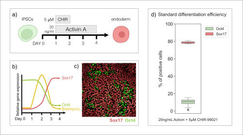
To study the differentiation to endoderm, I cultured iPSCs for four days in differentiation media with addition of CHIR-99021 (5 µM) for 24h, and Activin A (20 ng/mL) for 72h, following a standard protocol (Fig. 1a). Based on previous publications (Diekmann and Naujok, 2015), I expected that throughout this process, cells would follow certain patterns of gene expression (Fig. 1b). After 72h of differentiation (Day 4), I fixed and stained the cells with antibodies recognising key markers of pluripotency (Oct4) and endoderm (Sox17). I imaged the cells (Fig. 1c), quantified the results and was able to establish that the protocol of interest results in about 80% of stem cells differentiating to endoderm cells (Fig. 1d), confirming what has been observed in published articles.
Fig. 1 Standard differentiation efficiency from iPSCs to endoderm cells in vitro. (a) Schematic of the 4-day protocol used for differentiating pluripotent stem cells to endoderm cells. CHIR99021 functions as a Wnt pathway activator. Wnt signalling is required to reduce cell pluripotency, and promote mesendodermal (TBXT, EOMES) differentiation (Zhao et al., 2019). Activin A, a member of the TGF-β superfamily, is an activation factor for the Nodal pathway. High Nodal signalling gradient leads to further differentiation into endoderm (SOX17, GSC) (Richardson et al., 2023 ; Silva et al., 2022). (b) Predicted patterns for the expression of markers (Sox17, Oct4, Brachyury) throughout the differentiation process (Diekmann and Naujok, 2015). (c) Nuclei of cells fixed after carrying out the standard protocol (a), stained with Oct4 (green) and Sox17 (red) antibodies. Imaged using confocal microscopy. (d) Quantified results (n=8) of the percentage of differentiated cells expressing Sox17 (red) vs cells remaining pluripotent and expressing Oct4 (green).
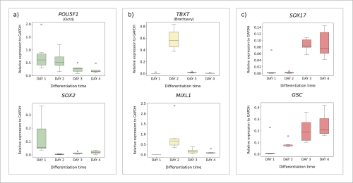
Having established the standard differentiation protocol, I wanted to understand the process itself in more detail. I set out to investigate the gene expression patterns throughout the transition of cells from pluripotency to endoderm. I collected cell samples on each day of the protocol. After extracting RNA, I synthesised cDNA to be used in quantitative polymerase chain reaction (qPCR) analysis. qPCR allows for the amplification of target DNA sequences, with simultaneous quantification of their concentration throughout the process. Thus, I was able to obtain and visualise the levels of expression of endoderm differentiation marker genes on each day of the protocol (Fig. 2). I was able to prove that cells transitioning from pluripotency to endoderm follow predicted patterns of gene expression (Fig. 1b). Pluripotency genes (POU5F1, SOX2) gradually decline as the differentiation continues (Fig. 2a), while endoderm markers (SOX17, GSC) are expressed more substantially towards the end of the process (Fig. 2c). I was also able to confirm that the cells go through an intermediate stage, with mesendoderm genes (TBXT, MIXL1) being expressed transiently on Day 2 (Fig. 2b).
Fig. 2 Relative expression of marker genes throughout differentiation of iPSCs to endoderm. (n=3) Expression levels of all genes of interest were normalised to that of a housekeeping gene GAPDH. Obtained results have been quantified and visualised using Python. (a) Relative expression levels of pluripotent marker genes POU5F1 and SOX2. (b) Relative expression levels of gene markers (TBXT and MIXL1) for the intermediate stage of mesendoderm, peaking at day 2. (c) Relative expression levels of endoderm marker genes SOX17 and GSC.
To better understand the signalling pathways involved in the process of cell differentiation to endoderm, Dr Berta Font Cunill screened a library of over 1,000 small molecules of diverse molecular structure that could possibly affect the process by interacting with proteins important for endoderm differentiation. She identified one small molecule (compound “953”) that increases the differentiation efficiency to endoderm (from 80 to 90% approximately).
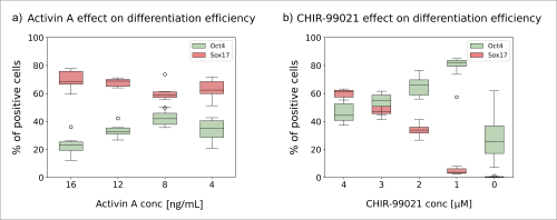
As I previously mentioned, after following the standard protocol the rate of differentiation to endoderm cells reaches about 80% (Fig. 1d). This high efficiency leaves a small margin for improvement. My goal was to modify the standard differentiation protocol to achieve a lower differentiation efficiency, and therefore increase the margin for improvement upon addition of compound “953”. I seeded pluripotent cells in media with varying concentrations of CHIR-99021 and Activin A (Fig. 3) and carried out the differentiation protocol for three days. I observed that even when the amount of Activin A was lowered from 20 ng/mL to 4 ng/mL, the differentiation proceeded only with minor changes in efficiency (Fig. 3a). However, lowering the amount of CHIR-99021 in just 1 µM increments hindered the process considerably (Fig. 3b). When CHIR-99021 was not present at all, most cells didn’t survive. These results show that CHIR-99021 is vital for endoderm differentiation.
Fig. 3 The effect of Activin A and CHIR-99021 concentration on differentiation efficiency. (n=8) The differentiation efficiency was measured on Day 3 based on the levels of expression of pluripotent (Oct4, green) and endodermal (Sox 17, red) marker genes. Obtained results were quantified and visualised using Python. (a) Pluripotent cells were cultured in conditions dictated by the standard protocol but with varying concentrations of Activin A. (b) Pluripotent cells were cultured in conditions dictated by the standard protocol but with varying concentrations of CHIR-99021.
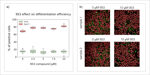
Based on the obtained results, I decided that the best condition to test the effect of compound “953” was 20 ng/mL of Activin A (48h), and 3 µM of CHIR-99021 (24h), which resulted in around 50% of differentiation efficiency, leaving a large margin for improvement. I cultured the cells for three days using these new conditions and different concentrations of compound “953”, after which I stained for Oct4 and Sox17 (Fig. 4). I observed that cells grown with 10 µM of compound “953” in the media, reached 10-15% higher differentiation efficiency than those grown without the compound (Fig. 4a). However, the final total number of cells was lower than in control groups (Fig. 4b). These results suggest that while molecule “953” pushes differentiation from pluripotency into endoderm cells, it is also possibly mildly toxic or hinders cell proliferation. The results also suggest that even if differentiation is hindered, compound “953” is not able to increase the differentiation efficiency beyond 10-15%.
Fig. 4 The effect of compound “953” on differentiation efficiency. (n=8) (a) boxplots representing quantified results of differentiation efficiency for varying concentrations of “953”. (b) Nuclei of cells fixed after carrying out the modified protocol, stained with Oct4 (green) and Sox17 (red) antibodies. Imaged using confocal microscopy. The represented samples suggest that while the amount of pluripotent cells (green) is lower when 10 µM of “953” is present in the growth media, and therefore the differentiation rates are higher, the observed total number of cells is lower than in control groups.
The future of this project will focus on identifying the protein impacted by compound “953” through extensive proteomic analysis. This will allow for a better understanding of the signalling pathways involved in cell differentiation to definitive endoderm, which is a necessary step for the successful differentiation of downstream endoderm-derived tissues and organs to develop novel solutions in regenerative medicine. I am incredibly grateful for the opportunity to contribute to such inspiring scientific advancements. It has been an honour to be supported by the Rosa Beddington Fund. This experience has been a defining moment for my academic and professional development, and I have made the decision to pursue similar research through a PhD studentship and the rest of my scientific career. I would like to thank the Hill Lab, where I had the pleasure of working with incredible scientists, for their support and for welcoming me as a valued team member. I am especially grateful for the guidance and expertise of my supervisor, Dr Berta Font Cunill.
SOURCES
Diekmann, U., Naujok, O. (2015). Generation and Purification of Definitive Endoderm Cells Generated from Pluripotent Stem Cells. Methods in Molecular Biology, 1341, 157-72. https://doi.org/10.1007/7651_2015_220
Fang, Y., Li, X. (2022). Metabolic and epigenetic regulation of endoderm differentiation. Trends in Cell Biology, 32(2), 151-164. https://doi.org/10.1016/j.tcb.2021.09.002
Richardson, L., Wilcockson, S.G., Guglielmi, L. et al. (2023). Context-dependent TGFβ family signalling in cell fate regulation. Nature Reviews Molecular Cell Biology, 24, 876–894. https://doi.org/10.1038/s41580-023-00638-3
Silva, I.B.B., Kimura, C.H., Colantoni, V.P. et al. (2022). Stem cells differentiation into insulin-producing cells: recent advances and current challenges. Stem Cell Research & Therapy, 13, 309. https://doi.org/10.1186/s13287-022-02977-y
Zhao, M., Tang, Y., Zhou, Y. et al. (2019). Deciphering Role of Wnt Signalling in Cardiac Mesoderm and Cardiomyocyte Differentiation from Human iPSCs: Four-dimensional control of Wnt pathway for hiPSC-CMs differentiation. Nature Scientific Reports, 9, 19389. https://doi.org/10.1038/s41598-019-55620-x
[This post is co-written by Joan-Josep Soto Angel and Pawel Burkhardt.]
Timelapse showing reverse development in a lobectomized individual of M. leidyi. Note the progressive reduction in size and reabsorption of lobes and auricles typical of the lobate phase (absent on Day 41), followed by a normal cydippid morphology, showing long, functional tentacles (Day 48). https://doi.org/10.1073/pnas.2411499121
What is this?
The video depicts the process of reverse development in the ctenophore Mnemiopsis leidyi over several weeks. Adult and larval M. leidyi are anatomically different: lobate adults have lobes and auricles that are not yet developed in the larval stage. Cydippid larvae have a rounded body and tentacles that get reabsorbed during the lobate adult stage. The timelapse video shows an adult comb jelly slowly transitioning to a larval form over time, with lobes and auricles disappearing, and tentacles being regained. This is a process called reverse development.
Where can this be found?
This footage was obtained in our Ctenophore Facility at the University of Bergen, under controlled laboratory conditions, and following the same individual over time. Whether or not these comb jellies are equally capable of doing this in the ocean still remains a mystery, but the potential is definitely there!
The timelapse is made out of 24 individual pictures, each taken every 2-3 days, and shows the animal in the same position for comparison purposes. We used a Canon 5D Mark IV coupled to a Canon MP-E 65mm f/2.8 1-5x Macro Photo, usually known as an extreme macro lens. As the animals are very transparent, and the details are difficult to observe, we used a black background and added an extra light source from the side using a Canon speedlite strobe.
What causes M. leidyi to reverse develop?
Reverse development in M. leidyi is triggered by improved environmental conditions after a period of stress. Stress was simulated by either removing the lobes (lobectomy) or by prolonged starvation. Mnemiopsis leidyi is able to efficiently regenerate any missing body part, as well as shrinking considerably when starved. However, when adequately fed after shrinking to a size of just a few millimetres, instead of growing back the lobes, they grew tentacles typical of the larval stage.
Why should people care about this?
So far, reverse development was thought to be restricted to a few cnidarian species and one cestode. Our study is the first to report the occurrence of this peculiar feature in ctenophores, suggesting that reverse development may be more widespread than previously thought. The occurrence of reverse development in a lineage that originated prior to cnidarians can help to better understand central aspects of life cycle plasticity and evolution in early animals. The ability to rejuvenate in harsh conditions also provides further research opportunities for ecological studies aiming to explain, among others, the high invasive success of this comb jelly. Our study highlights Mnemiopsis leidyi as a potential model species to study life cycle plasticity, aging and rejuvenation.
How would you explain this to an 8-year-old?
Aging is a one-way route for a great majority of animals. However, there are a few that seem to be able to escape the fate of getting old. An adult transforming into a baby was only known for a species of jellyfish, usually called the immortal jellyfish (its scientific name is Turritopsis dorhnii). We found this capacity in an entirely new group of animals: the comb jellies. When Mnemiopsis leidyi (also known by its common name as sea walnut) becomes an adult, it grows two lobes and four pointy finger-like structures called auricles that they use for feeding. Baby sea walnuts do not have these body parts. Instead, they use two long tentacles to trap their prey and direct it to the mouth. The tentacles are lost once these animals reach their final adult form. We discovered that adult sea walnuts can rejuvenate when they eat properly after going through a period of stress.
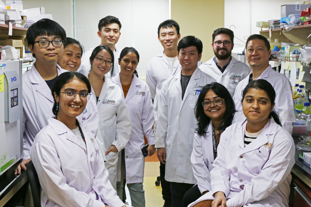
Joe: Our lab aims to understand how developmental mechanics regulates mammalian folliculogenesis and ovarian dynamics. We focus on three overarching themes: First, how mechanical signals such as tissue pressure and hydraulics impact folliculogenesis and oocyte growth. Second, how the mechanical environment around the follicles influence collective dynamics of follicles and ovarian functions. Third, we are interested to understand how changes in tissue mechanics and misregulated mechano-signaling impacts follicle functions and ovulation during ageing and infertility. To address these questions, we develop multidisciplinary approaches based on novel (bio)mechanics, (bio)photonics and (bio)physical tools, using ex vivo assays.
Group photo of the lab
Can you give us a lab roll call?
Arikta Biswas: I am a postdoctoral research fellow: I study how mechanical interactions are generated, controlled, and transmitted within ovarian follicles at the secondary stage of development using mice as model systems.
Kim Whye Leong: I’m a postdoctoral fellow investigating the process of antrum formation and how its mechanochemical functions shape ovarian follicle development. Using advanced 3D microscopy (my favorite technique) and cutting-edge tools for force measurement and manipulation, I’m exploring the forces and dynamics that drive this crucial stage of development.
Jake Turley: I am a postdoc in the lab and I work on the biophysics of ovulation combining advanced microscopy, biomechanical tools and machine learning.
Huan Ting Ong: I am a Postdoctoral Research Fellow co-advised by Jennifer Young and Joe, studying mechanoregulation during ovarian ageing driven by the extracellular matrix and stroma.
Boon Heng Ng: A fourth-year student, trying to study how ovarian theca cells could sense mechanical cues and contribute to ovarian follicle development.
Kosei Tomida: I am a second year PhD student in the lab and I am studying follicle-follicle interactions through mechanochemical feedback interactions.
Kelly Tan: I am an incoming first-year graduate student, hoping to investigate a cool question on how the somatic (granulosa) cells contribute mechanically to follicle growth, apart from its classical signalling pathways.
Apoorva Shivankar: I am a Research Assistant in the Chan Lab group. In addition to my administrative duties, I have the opportunity to engage in research. I’m currently investigating the functional and mechanical differences between the immune cells in young and aged ovaries.
Le Mai Tan Dat (Daniel): I’m an undergraduate student doing a semester-long research project, mentored by Jake. We are studying the biophysics of ovulation, applying machine learning to quantify tissue dynamics.
Favourite technique, and why?
Joe: I adore any new biophysical tools that allow us to gain new insights into tissue mechanobiology. One of my favourite tools is micropressure probe, which allows us to measure and manipulate fluid or cytoplasmic pressure in cells and tissues. I am excited to expands its use to study hydraulic control in development, physiology, disease and ageing.
Apart from your own research, what are you most excited about in developmental and stem cell biology?
Joe: I am a firm believer that new technology often leads to new discoveries, so any biophysical approaches to probe mechanics in living tissues excite me! I also think the application of machine learning approach will not only help us gain quantitative understanding of tissue dynamics during development, it also harbours great potential to uncover new physics regulating biological processes. I am particularly excited about ongoing work to apply these approaches to address ageing and diseases.
How do you approach managing your group and all the different tasks required in your job?
Joe: As an assistant professor, it can indeed be daunting to handle multiple tasks like teaching, research and administrative duties. I hold regular group and 1-1 meetings with lab members to stay connected with their projects and give timely feedback. Given the highly interdisciplinary nature of our research, I do my best to foster exchange amongst lab members, often teaming them up in joint projects, while maintaining autonomy in their own projects. Recently I realised that too many back-to-back meetings can negatively impact the quality of meetings, and moving on I might try to cut down non-essential meetings. I also work on annual individual development plans with my team, which is a great platform to provide mutual feedback. This also helps to keep track of what’s going on at the ground and calibrate my management style. I recently shared this in more detail in a People & Ideas article in Journal of Cell Biology.
Something I wish to do, but haven’t got a chance to, is to hold an annual lab retreat or a joint retreat with other labs – those are great opportunities to share blue-sky research ideas and learn from each other!
What is the best thing about where you work?
Joe: I am super grateful to have a family-like lab-mosphere! The various core facilities and admin team at MBI are doing an amazing job in supporting the research. Being at Singapore is unique in the sense that we can serve as a focal point in Asia pacific for scientific exchange and collaborations with people from Japan, China, Hong Kong and Australia.
Arikta: It is not often that researchers have dedicated core facilities who work tirelessly and silently ‘behind-the-scenes’ to make our science go smoother, but it is the case here and I am very appreciative of their strong yet quiet efforts. Also, I thoroughly enjoy the freedom to think and brainstorm ideas with my lab-mates without hesitation.
Jake: My labmates are very supportive and generous with their time and help. This allowed me to quickly settle in and it also makes working in the lab a more enjoyable experience.
Kim Whye: The best thing about where I work is the collaborative atmosphere. Everyone, from researchers to staff, is incredibly supportive, and it fosters a real sense of teamwork. We also have a lot of microscopes and cutting-edge biophysical tools! It’s exciting to work in a place where you’re empowered to push the boundaries of what’s possible.
Huan Ting: The multidisciplinary environment the institute has, which encourages collaborations and cross-fertilisation of new ideas!
Boon Heng: Everyone is very supportive and collaborative with the different projects! If we are unsure about anything, we never hesitate to consult anyone in the lab. We also try to have lunch together every day, like a ‘family’ daily lunch?
Kosei: I appreciate the availability of resources in core facilities, items, and people from different backgrounds. I was lucky to have been blessed with a wonderful mentor.
Kelly: I really like how the lab fosters a welcoming environment for anyone to ask all sorts of questions, and to suggest unconventional (crazy) ideas. Everyone is more-than-merely-willing to help one another, and there has been no shortage of encouragement (and laughter) to enjoy pursuing great science even amidst the most stressful and busiest of days.
Apoorva: To me the four cornerstones of our lab are the 1) freedom to explore topics, 2) supportive and friendly atmosphere, 3) access to cutting-edge techniques, and 4) guidance from supportive colleagues.
Daniel: I enjoy the conversations that spontaneously arise, especially in the weekly lab meeting. Sometimes, we just share about our projects and scientific discussions will follow. Everyone is very experienced and receptive to comments and opinions, making it a great opportunity for me to learn more about the sciences.
What’s there to do outside of the lab?
Joe: I enjoy hiking and exploring the beautiful nature reserves across the island with my family. After sending kid to school in the early morning, I cherish the quiet moments where I get to hang out at the coffee shop in the hawker center to do some personal reading. Watching sci-fi movies (even better, combined with horror elements like Alien series), is also one of my favourite hobbies.
Arikta: I love reading outside of work, mostly fiction though, and spend an incredibly large amount of my free time at the Kinokuniya bookstore. When I am not strolling through the aisles of books, I am usually watching any Southeast Asian content on the internet; and then messaging my lab members to bug them about the accuracy of the cultures being represented onscreen.
Jake: I mainly enjoy eating out at the massive variety of food places available in Singapore and watching live gigs.
Kim Whye: When I’m not deep in follicle research, I enjoy reading up on bottom-up approaches in synthetic developmental biology and staying on top of the latest biotech innovations. Singapore may not be renowned for its pub culture (though we do indulge occasionally), but what we lack in that area, we more than make up for with our incredible food scene. There’s always something new and delicious waiting to be discovered.
Huan Ting: Despite the hot and humid weather, Singapore has plenty of natural trails featuring the flora and fauna unique to the tropical regions, just around the neighbourhood. Coming from Australia, one of my favourite things to do outside the lab is to explore these hiking trails with my family.
Boon Heng: While Singapore might lack nature reserves, there are still nice hiking trails and various outdoor activities. Maybe just a little bit hot and humid. As a Singaporean, I like to explore interesting food places from different races/cultures. You can’t go wrong with nice food!
Kosei: Swimming became my routine in Singapore. I often go swimming outside throughout the year because it’s warm and sunny every day, making you feel refreshed!
Kelly: I like to stay at home… but otherwise, targeting the stereotyped marathon-running scientist, there are many scenic running routes at every corner of Singapore, so you can get your daily dose of running endorphins easily.
Apoorva: Outside of the lab, I enjoy exploring the beautiful spots in the city, such as Gardens by the Bay, Marina Bay Sands, Sentosa and Singapore Botanic Gardens. I also love to check out the charming small islands surrounding Singapore.
Daniel: I enjoy reading books, especially classic novels and biographies. I enjoy the stories that are to unfold on each page of the books, with a plethora of emotions attached to each story. Books allow me to appreciate life in another form. “When Breath Becomes Air” by Paul Kalanithi is one of my favourites.
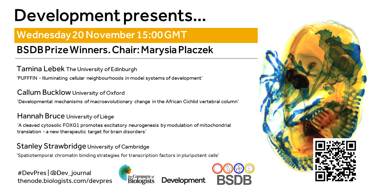
This special edition of Development presents… features the four prize winners from the 2024 BSDB Spring meeting. Hosted by BSDB Chair, Marysia Placzek (University of Sheffield).
Wednesday 20 November – 15:00 GMT
Tamina Lebek (The University of Edinburgh) ‘PUFFFIN – Illuminating cellular neighbourhoods in model systems of development’
Callum Bucklow (University of Oxford) ‘Developmental mechanisms of macroevolutionary change in the African Cichlid vertebral column’
Hannah Bruce (University of Liege) ‘A cleaved cytosolic FOXG1 promotes excitatory neurogenesis by modulation of mitochondrial translation – a new therapeutic target for brain disorders’
Stanley Strawbridge (University of Cambridge) ‘Spatiotemporal chromatin binding strategies for transcription factors in pluripotent cells’
At the speakers’ discretion, the webinar will be recorded for viewing on demand. To see the other webinars scheduled in our series, and to catch up on previous talks, please visit: thenode.biologists.com/devpres
This post is co-written by Christopher Thomas and Tabea Marx.
What is this?
This video shows ovulation live! Watch as an egg is released from an isolated ovarian follicle from a mouse. Our latest research uses live microscopy to visualise this fascinating event at the start of life.
Where can this be found?
Ovulation takes place in the ovaries, the female reproductive organs responsible for growing and releasing eggs. The eggs are stored inside fluid-filled sacs called follicles. To make the process visible, we cultured these follicles outside of the ovaries and induced ovulation ex vivo.
How was this taken?
This video was captured using a Zeiss LSM-800 confocal microscope. While working together in the Schuh lab, we developed an imaging setup that allowed us to observe ovulation in real time using isolated follicles from transgenic mice expressing fluorescent markers for histones (H2B-EGFP) and cell membranes (Myristoylated tdTomato). We also injected dextran into the follicular antrum, an extracellular space surrounding the egg, to label the follicular fluid.
What does the follicle do during ovulation?
The follicle goes through three phases during ovulation: it expands, contracts, and then ruptures, releasing the egg for fertilisation. In our paper, we show that expansion is mediated by fluid influx into the follicle, driven by hyaluronic acid secretion, while contraction is controlled by smooth muscle cells in the outer layers of the follicle.
Why should people care about this?
Ovulation is at the heart of reproduction. Understanding this fundamental process better could help treat infertility caused by disorders that cause ovulation to fail, such as PCOS (Polycystic ovary syndrome).
How would you explain this to an 8-year-old?
This video shows how a tiny egg moves from inside to outside of the ovary, a part of the body that helps make babies. The egg’s home in the ovary, called the follicle, bursts like a balloon to release it. Scientists really wanted to see how this happens, so they took some of these tiny bubbles and put them under a special camera that can see very tiny things. Once outside, if the egg meets a sperm, they can join together to make a baby.
Where can people find more about it?
Want to know more about how we filmed this amazing process? Check out our paper in Nature Cell Biology to learn all the details! You can find it here:
This is part of the ‘Lab meeting’ series featuring developmental and stem cell biology labs around the world.
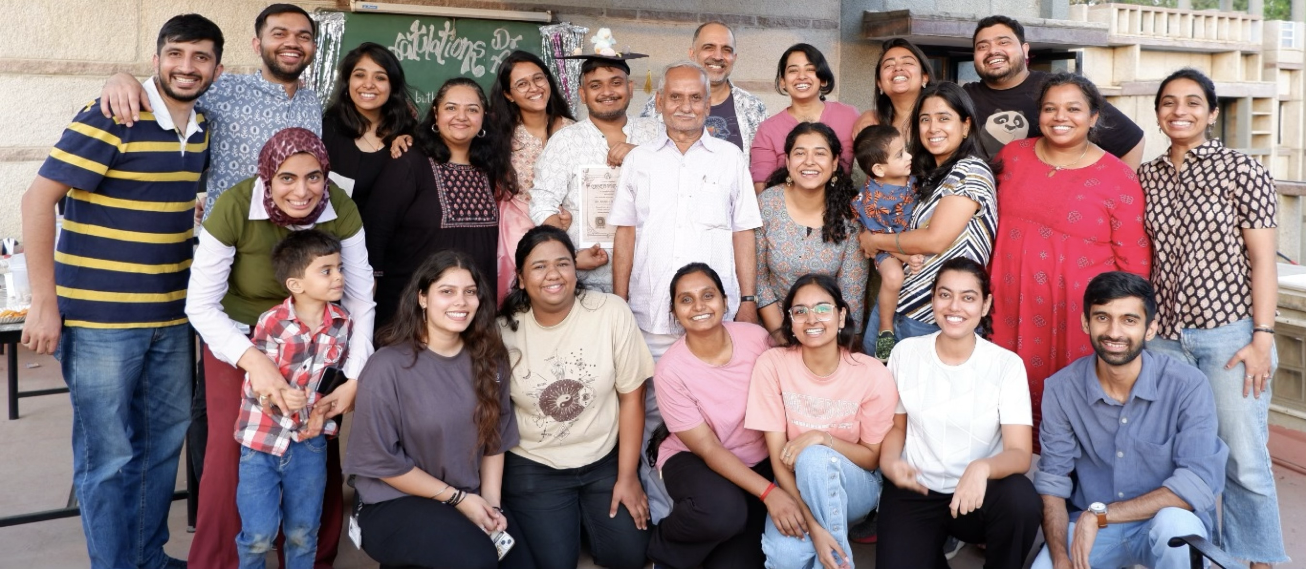
Can you briefly introduce your lab?
My name is Raj Ladher at the National Centre for Biological Sciences (NCBS) in Bangalore, India. I am interested in how developmental programmes subvert cellular processes to shape and specialise tissues, and we use the inner ear as our model system. We look at the inner ear from induction (what the signals are, what changes does induction elicit), differentiation (how sensory precursors are picked out) and morphogenesis of the cochlea and vestibular systems, and of the individual hair cells.
Raj: I have a lab of the most amazing students – they’re listed from oldest to newest here.
Nishant is looking at hair cell regeneration in the chick cochlea.
Varsha is investigating epithelial fusion during the closure of the otic vesicle.
Surjit is looking at how the birth order of neuroblasts affects where they end up in the inner ear ganglion.
Anubhav is investigating the morphogenesis of the cochlea and how planar polarity is generated.
Raman is working on non-canonical functions of protocadherin-15 in polarity generation.
Mona is working on the role of mitochondrial dysfunction in development.
Sukanya is looking at the downstream roles of Sox2 in the cochlea.
Neelanjana is looking at the signalling pathways involved for sensory fate specification.
Fenil is interested in understanding the molecular processes by which stereocilia acquire their shape.
Rridhi is looking at how inner ear progenitors acquire polarity.
Palak is investigating sensory cell differentiation.
Shivangi investigates how the otic placode thickens and what that means for lineage commitment.
We also have some interns who join our lab to do, for example, their Masters dissertation with us. Sardha, from IISER Mohali, is currently completing her MSc project on actin polymerisation in hair cells with us.
Favourite technique, and why?
Raj: I did my PhD on Xenopus mesoderm induction (with Jim Smith), and “grew up” doing the original organoid experiment – animal caps. Those kinds of classic embryological-type experiments have lots of mileage. I like heterotopic grafts (grafting pieces of tissue from one region of the embryo to another) and love quail-chick chimaeras.
Apart from your own research, what are you most excited about in developmental and stem cell biology?
Raj: Shape interests me, particularly of the face. I am interested in how natural variation tweaks mechanisms that would result in different morphologies of the face.
How do you approach managing your group and all the different tasks required in your job?
Raj: Labs in India run on students, and I have been amazingly fortunate with the students I have. They are amazing – creative, ambitious and fearless. Managing really means making sure that I am accessible or that I can point them to experts who can help them. We have regular lab meetings, and my office is in the lab, and my door is always open. It is also important to ensure they can attend conferences, workshops and courses that help them become better scientists. We have many interns from other institutes come through, giving the lab a chance to be mentors themselves. This holds enormous value, and my lab members are better researchers because of it.
What is the best thing about where you work?
Raj: I have great colleagues doing this fabulous range of topics, so it broadens my horizons about biology. We have 7 cafes on campus, and many places to sit and chat. The campus is gorgeous.
Varsha: The exposure to science at different scales, the facilities that we have access to use and the community.
Anubhav: The place thrives on the support of interdisciplinary exchange of ideas. These discussions over coffee give a fresh perspective on work as well as the outside world.
Raman: The chance to meet people from different culture and discussing the food interests with them and in the meantime science as well.
Neelanjana: The opportunity to discuss our work in progress with the community and the scope of collaborations between various labs on campus is one of the best things about this campus.
Fenil: Clean air, green cover, a bustling cafeteria.
Rridhi: The exposure to different kinds of aspects of biology and how any student can discuss science with anyone on campus.
Palak: The exposure and freedom to explore new projects. Although, it does have its ups and downs. However, the satisfaction of starting something new, making progress, and learning and evolving along the way is incomparable. Over the past two years, I have seen my perspective changing. And, I indeed consider it as a big growth. It’s great to learn from my seniors and gain insights into their journey and how they all have overcome these phases. It gives me hope and motivation to keep on trying and making new mistakes.
What is there to do outside of work?
Raj: There are places to eat and drink, old bazaars and markets to explore, and there’s a pretty decent second-hand book selection in town. Bangalore is surrounded by the Western Ghats, so there are wildlife, rainforests and jungles around us. It is also a wine-growing region, and the wine grown about an hour from the campus is actually very good.
Varsha: There are extracurricular clubs on campus, including movie and book clubs, dance and music clubs, concerts and dance shows arranged by the institute regularly. We also have a very good gym and sports facility.
Anubhav: The campus is at the intersection of the tall building, serving nice coffee and beer and hills, giving you a scenic drive, trek, and wines.
Raman: I like to play all the sports, especially cricket, badminton and sometimes swimming. I also watch different sports. And I love watching movies.
Neelanjana: There are various talks and exhibitions organised by the Archives in NCBS about history of science and its influence on general society which are always interesting to hear. Being a very green campus, a quick walk around is very refreshing to experience. Also taking clicks from the SLC terrace during sunset time.
Fenil: Outside of lab, I love to attend talks, concerts, and playing sports.
Rridhi: There are groups on campus that organise treks, stargazing, observing and photographing birds/butterflies/fireflies.
Palak: There are many things that you can do outside of the lab. But what I like the most is chilling in my room while watching some show, dancing to random songs, making mandalas (it brings so much serenity), going for night walks, exercising or probably having a small catch up session with my friends. It’s fun to hang out with them and listen to their gossip or have game nights with them.
[This October newsletter was originally sent out to Development’s journal news mailing list. This edition features, amongst other items, news of our upcoming celebratory conference and an update from our Pathway to Independence programme.]
Biologists @ 100
2025 marks 100 years since the founding of our publisher, The Company of Biologists. To mark this occasion, we are excited to invite you to The Company of Biologists’ 100-year anniversary conference Biologists @ 100, which will take place 24-27 March 2025 in Liverpool, UK.
The conference will bring together a range of scientists in developmental biology, cell biology, comparative physiology and disease biology. It will incorporate the 2025 Spring Meeting of the British Society for Developmental Biology, alongside other scientific strands, and plenary sessions that cover topics of importance to the whole biological community. The cell and developmental biology strand includes a list of outstanding speakers, including Development’s former Editor-in-Chief Olivier Pourquié.
To find out more, read this Node post by Development’s Reviews Editor Alex Eve and visit the conference website, from where you can now register for the meeting. Our community site the Node is also looking for a meeting reporter for this exciting event.
You’ll be hearing much more about how the Company has evolved over the past century, and our perspective for the future, throughout the course of next year.
Pathway to Independence programme
Last month, we were delighted to welcome our 2024 cohort of ‘Pathway to Independence’ (PI) fellows to our office in Cambridge for a three-day meeting – a key part of the support we’re providing them over the course of this year.
During this time, each fellow had the opportunity to present their ‘research vision’ to the rest of the group, as well as to some of last year’s PI fellows and the Development in-house team, and to gather feedback on how best to articulate their future research plans. This was followed by a two-day leadership training course run by hfp consulting, described by one of our fellows as “a very positive, enriching and informative experience that allowed me to learn tools and tricks I will be able to use in all spheres of my life”. We’re delighted to be supporting these talented postdocs as they apply for independent positions, and hope to grow this network of young leaders in the developmental biology field in coming years – look out for an announcement about the next call for applications later this year.
Find out more about our PI fellows’ research interests and thoughts on the future of the field in this Perspective article.
The 2024 cohort of PI Fellows visited The Company of Biologists office in September.
Special issues
Our 2024 special issue, Uncovering Developmental Diversity, is now being finalised and will be complete by the end of this month. Featuring over 30 different species, and covering topics ranging from axis determination in kelp to body size plasticity in sea anemones, this issue showcases some of the cutting edge research now possible in non-classical experimental systems.
Do also look out for the upcoming formal announcement of our 2025 special issue on ‘Lifelong Development: the Maintenance, Regeneration and Plasticity of Tissues’.
Featuring Alice Roberts, Professor of Public Engagement at the University of Birmingham, and a host of researchers working across the spectrum of the developmental biology field, we hope this video will help to promote our field and inspire the next generation of developmental biologists.
Available in both full-length and short versions, we invite you to watch, enjoy and – most importantly – share these movies.
The Forest of Biologists
By publishing in one of the journals of The Company of Biologists, you not only contribute to science, but you also directly contribute to the natural world. For each published Research or Review article, we plant a tree in The Forest of Biologists. And to acknowledge our peer reviewers, for each completed peer review we protect a tree in an ancient woodland. Since the launch of this project we have already planted over 2,000 new trees and protected 6,667 existing trees. Visit our virtual forest to check out your tree, or to read more about our forests.
The Company of Biologists’ Grants and Workshops: upcoming deadlines
Heterotypic interaction promotes asymmetric division of human hematopoietic progenitors Manuel Théry, Stéphane Brunet and colleagues Use of microfabricated niches reveals that interactions between human hematopoietic progenitors and stromal cells promote asymmetric division of progenitors and boost siblings’ heterogeneity, thus contributing to the plasticity of the early steps of hematopoiesis.
Sign up to Development’s email alerts (such as table of contents alerts) and the journal’s newsletter, to keep up to date on news, including special issues, calls for papers, content highlights/updates, journal meetings and more.
 (No Ratings Yet)
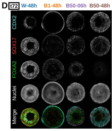
 (2 votes)