Report on GUDMAP Outreach at ISN World Congress of Nephrology (ISN WCN)
Posted by Simon Harding, on 30 March 2015
Report on GUDMAP Outreach at ISN World Congress of Nephrology (ISN WCN)
March 13-17, 2015
Cape Town, South Africa
Author: Chris Armit
Date: 23rd March 2015
Introduction
The International Society of Nephrology (ISN) holds biennial meetings throughout the world, and this was the first ISN WCN to be held in Africa. There was attention brought by the scientific program to maternal and child renal health, both of which are key problems in the developing world. There was additional emphasis on translational and clinical nephrology, with the session entitled ‘Congenital anomalies of the kidney and urinary tract: Prenatal through adulthood’ being of particular relevance to developmental biologists.
In addition, for the first time particular attention was paid to renal nurse involvement through their involvement both as speakers with the support of the Renal Care Society of South Africa and similar nurse associations around the world. It was anticipated by the ISN Committee that this would have great relevance, and is potentially of enormous impact on renal care in resource-poor settings of Africa where nephrologists and even non-specialised physicians are very scarce, and where life-death decisions for people with kidney disease often depend on well-trained nursing staff.
Congenital anomalies of the kidney and urinary tract: Prenatal through adulthood
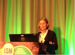
This session opened with a talk entitled ‘Organogenesis of the kidney’ by Melissa Little (University of Melbourne). Melissa introduced some of the key events in nephrogenesis and collecting duct formation, and provided an insightful overview of some of the key differences in kidney development between human and mouse. Following this, Melissa introduced the GUDMAP Project and GUDMAP Database as invaluable resources for researchers in the field wishing to know more about genitourinary development and the molecular anatomy of the developing kidney. There followed a more detailed discussion on GUDMAP, with reference to compartment isolation by LCM/FACS, expression profiling by arrays and RNA-seq, spatial validation by ISH, and the generation of transgenic reporter mice. Gene expression profiles of anchor genes defining nephron subcompartments were introduced as an illustrative way of identifying molecular anatomical profiles in GUDMAP sample data. In addition, comparative analysis of molecular markers of stage III and stage IV nephron were used to demonstrate the use of GUDMAP in identifying proximodistal expression patterns in GUDMAP SISH expression data. In addition, Melissa very kindly advertised the presence of a GUDMAP Exhibitor Stand at the ISN WCN where more information about the project could be obtained.
Recent data from Melissa’s lab highlighted the importance of hypoxia in congenital anomalies during embryonic development with Beta-catenin as a potentially important prognostic factor. Specifically, downregulation of the Beta-catenin BATGAL reporter is observed following hypoxia and this precedes the medullary collapse that is observed following hypoxic insult. Consequently, there is great interest in the role of Wnt/Beta-catenin signalling in the response to hypoxia. Of interest, medullary collapse was additionally observed in Cited1 knockout mouse embryos. The Cited1 knockout shows placental vasculature abnormalities, but not metanephros vascular abnormalities, and this highlights a potential role of systemic hypoxia in medullary collapse and may be relevant to understanding congenital kidney anomalies in clinical scenarios where systemic hypoxia may be important (e.g. pre-eclampsia).
The second speaker Nine Knoers (UMC Utrecht, Netherlands) spoke on ‘The next generation sequencing revolution’ and introduced this topic to a primarily clinical audience who were less aware of recent developments in this technology. Using renal dysplasia as a specific clinical example, Nine Knoers pointed out that Pax2 and Hnf1b are mutated in 10-15% of kidney dysplasia patients. However, in the majority of cases of renal dysplasia a gene association has not been identified. There was emphasis on how next-gen sequencing of cohorts of patients could be used to find genes associated with specific congenital disorders.
There was a change to the advertised program, and the third speaker was Christel du Buisson (Stellenbosch University, South Africa) who provided clinical case studies of antenatal hydronephrosis. In the first example of antenatal hydronephrosis, a case was presented where foetal pelvic ureteric junction obstruction was demonstrated by ultrasound. In this case study it was shown that a diuretic renogram did not differentiate between a functional abnormality or an obstruction. In a second case study of antenatal hydronephrosis, a case of primary vesico-ureteric reflux was presented where the developmental anomaly reflects abnormal insertion of the ureters into the developing kidney. Taken together these case studies highlight the inherent difficulties of diagnosing kidney disease in a pre-term clinical environment.
The final speaker in this session was Tong Zhang (Changzheng Hospital, Shanghai, China) who spoke on an ‘Interaction between Polycystin-1 and the tuberous sclerosis complex (TSC1/2) gene products and its role in regulating canonical WNT signaling’. It is known that TSC1/TSC2 activates mTor and disrupts PI3Ksignaling (Zhang et al., J Clin Invest. 2003 Oct;112(8):1223-33) but a direct role of these gene products in inducing Beta-catenin/WNT signalling has not been reported previously. I considered it appropriate to point out some recent work by Nils Lindstrom, Neil Carragher, and Peter Hohenstein of the University of Edinburgh and Roslin Institute who have shown that the PI3K pathway and the Beta-catenin pathway have opposing roles in self-renewal and differentiation of nephron progenitor cells (Stem Cell Reports, 2015, http://dx.doi.org/10.1016/j.stemcr.2015.01.021), with PI3K signaling triggering premature differentiation of the nephron progenitors. It is unclear whether TSC1 and TSC2 have a dual role in regulating in both these pathways but I consider this to be of potential interest.
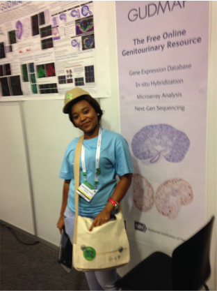
GUDMAP Exhibitor Stand
The GUDMAP Exhibitor Stand promoted GUDMAP as ‘The Free Online Genitourinary resource’ and was used to invite discussion on the scope of the GUDMAP Project and to provide live demos on how to use the GUDMAP web resource. Dr Simon Harding and Dr Frances Wong of the GUDMAP Editorial Office provided excellent exhibitor material in the form of banners and leaflets. Whereas there was some interest at the Exhibitor Stand on the Saturday afternoon, this increased following Melissa Little’s talk on the Sunday morning and there was a steady flow of interested parties to the GUDMAP Exhibitor Stand for the remainder of the Congress.
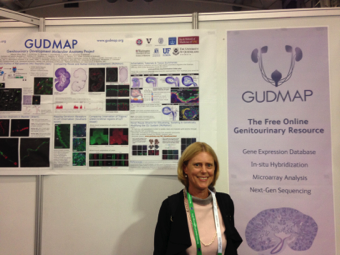
At the Exhibitor Stand, there was great interest in the scope of the GUDMAP Project and how it was funded. Following the session on ‘Congenital anomalies of the kidney and urinary tract: Prenatal through adulthood’ there was a lot of interest in the compartment-specific analysis that Melissa had mentioned in her talk. The GUDMAP Poster served as an excellent overview of the project, with online demos being used to show users how to find these data using the GUDMAP Database. There was additional explanation of using SISH to validate gene expression patterns following cDNA microarray analysis, and of using gene strips as a means of finding OMIM disease associations, in situ expression images, and array and RNA-seq data for a gene of interest. Andy McMahon’s ‘Comparing Mouse and Human Kidney Development’ section of the GUDMAP poster was particularly relevant for a clinical audience and highlighted the similarities between human and mouse at early stages of kidney development. A number of clinicians that visited the stand were surprised by how similar the Six2 and Cytokeratin gene expression patterns were between human and mouse, and this highlighted the importance of the mouse as a model organism for investigating kidney development and disease.
There was additional interest from clinicians who had attended the plenary talk by Barry Brenner (Harvard Medical School) entitled ‘Hypertension and kidney disease: The fault is not in our stars, but may be in our embryos’. In this talk, Barry Brenner emphasised the striking clinical correlation between low birth weight and low nephron number/kidney mass, and used as case studies two twin studies whereby the identical twin with low birth weight developed hypertension and kidney disease in later life. Clinicians who had attended this talk wished to know more about kidney development, and the GUDMAP poster and the GUDMAP online tutorials were particularly illustrative in this respect. On several occasions I was asked whether GUDMAP has any data on hypertension, to which I replied that the GUDMAP Database archives gene expression data on normal genitourinary development, and does not explore specific disease models. However, from the feedback I received I believe that if GUDMAP were to host primary data of mouse models of kidney disease then this would be of wide interest to the ISN community. There was additional interest as to whether GUDMAP had primary data relating to cell proliferation and cell death as this could be a means of addressing the phenomenon of nephron number/kidney mass as outlined in Barry Brenner’s talk. This may also be of wide interest to the nephrology community.

There was considerable interest in epigenetics following the plenary talk by Katalin Suszstak (Perelman School of Medicine, University of Pennsylvania) entitled ‘Epigenetics: Finding the missing heritability of complex diseases’. Katalin specifically discussed 5-mC methylation in the context of chronic kidney disease, and reported on some major findings over the last 5 years, including the seminal work by Bechtel et al (Nature Medicine 2010; 16, 544–550) to demonstrate that Dnmt1-dependent methylation determines fibroblast activation and fibrogenesis in the kidney. There was additional discussion on the role of Tet proteins in renal fibrosis, with tubule-specific Ksp2-Cre deletion of Tet2 being shown to alter regeneration after kidney injury and induce renal fibrosis. Katalin additionally pointed out that from a clinical perspective, 5-azacytidine has been used as a FDA-approved DNA methyltransferase inhibitor since 2004 for the treatment of myelodysplastic syndromes, however there is recent interest in its use in the treatment of renal fibrosis. Following this plenary session, there was interest at the GUDMAP Exhibitor Stand as to whether GUDMAP will host primary data relating to epigenetic modifications during kidney development.
As mentioned in the introduction, there was an emphasis on renal nurse involvement at the ISN WCN. At the GUDMAP Exhibitor Stand there were additional visits from nursing and technical staff who were interested in understanding more about the developing kidney and its role in health and disease. The GUDMAP web resource, and in particular the schematics and online tutorials that are made freely available were particularly welcomed by these health professionals.
 (2 votes)
 (No Ratings Yet)
 (6 votes)
 The medium-sized spiny neurons, the main projection neurons of the striatum, are generated in the lateral ganglionic eminence (LGE) and degenerate in the early stages of Huntington’s disease (HD) – for which no pharmacological treatment is yet available. Hence, an efficient way to derive striatal neurons is crucial for disease modelling, drug development and cell-replacement therapy. Striatal neurons have previously been generated from human pluripotent stem cell (hPSC)-derived neural progenitors treated with sonic hedgehog (SHH), or SHH plus Wnt pathway inhibition. Now, Meng Li and co-workers (p.
  Neural tube closure occurs through highly orchestrated cell shape changes mediated by actin dynamics. Its failure results in some of the most common and severe human congenital malformations. Cofilin 1, an actin-depolymerising protein, is known to be involved in neural tube closure but its precise functions had not been elucidated. In this study (p.
  On p.
On p.  In the second study (p.
  The two most influential ideas in the field of pattern formation are those of Alan Turing’s ‘reaction-diffusion’ and Lewis Wolpert’s ‘positional information’. Much has been written about these two concepts but some confusion still remains, in particular about the relationship between them. Here, Jeremy Green and James Sharpe address this relationship and propose a scheme of three distinct ways in which these two ideas work together to shape biological form. See their Hypothesis article on p.
 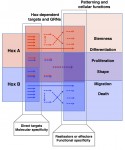 Hox genes encode homeodomain transcription factors that control morphogenesis and have established functions in development and evolution.Here, Yacine Graba and colleagues discuss the molecular and cellular mechanisms underlying the diverse and context-dependent functions of Hox transcription factors during morphogenesis and organogenesis. See the Review article on p.