The community site for and by
developmental and stem cell biologists

Tissue-specific genome editing in Ciona embryos by CRISPR/Cas9

Posted by , on 31 October 2014

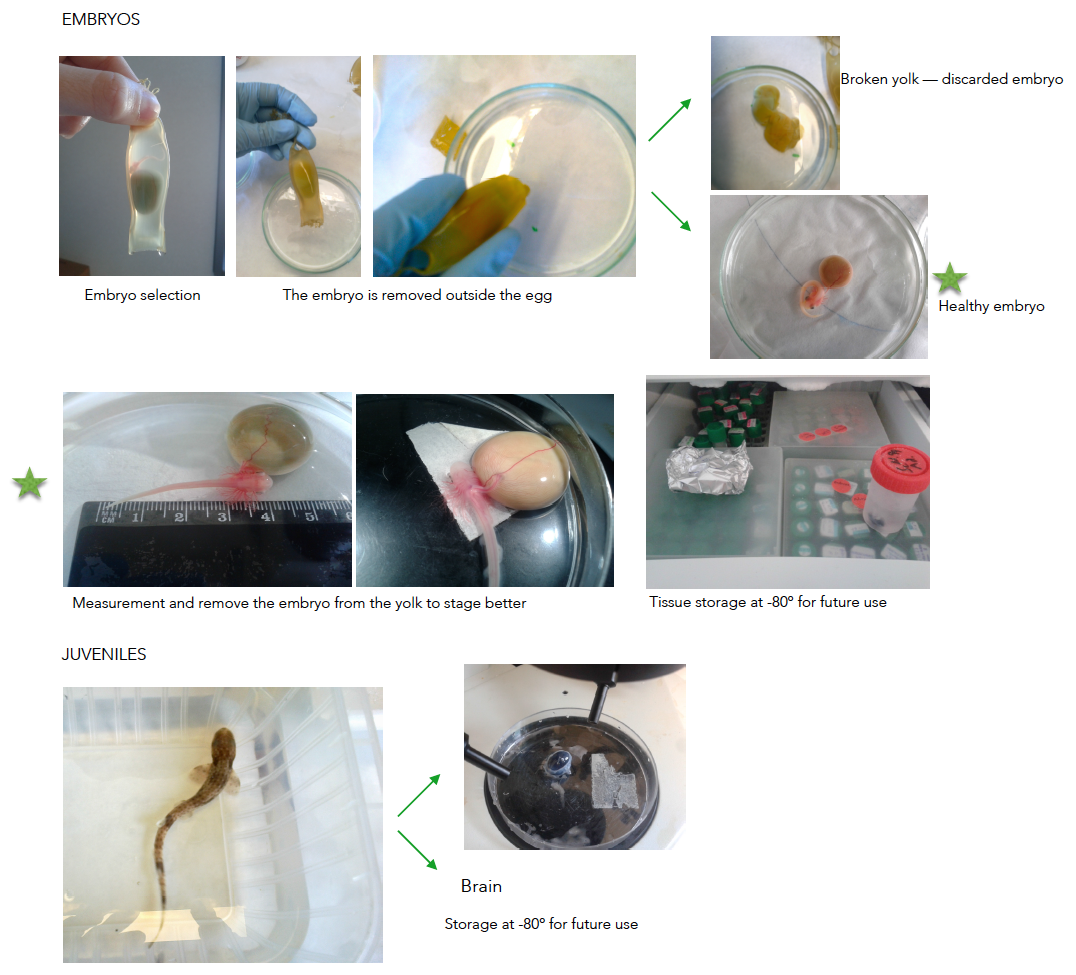
Researchers have always been interested in tissue-specific loss of function to probe the role of specific genes in embryonic development, cell physiology and disease conditions. Migration of lateral plate primordial germ cells in zebrafish, border cell migration during oogenesis in drosophila, interaction of T-cells with their target, and numerous other cases have continued to give us insights about how cells behave differently depending on their position and function in the animal. Until recently, genome-editing approaches were limited to non-specific mutations, induced by chemical or transposable elements. Homologous recombination for precise genome editing has only been achieved in mouse embryonic stem cells. Our lab studies the embryonic development and gene expression in Ciona intestinalis, a hermaphroditic tunicate. Phylogenetic studies have shown that tunicates are the closest living relatives of vertebrates. The Ciona larva possesses the basic body plan of chordates. A small sequenced genome, low cell number (~2600 at the larval stage), well-defined cell lineages, and easy methods for transient transfection assays are some of the experimental advantages offered by this model chordate. Previous studies have reported high rate of mutagenesis and tissue-specific gene knockouts in Ciona intestinalis using TALENs and ZFNs, but the application of these methods has been limited because of expensive and tedious cloning procedures.


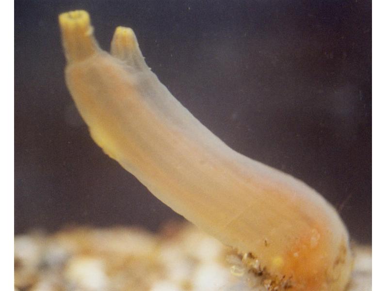
An adult Ciona intestinalis (Copyright: John Rundle)


Significant technical breakthroughs have been achieved in the field of genome engineering by harnessing the CRISPR (Clustered, Regularly Interspaced, Short Palindromic Repeats)/Cas (CRISPR Associated) system, an important part of the prokaryotic immune response. In this system, a short sequence, known as protospacer guides the endonuclease Cas9 to specific sites on the genome where it induces double stranded breaks. A broad range of applications, like genome editing, knock-in of exogenous DNA fragments, and regulation of transcription of endogenous genes, in a wide variety of model organisms, have demonstrated the versatility of this system. In our Development paper, we have established the CRISPR/Cas9 system for tissue specific genome editing in Ciona embryos using a simple electroporation-based transfection technique to induce site-specific double stranded breaks (DSBs) in the Ciona genome. High mutation efficiencies observed in our study are in sync with the results reported by Sasakura et al (2014). The main highlights of our paper include tissue-specific mutagenesis obtained using CRISPR/Cas system, and the phenotypic effects observed as a result of double stranded breaks in Ciona embryos.

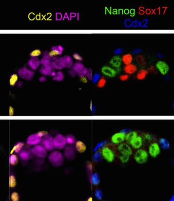
We have demonstrated that electroporation-based usage of the CRISPR/Cas system is sufficient to disrupt the activity of Ebf (previously known as COE), the sole C. intestinalis homolog of vertebrate EBF1, -2, -3 and -4. Ebf has been reported to play critical roles in both the ectoderm-derived motor neurons and in the cardiogenic mesoderm-derived Atrial Siphon Muscles (ASM), for their specification at the expense of heart development in Ciona. Ebf was thought to specifically up-regulate Islet expression in both the motor neurons and ASM precursors. To drive the expression of Cas9 in the ectoderm, we used the upstream cis-regulatory sequences of the C. intestinalis Sox1/2/3 gene. We observed a down-regulation of Islet reporter activity in motor ganglion neurons and ASM precursors, validating that Ebf is required for the specification and differentiation of motor ganglion neurons and pharyngeal muscles in Ciona intestinalis. Expression of a CRISPR/Cas9-resistant form of Ebf (Ebf>Ebfm774) was sufficient to rescue Islet expression, suggesting that the observed phenotypes were specific consequences of Cas9-mediated mutagenesis of Ebf gene. Moreover, when Ebf was targeted in the cardiogenic mesoderm by Mesp enhancer driven Cas9, Islet reporter expression was lost in the ASM but unaffected in motor ganglion neurons, confirming the tissue-specificity of CRISPR/Cas9 system in Ciona.

Spatial variation in expression of exogenous DNA associated with electroporation, known as mosaicism, is commonly associated with transient transfection assays. It has been shown to result in electroporated embryos containing both transfected and non-transfected cells. The latter would not experience CRISPR/Cas9-mediated mutagenesis, thus potentially leading us to underestimate mutagenesis efficacy. To address this problem, we used a simple cell-enrichment technique called MACS (Magnetic Activated Cell Sorting). Transfected cells expressed the guide RNA vector and Cas9 alongside the membrane-bound reporter hCD4::mCherry, which served for affinity purification using anti-hCD4 antibodies conjugated to magnetic nano-beads. Following one-step incubation with the beads, the sample was applied to a MACS column. The magnetically labeled cells were retained within the column, and were later eluted, whereas the unlabeled cells passed through. The sorted cells exhibited 66.2% mutagenesis when compared to unsorted cells from the same pool of embryos, which had 45.1% mutagenesis. Hence, a significant increase in the estimation of mutagenesis rates was observed using this simple technique. Furthermore, studying the temporal dynamics of Ci-EF1α promoter revealed the onset of its activity at 5 hours post fertilization (hpf) at the earliest, suggesting that the microinjection of mRNA transcripts into Ciona embryos might be necessary in order to target genes that are expressed before the gastrula stage.

Recently, the Sasakura lab at the University of Tsukuba published a paper demonstrating CRISPR/Cas9-mediated knockout of the Ci-Hox3 and Ci-Hox5 genes in Ciona intestinalis. They compared microinjection of in-vitro transcribed single guide RNA (sgRNA) and Cas9 mRNA transcripts with an electroporation-based approach to induce double stranded breaks in the Ciona genome. They observed an increase in the rate of mutagenesis when more RNA transcripts were microinjected into the embryos, although, the authors have not addressed questions related to tissue specificity and the efficacy of CRISPR/Cas9 induced mutations to cause specific phenotypes. One of the key findings of their paper was the high sensitivity of CRISPR/Cas9 mediated mutagenesis to the sequence of sgRNA vectors and mRNA transcripts. They observed a complete loss in mutation frequency with even a single nucleotide difference in the protospacer sequence, albeit reports of significant off-target effects of Cas9 in various model organisms. The authors of the paper have used instability of the Cas9-sgRNA complex formed on the genomic DNA as an explanation for this observation. However, we believe that this observation could be a result of using an unstable sgRNA backbone, an issue that has been addressed in our article. Two modifications, namely Flip (F) and Elongation (E), have been suggested in the guide RNA backbone to increase its in-vivo transcription efficiency, at the same time, promoting its ability to form a stable complex with Cas9. Using a modified backbone, we were able to achieve mutations in the 5’ flanking regions of Foxf and Hand-related, suggesting that CRISPR/Cas9 could be used for targeted mutagenesis in a wide variety of loci in the Ciona genome.

In conclusion, both papers report the successful application of the CRISPR/Cas system for effective genome editing in Ciona intestinalis. An expression vector-mediated electroporation method enables validation and extensive screening of sgRNA vectors, which is essential in the absence of a well-defined designing criterion to obtain high mutational activity. That being said, a microinjection-based approach might be necessary to construct loss-of-function sgRNA library to target genes that are expressed at an early stage during embryogenesis. Either ways, the CRISPR/Cas system has the potential to serve for the rapid scaling of genome editing in this model chordate.


Reference:

Stolfi, A., Gandhi, S., Salek, F., & Christiaen, L. (2014). Tissue-specific genome editing in Ciona embryos by CRISPR/Cas9 Development, 141 (21), 4115-4120 DOI: 10.1242/dev.114488

Thumbs up (6 votes)
Loading...

Tags: , , , ,
Categories: Research

Navigate the archive

Use our Advanced Search tool to search and filter posts by date, category, tags and authors.

A day in the life of a shark lab

Posted by , on 31 October 2014

Hi!

I am Nuria, a PhD student. I work in the Department of Cell Biology at the University of Santiago de Compostela (Spain).

Our group, the BRAINSHARK group, has been working in the evolutionary changes occurring during the development of the nervous system of a small shark (Scyliorhinus canicula) for many years. Our work mostly focuses on studying the development and the gene expression in this cartilaginous fish comparing with other vertebrates to gain insights in common processes mainly during the early development of the brain,  but also in other parts of the nervous system such as the olfactory system or the retina.

When I was an undergrad in Biology, I have never thought that I would work with sharks in a lab! Yes, I know it is a small one, but still a shark! Now, I am using this friendly fish to study the development of the retina.


Sharks as a model for developmental studies

The lesser-spotted dogfish, S. canicula, is an increasingly used model organism for studying embryonic nervous system development.

But why a shark? Chondrichthyans (cartilaginous fishes) constitute a monophyletic group, an ancient lineage of gnathostomous vertebrates characterized by having a cartilaginous endoskeleton, and placoid scales (dermal denticles) covering their body.  This group includes two major radiations that diverged over 250 million years ago. Elasmobranchs are one of these groups, including sharks, skates and rays, characterized by having articulated jaws. Its phylogenetic position makes chondrichthyans a key animal group to study the ancestral vertebrate condition of brain organization, because they provide a comparative reference for distinguishing between ancestral and derivative characters (Figure 1). Despite the fact that chondrichthyans represent an ancient vertebrate radiation, they don’t have primitive or unspecialized brain structures. Instead, they present well-developed, large brains, comparable in size to those of birds and mammals. In addition, the lesser-spotted dogfish offers great advantages as (1) the access to a unique collection of genomic resources, (2) the access to the embryonic development in ovo, (3) the possibility of maintaining embryos outside the eggshell for several days, and (4) the extremely slow growth and the big size of embryonic brain, which is really important to make detailed studies of particular regions.


Figure 1. Comparison of a mouse and shark embryo.


In sharks, vision, together with other sensory systems such as the lateral line, play important roles in habitat selection and in predatory and reproductive behaviour.  Sharks have conspicuous eyes, generally positioned laterally on the head, thus providing some binocular overlap in the visual field (Figure 2).


Shark New Fig 2

Figure 2. In sharks the eyes are generally positioned laterally on the head.


As an accessible extension of the brain, the retina offers an exceptional model to extend our knowledge on the development of the nervous system. Wherefore, several morphological and physiological studies have characterized the retinal circuitry and cells types in different elasmobranchs species. Fish retina has been found to be useful for studying retinogenesis as it contains retinal stem cells, which give rise to all cell types throughout the entire life of the animal. Our group has contributed significantly with developmental studies dealing with retinal morphogenesis.


Animal maintenance

Embryos of sharks are supplied by the Marine Biological Station of Roscoff (France). They are very young travelers that arrive to our lab packed in plastic bags. Apart form the eggs, sometimes we receive juveniles kindly provided by the Acuaria of Gijón, O Grove and A Coruña (Spain). Then the embryos from different broods and juveniles are raised in a tank of fresh sea water under standard conditions of temperature (16-18º) and 12:12 hours day/night cycle. They are introduced inside plastic bags to facilitate the acclimatization to their new home. The eggs are easily maintained under laboratory conditions until hatching, and the transparency of the egg shells makes it possible to select the required stage of development. The eggs have tendrils that allow their to attach to a substrate such as corals or seaweed. We use the tendrils to anchor the eggs to floating rods speeding up their development. In oviparous shark species as S. canicula, the embryos get their nourishment from a yolk sac. They may take several months to hatching, facilitating the study of developmental processes (Figure 3).


Shark New Fig 3

Figure 3. Our shark tanks. Tendrils of the eggs (asterisk). Click to see bigger version.


A typical day in our lab

When we arrive in the morning, we usually do the maintenance of the tank. It is necessary to test the pH, the concentration of nitrites and nitrates, check the temperature, and remove some dirty water and introduce some cleaning new. If we have juveniles, we feed them with frozen shrimp or squid. Finally, we check the embryos one by one, removing embryos appear damaged (when the yolk is broken or when the embryo does not move inside the egg). We can do it because of the transparency of the eggs! This allows us in addition to check the eggs, observe their development without removing the embryo from the egg (the video shows a healthy embryo inside the egg).


Staging embryos

To perform developmental experiments the first thing I have to do is to select the embryos on the stage I need to study. I stage the embryos on the basis of their external features according to Ballard et al. (1993) using a stereoscopic microscope (Figure 4).


Shark Fig3

Figure 4. External features during S.canicula development 


What do I do with sharks?

Tissue preparation

Little embryos up to stage 32 are deeply anesthetized in seawater commensurate with the adequate measures to minimize animal pain or discomfort. Soon after, I separate the embryo from the yolk before fixation. For embryos from stage 33 onwards and juveniles, I anesthetize in the same way and then, I perfuse their intracardially. Afterward, I remove the eyes and the brain. My colleagues work in other parts of the nervous system, so no rest is wasted (Figure 5).


Shark New Fig 5

 Figure 5. Tissue preparation. Click to see bigger version.


I usually cut the eyes on a cryostat (sometimes a use a microtome, depending on the further use).

Our group works with the typical developmental techniques such immunohistochemistry, in situ hybridization, tracing, etc. Finally, we study the sections and take the photomicrographs using different types of scopes (Figure 6).


Shark New Fig 6

Figure 6. My work place in the lab. and the confocal microscopy station. 


Future expectations

We are moving towards a more functional approach, trying to set-up new techniques that allow us to finding molecular determinants that can stabilize the neural stem stage, serve as fate determinants towards the neuronal lineage or can reverse a glial precursor into a neuronal precursor. However genetic manipulation in S. canicula is far from being optimized, it would be the next step to establish testable hypothesis for our descriptive data. To learn the techniques we needed in our lab, I am currently doing a research stay at the Center for Regenerative Therapies (CRTD) of Dresden, founded by Disease Models & Mechanisms (The Company of Biologists).


Acknowledgements:  Thanks to Dr. Idoia Quintana to encourage me to write this post and for her kindly review and suggestions, and my college Santiago Pereira for his help in changing the format of the images.


Node day in the life new doodle squareThis post is part of a series on a day in the life of developmental biology labs working on different model organisms. You can read the introduction to the series here and read other posts in this series here.


Thumbs up (16 votes)
Loading...

Tags: , , , , ,
Categories: Lab Life

A mechanism that allows a differentiated cell to reactivate as a stem cell revealed

Posted by , on 29 October 2014

The study, performed with fruit flies, describes a gene that determines whether a specialized cell conserves the capacity to become a stem cell again.

Unveiling the genetic traits that favour the retention of stem cell properties is crucial for regenerative medicine.

Published in Cell Reports, the article is the fruit of collaboration between researchers at IRB Barcelona and CSIC.

One kind of stem cell, those referred to as ‘facultative’, form part—together with other cells—of tissues and organs. There is apparently nothing that differentiates these cells from the others. However, they have a very special characteristic, namely they retain the capacity to become stem cells again. This phenomenon is something that happens in the liver, an organ that hosts cells that stimulate tissue growth, thus allowing the regeneration of the organ in the case of a transplant. Knowledge of the underlying mechanism that allows these cells to retain this capacity is a key issue in regenerative medicine.

Headed by Jordi Casanova, research professor at the Instituto de Biología Molecular de Barcelona (IBMB) of the CSIC and at IRB Barcelona, and by Xavier Franch-Marro, CSIC tenured scientist at the Instituto de Biología Evolutiva (CSIC-UPF), a study published in the journal Cell Reports reveals a mechanism that could explain this capacity. Working with larval tracheal cells of Drosophila melanogaster, these authors report that the key feature of these cells is that they have not entered the endocycle, a modified cell cycle through which a cell reproduces its genome several times without dividing.

“The function of endocycle in living organisms is not fully understood,” comments Xavier Franch-Marro. “One of the theories is that endoreplication contributes to enlarge the cell and confers the production of high amounts of protein”. This is the case of almost all larval cells of Drosophila.

The scientists have observed that the cells that enter the endocycle lose the capacity to reactivate as stem cells. “The endocycle is linked to an irreversible change of gene expression in the cell,” explains Jordi Casanova, “We have seen that inhibition of endocycle entry confers the cells the capacity to reactivate as stem cells”.

Cell entry into the endocycle is associated with the expression of the Fzr gene. The researchers have found that inhibition of this gene prevents this entry, which in turn leads to the conversion of the cell into an adult progenitor that retains the capacity to reactivate as a stem cell. Therefore, this gene acts as a switch that determines whether a cell will enter mitosis (the normal division of a cell) or the endocycle, the latter triggering a totally different genetic program with a distinct outcome regarding the capacity of a cell to reactivate as a stem cell.

The first co-authors of the study are postdoctoral fellows Nareg J.-V. Djabrayan, at IRB Barcelona, and Josefa Cruz, at the Instituto de Biología Evolutiva (CSIC-UPF).


Reference article:

Specification of Differentiated Adult Progenitors via Inhibition of Endocycle Entry in the Drosophila Trachea

Nareg J.-V. Djabrayan, Josefa Cruz, Cristina de Miguel, Xavier Franch-Marro, Jordi Casanova

Cell Reports (2014) DOI: http://dx.doi.org/10.1016/j.celrep.2014.09.043


This article was first published on the 29th of October 2014 in the news section of the IRB Barcelona website

Thumbs up (2 votes)
Loading...

Tags: , ,
Categories: Research

Barcelona BioMed Conference: Drosophila as a model in cancer (15 Jun 2015 to 17 Jun 2015)

Posted by , on 29 October 2014

IRBCN_2Hori_ANG_CMYK_PASPIRBCN_2Hori_ANG_RGB_PASP

Barcelona BioMed Conference: Drosophila as a model in cancer

15 – 17 Jun 2015

Cancer is a multi-hit process involving mutations in oncogenes and tumour suppressors, as well as interactions between the tumour cells and the surrounding normal tissue.

The fruit fly, Drosophila, is an excellent, genetically-tractable system for modelling the development of cancer, due to the conservation of signalling pathways, cell proliferation and survival genes between fly and humans. Drosophila also presents an excellent system to study the importance of stem cells, which have pluripotent potential, in the development of cancer.

This conference presents a forum for the discussion of cutting-edge models of tumourigenesis in Drosophila and their use in high-throughput screening of small molecule inhibitors that can be developed to combat human cancer.

Organizers: Cayetano González and Marco Milán, IRB Barcelona (Barcelona, Spain)

Organised by the Institute for Research in Biomedicine (IRB Barcelona) with the collaboration of the BBVA Foundation.


Marco

Registration deadline: April 15, 2015

There is no registration fee for this conference, but the number of participants is limited.

Participants are invited to submit abstracts, a number of which will be selected for short talk and poster presentations. Abstracts should include a title, authors, affiliations, summary (max 250 words) and references.

The BARCELONA BIOMED CONFERENCE on Drosophila as a model in cancer will be hosted by the Institut d’Estudis Catalans (IEC) in the heart of downtown Barcelona. Talks will take place in the Sala Prat de Riba.

Institut d’Estudis Catalans
C. del Carme, 47
08001 Barcelona

Register at: http://www.irbbarcelona.org/en/events/drosophila-as-a-model-in-cancer


Thumbs up (No Ratings Yet)
Loading...

Tags: , ,
Categories: Events

4 Yr PhD Programme in Developmental Mechanisms

Posted by , on 27 October 2014

Closing Date: 15 March 2021

Applications are open for the Wellcome Trust funded four year PhD programme in Developmental Mechanisms at Cambridge. We are looking for talented, motivated graduates or final year undergraduates, and are keen to attract outstanding applicants in the biological sciences, who are committed to doing a PhD. We are able to fund both EU and non-EU students. Closing Date: 9th January 2015. For more details about the application process and the programme please see the website: http://www.pdn.cam.ac.uk/phd/


4yrPhD_DevMech_Cambridge


Thumbs up (No Ratings Yet)
Loading...

Categories: Jobs

Biology and maths partner to understand life decisions

Posted by , on 26 October 2014

Nanog image

Starting with the one fertilized egg that we all once were, embryonic development is made of cell divisions and most importantly of cell decisions. These first life decisions are the first steps of the development of various cell types, which will further divide, decide, specialize, organize, form specialized organs and ultimately an entire very complex human being. Being able to understand how these very early decisions are made is of course a matter of scientific fascination.

One of the very first specialization steps is the formation of the trophectoderm versus the inner cell mass. The trophectoderm (marked by the protein cdx2) will form the extra-embryonic tissues such as the placenta. The inner cell mass (from which are derived embryonic stem cells) will become the fetus itself. Within the inner cell mass, further cell divisions and decisions will form the epiblast (marked by the protein nanog) and the primitive endoderm (marked by the protein Sox17). The epiblast will become the fetus itself and the primitive endoderm will play a key role in supporting the next steps in development of the epiblast.

In a recent study published in Development, Bessonard and colleagues showed that the protein Gata6 is necessary for the formation of the primitive endoderm. They combined these observations with additional published data to develop a mathematical model that can recapitulate cell behaviors observed experimentally and describes the temporal dynamics of inner cell mass differentiation.

In the upper row of this picture, you can observe a 3.75days old normal mouse embryo. In the lower row, you can observe a 3.75days old mouse embryo in which the protein Gata6 was removed. Cdx2 (in yellow on the left and in blue on the right) mark cells of the trophectoderm. Violet cells (DAPI, which marks DNA) are cells of the inner cell mass. These can be divided in cells of the epiblast (marked by nanog, green) or cells of the primitive endoderm (marked by Sox17, green). You can observe that in the absence of Gata6 (bottom row), only epiblast cells that express the protein nanog are formed, confirming the current hypothesis that Gata6 is necessary for the formation of the primitive endoderm.

Interestingly, Bessonard and colleagues used these observations, went further and pulled forces with mathematicians to create a mathematical model with which they could describe and predict the early inner cell mass fate decisions. Beautifully, that highlights how joining strengths of different scientific fields is becoming key to further the understanding of the most fascinating biological process of all…development.


Picture credit:

Bessonnard, S., De Mot, L., Gonze, D., Barriol, M., Dennis, C., Goldbeter, A., Dupont, G., & Chazaud, C. (2014). Gata6, Nanog and Erk signaling control cell fate in the inner cell mass through a tristable regulatory network Development, 141 (19), 3637-3648 DOI: 10.1242/dev.109678

Thumbs up (2 votes)
Loading...

Tags: , ,
Categories: Research

Public Engagement Officer

Posted by , on 24 October 2014

Closing Date: 15 March 2021

Department/Location: Wellcome Trust – Medical Research Council Cambridge Stem Cell Institute

Salary: £28,695-£37,394

Reference: PS03788

Closing date: 16 November 2014

Fixed-term: The funds for this post are available until 30 June 2017 in the first instance.

The Stem Cell Institute is a world-leading centre of excellence in stem cell biology and regenerative medicine, supported by a strategic funding partnership between the Wellcome Trust and the Medical Research Council (www.stemcells.cam.ac.uk).

Increasing awareness and understanding of the promise and challenges of stem cell research is embedded in the Stem Cell Institute vision; “deep understanding of stem cell biology for the prevention and treatment of human disease”. The Institute’s 5 strategic goals include “Communication and public engagement; providing reliable information, useful resources, and dialogue opportunities for a range of audiences including schools, policy makers, patient groups, professional bodies and the media”.

The central role of the Public Engagement Officer (PEO) is to foster a community of scientists who recognise the importance of dialogue with the public and who have the skills and opportunities to undertake public engagement activities. The PEO will build networks with local schools, arts groups and businesses to develop activities/events with/for them. The PEO will liaise with other Public Engagement officers in the University, Wellcome Trust Centres and Medical Research Council Institutes/Units and will participate in international networks via EuroStemCell, ISSCR and other trans-national initiatives.

We are seeking a dynamic, innovative and self-motivated individual who will bring expertise and leadership for the Institute’s outreach activities. Experience of working in the HE or research sector would be an advantage. Educated to degree level (or equivalent) in a scientific discipline, you will be responsible for growing a community of researchers who are enthusiastic about dialogue with the public and who have the skills and confidence to deliver stimulating interactive events.

The Stem Cell Institute, is currently spread across several sites in Cambridge, you will organise training, logistics and activities with the aim that all groups in SCI will contribute to at least one event each year. The PEO will expand the existing Public Engagement strategy and design and implement innovative public engagement activities that will interconnect the SCI with target audiences. You will develop measures for evaluating the effectiveness and impact of public engagement activities. You will submit reports to the Institute steering committee and sponsors and will prepare funding applications for public engagement activities.

You must demonstrate a proven track record in relationship building, event organisation, report writing, and data management. You will have outstanding organisational and administrative experience and be comfortable working to tight deadlines with minimal supervision. You should have demonstrable experience in web-based/social media communication and you should have excellent written and verbal communication and negotiation skills. You will be IT literate and able to work both on your own and as part of a team.

The post will report to the Institute Director. You will have a degree (or equivalent).

Once an offer of employment has been accepted, the successful candidate will be required to undergo a health assessment.

To apply online for this vacancy and to view further information about the role, please visit: http://www.jobs.cam.ac.uk/job/4403. This will take you to the role on the University’s Job Opportunities pages. There you will need to click on the ‘Apply online’ button and register an account with the University’s Web Recruitment System (if you have not already) and log in before completing the online application form.

The closing date for all applications is the Sunday 16th November 2014.

Please upload your Curriculum Vitae (CV) and a covering letter in the Upload section of the online application to supplement your application. If you upload any additional documents which have not been requested, we will not be able to consider these as part of your application.

Informal enquiries about the post are also welcome via email: jrw46@cam.ac.uk.

Interviews will be held Wednesday 26th November 2014. If you have not been invited for interview by 21st November 2014, you have not been successful on this occasion.

Please quote reference PS03788 on your application and in any correspondence about this vacancy.

The University values diversity and is committed to equality of opportunity.

The University has a responsibility to ensure that all employees are eligible to live and work in the UK.

Thumbs up (1 votes)
Loading...

Categories: Jobs

Post-doc and research assistant positions in Drosophila planar polarity

Posted by , on 22 October 2014

Closing Date: 15 March 2021

Two Wellcome Trust-funded positions are available for candidates with a background in cell and/or developmental biology to join an interdisciplinary team investigating cell signalling and coordinated cell polarisation using Drosophila epithelial development as a model system. The Strutt lab (http://www.shef.ac.uk/bms/research/strutt) studies cell signalling and coordinated cell polarisation in animal tissues via analysis of the “core” and Fat/Dachsous planar polarity/PCP pathways, using a range of molecular genetic, cell biological and computational techniques. Our current research programme aims to dissect the molecular feedback interactions underlying cell polarisation, and has a strong emphasis on studying in vivo protein dynamics, using high resolution (including super resolution) live imaging, and integrating experimental results with computational modelling.

Informal enquiries may be directed to David Strutt (d.strutt@sheffield.ac.uk). Formal applications should be made directly to the University of Sheffield (http://www.sheffield.ac.uk/jobs Job Refs: UOS009537 and UOS009538) by no later than the 21st November 2014.

Thumbs up (No Ratings Yet)
Loading...

Categories: Jobs

Human Stem Cell research: new ethical questions?

Posted by , on 21 October 2014

The Ethics Session at the Company of Biologists “From Stem Cells to Human Development” workshop

Last September the Company Of Biologists organized an exciting three-day dive into the biology of human stem cells and their use to study human development and regeneration (look out for the full meeting report in Development, coming soon, and Katherine Brown’s post on the Node). Some of the scientific work presented at the meeting was extremely compelling. With the induced Pluripotent Stem Cell (iPSC) technology that allows the generation of human stem cells from somatic cells without destroying an embryo, experiments that were confined to science fiction not so long ago are now being carried out rather routinely in labs. For example, in Dr Nakauchi’s lab rat organs are grown in mice and vice-versa, the ultimate goal being to grow human organs in large vertebrates such as pigs to transplant them into patients requiring organ transplants. But such ground-breaking work challenges our current biological and moral definitions. Is it ethical to break the species barrier? Do human/non-human animal chimeras blur the distinction between humans and animals? How does that affect human dignity and the dignity of other species? This pioneering line of research also challenges our regulatory codes. As the 2006 and 2008 ISSCR’s guidelines for stem cell use are both currently being revised, it would seem particularly timely to think about what comes next in terms of regulations to accommodate these new technical and conceptual advances. What particular techniques and topics require our attention? Can we foresee what these new techniques will allow us to do and how to leave room in the regulations for that?

To discuss the ethical questions surrounding this emerging field, Dr Hermeren and Dr Hyun held a very interactive session entitled “Ethical Aspects of Stem Cell Research”, chaired by Dr Austin Smith. In this piece, I will try to convey some of the points of debate that arose from the speaker’s presentations, the stories shared by the panel and the questions from the audience.

Ethics_5

One of the points that raised particular attention was crossing the species barrier, especially in light of the fascinating work conducted by Dr Nakauchi, who joined the discussion panel during the session. Another example is the work of Dr Goldman, who engrafted human glial progenitors in mice, altering their cognitive ability. What is the status of such humanized chimeric animals? As Dr Hyun appropriately put it: if we build a gas station with stones from a cathedral, does it make it a cathedral? Is there a distinction between biological humanization and moral humanization? How does that alter animal welfare? What are the biological and moral significance of the presence of animal cells in human organs grown in non-human hosts if transplanted back to a patient?

Related to Dr Nakauchi’s work, a question from the audience sparked a very interesting discussion: how is his research perceived by the general public in Japan? He explained that at first, the experiments he was carrying out in his lab, namely generating cross-species chimeras in the hope of growing human organs in large mammals, were regarded with circumspection. Then, he started communicating his research to the general public and explaining the ultimate goal: generating human organs using the patient’s own cells to compensate for the shortcomings of organ donations and allow organ transplants without the need of immunosuppressive therapy. Communication really changed the public’s perception of his experiments for the better. After a few questions and answers, a consensus arose in both the audience and the panel: it is crucial to actively communicate, not merely react, about the new lines of research involving human stem cells, in order to make the public appreciate their use and dissipate misconceptions.

Another point raised during the discussion with the speakers was the availability of human embryos. Dr Rossant, also part of the panel, explained during her research presentation that early human development is mainly studied with embryos obtained by in vitro fertilization (IVF). However, these embryos are not an ideal material since they have exclusively seen an in vitro environment. It was therefore discussed whether we should consider creating human embryos exclusively for research use. Also, for how long should these embryos be allowed to grow in vitro? Current regulations allow a period of 14 days but this significantly restricts the developmental stages that can be studied. This also brings more basic questions such as the real definition of an embryo both morally and biologically.

Ethics_2

Another novel ethical question that arises with iPSCs is informed consent from the patient. Numerous tissue banks have been created over the years, many before it was even imaginable to create human stem cells from somatic cells. Patients gave up their rights on surgical discards that can now be used to create an immortal embryonic cell line that co-exists with the patient. Those embryonic cells could one day, and maybe not so far ahead in the future, be used to generate an organ or another human being carrying the same genetic information. Who is then the study subject? The patient? The immortalized cell line that carries his/her genome? It is also interesting to think about the rights of the donor. Could he/she forbid the use of the tissue he/she consented to give for the generation of an iPSC line? Furthermore, with the refinement and wider use of whole genomic studies, how to proceed when pathological genetic mutations are found in patients-derived cell lines? Should the donor be informed?

All the new ethical questions discussed above relate to how research on human stem cells is perceived and regulated from the outside. However, there is a more internal aspect to this ground-breaking research: how can researchers keep this line of work ethical? What is research integrity? In the light of recent reports of fraudulent research involving stem cells (most notably the STAP matter), it seems timely to reflect on what is good research practice and how common are the deviations. Why are researchers tempted to deviate? What really constitutes fraud? On that topic, Katherine Brown, Development’s executive editor, was asked about what exactly constitutes plagiarism: is reproducing our own published work plagiarism? What happens when there is only limited ways to describe a method?

From this meeting, it was compelling to see that the use of human stem cells has emerged as an incredibly powerful tool to study human development and human regeneration. Hence, ethics sessions where researchers, regulators and publishers can discuss the next steps appear crucial to define how they want to conduct their research, how regulations should accompany these new technical and conceptual advances, and how to communicate to dissipate general public’s misconceptions.

Thumbs up (8 votes)
Loading...

Tags: , , ,
Categories: Events

In Development this week (Vol. 141, Issue 21)

Posted by , on 21 October 2014

Here are the highlights from the new issue of Development:


Two top tips for angiogenesis

FigureThe widely accepted model of angiogenic sprouting proposes that a single cell – the tip cell – is found at the leading edge of vessel sprouts. Now, Victoria Bautch and colleagues describe an alternative blood vessel topology in which multiple endothelial cells (ECs) constitute the tip of sprouting blood vessels and polarize to promote lumen formation (p. 4121). By mosaically labelling ECs in both in vitro and in vivoangiogenesis assays, the researchers first demonstrate that sprouts often contain two cells at their tips, and that both cells extend filopodia. Live imaging studies show that, as reported previously, these tip cells switch positions, but the cell-cell overlap is largely maintained throughout switching. The researchers further report that tip cells polarize along their longitudinal cell-cell border; this border is characterized by apical polarity markers and is the site of lumen formation. Finally, the authors show that the loss of an atypical protein kinase C (aPKC) isoform disrupts EC overlap and lumen formation, highlighting a role for aPKC in angiogenic sprouting. Together, these observations suggest that the paradigm of a single cell at the tip of developing blood vessels requires revision.

Fat body shapes up the trachea

FigureDevelopment of the Drosophila tracheal system – the fly respiratory network – is known to require the chitin deacetylase serpentine (Serp), which is expressed in tracheal epithelial cells. Here, Shigeo Hayashi and co-workers show that Serp derived from the Drosophila fat body, which is functionally equivalent to the mammalian liver, is transported to the tracheal lumen and is required for tube morphogenesis (p. 4104). They first show that Serp is expressed in the fat body and that Serp levels accumulate in fat bodies of the rab9 and shrub vesicle trafficking mutants. By contrast, Serp levels in the tracheal lumen of these mutants are reduced, suggesting that Serp is secreted into the haemolymph and is taken up by tracheal cells. Importantly, the researchers show that fat body-derived Serp is able to rescue the tracheal defects observed in serp mutants. These studies suggest that Serp should be added to the expanding repertoire of proteins that the fat body supplies to other organs and highlight the role of the fat body in regulating the development of other organs.


Nf2: guiding axons across the midline

FigureThe cerebral hemispheres are connected by the largest axonal tract in the mammalian brain: the corpus callosum (CC). During CC development, axons from one hemisphere navigate across the midline, channelled along their way by guidepost cells that secrete guidance cues, to reach specific targets on the other hemisphere. Neurofibromatosis type 2 (Nf2/Merlin) mouse mutants show a complete absence of the CC but how Nf2, a signalling protein involved in various cellular processes and signalling pathways, controls axonal pathfinding is currently unclear. Using Nf2-conditional knockout mouse models, Xinwei Cao and colleagues show that, surprisingly, Nf2 is not required in callosal neurons or their progenitors but is required in midline neural progenitors that generate guidepost cells (see p. 4182). The authors reveal that Nf2 controls guidepost development and the expression of Slit2, a major signalling cue secreted by guidepost cells, through the suppression of YAP, an effector of the Hippo pathway. These findings represent an exciting step forward in the molecular understanding of midline formation and brain wiring, as well as uncovering an intriguing previously undescribed function for the Hippo pathway.

Getting to the heart of slow conducting cardiomyocytes

FigureDuring a heart beat, an electrical impulse leads to the contraction of the upper part of the heart: the atria. The electrical impulse slows down through the atrioventricular junction (AVJ) before resuming rapid propagation to induce contraction of the lower part of the heart: the ventricles. This contraction delay, combined with the presence of cardiac valves, is crucial for unidirectional blood flow in the heart and is altered in various heart diseases. How is this slow conducting property established and restricted to the AVJ? On p.4149, Takashi Mikawa and colleagues discover that, contrary to the current hypothesis, the AVJ does not maintain juvenile slow conduction; instead, AVJ conduction velocity is plastic and determined by its proximity to the endocardium (the inner lining of the heart). They further show that the cardiac jelly (an extracellular martix-rich deposit that accumulates during valve formation) acts as a crucial physical barrier separating the AVJ from endocardial signals that induce a fast conduction phenotype. The authors thus uncover an exciting mechanism whereby valve formation and the delay in chamber contraction are developmentally linked, and open new perspectives for understanding heart development and congenital diseases.


PLUS…


Development of the cerebellum: simple steps to make a ‘little brain’

DEV4031The cerebellum is a pre-eminent model for the study of neurogenesis and circuit assembly. In recent years, our understanding of cerebellar growth has undergone major recalibration, and insights from a variety of species have contributed to an increasingly rich picture of how this system develops. Here, Thomas Butts, Mary Green and Richard Wingate review these advances. See the Review on p. 4031


Making designer mutants in model organisms

DEV4042Recent advances in the targeted modification of eukaryotic genomes have unlocked a new era of genome engineering. From pioneering work using zinc-finger nucleases, to the advent of TALEN and CRISPR/Cas9 systems, we now possess the ability to analyze developmental processes using sophisticated genetic tools. Here, Stephen Ekker and colleagues summarize the common approaches and applications of these still-evolving tools. See the Review on p. 4042

Thumbs up (1 votes)
Loading...

Categories: Research