Vertebrates, including humans, possess a “head” comprising cranial bones, the central nervous system, and sensory organs. It is believed that the emergence of the vertebrate “new head” is closely linked with the evolutionary acquisition of two cell populations: neural crest cells (NCCs) and cranial placode cells. Therefore, understanding the evolutionary origin and history of NCCs and cranial placode cells is crucial for understanding the evolution of vertebrates.
In vertebrate embryos, both NCCs and cranial placode cells arise from the border region between the neural plate and the epidermis. NCCs are unique because they produce not only cell types of ectodermal origin, such as sensory neurons and melanocytes, but also cell types of mesodermal origin, including smooth muscle cells, osteocytes, and chondrocytes.
Ascidians: fascinating model organisms for evolutionary developmental biology
Ascidians, commonly known as sea squirts, belong to the subphylum Urochordata or Tunicata, the sister group of vertebrates. They have been providing key insights into chordate developmental mechanisms and their evolution (FIGURE 1). Recent studies suggested that ascidian embryos have cells that share an evolutionary origin with vertebrate NCCs[1-3]. For example, ascidian cells called a9.49, located in the neural plate border, likely share an evolutionary origin with vertebrate NCCs[1]. Indeed, this cell pair expresses orthologous genes that specify the neural plate border cells and NCCs in vertebrate embryos. Furthermore, a9.49 cells can be reprogrammed to migratory pigment cells by overexpression of Twist, which encodes a transcription factor for mesenchyme specification. However, unlike vertebrate NCCs, ascidian NCC-like cells identified thus far do not produce cell types that are commonly of mesodermal origin. Therefore, it is believed that the multipotency of NCCs has been acquired within the vertebrate lineage after the split from the ascidian lineage.
FIGURE1 The sea squirt Ciona robusta (Ciona intestinalis type A)
A key observation made nearly 40 years ago
In 1987, Nishida found that ascidian cells called b8.17 and b8.19 give rise to muscle cells, nerve cord cells, and endodermal cells near the tip of the tail of embryos[4]. Both b8.17 cells and b8.19 cells are located in the neural plate border, which abuts the neural plate cells that give rise to the central nervous system. These cells express many orthologous genes that specify the neural plate border cells and NCCs in vertebrates. Therefore, if b8.17 and b8.19 cells share an evolutionary origin with vertebrate NCCs and produce cell types that are commonly ectodermal and mesodermal origin, the potential of NCCs to produce cells of multiple germ layers may date back to the last common ancestor (LCA) of vertebrates and ascidians, contrary to the prevailing hypothesis explained above.
In light of this context, we have decided to investigate the possibility that ascidian b8.17 and b8.19 cells share an evolutionary origin with vertebrate NCCs. First, we confirmed that b8.17 cells indeed produced muscle cells, as Nishida showed previously[4]. Second, we showed that these ascidian cells expressed Msx, Zic, Pax3/7, and Snai, which encode orthologs of key transcriptional factors specifying neural plate border cells of vertebrate embryos. We indeed showed that these genes were involved in specifying these ascidian cells. The location and the gene circuit for specification indicate that this ascidian cell population shares an evolutionary origin with vertebrate NCCs.
Do neural plate border cells of ascidian embryos share the evolutionary origin with vertebrate neuromesodermal progenitors (NMPs)?
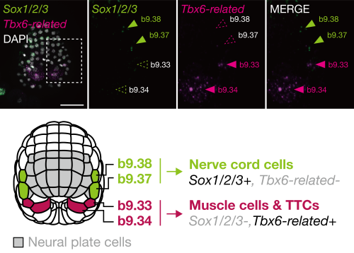
In the middle gastrula stage, the ascidian neural plate border consists of four cells: b9.34, b9.33, b9.37, and b9.38, in order from posterior to anterior. In later embryos, the anterior two cells (b9.37 and b9.38) give rise to nerve cord cells (commonly of ectodermal origin), and the posterior two cells (b9.34 and b9.33) give rise to muscle cells (commonly of mesodermal origin) and other cells near the tip of the tail region[4]. On the basis of this observation, we hypothesized that these cells may share an evolutionary origin with vertebrate neuromesodermal progenitors (NMPs).
In vertebrates, Tbx6 is expressed in NMP-derived mesodermal cells and Tbx6 negatively regulates Sox2, which is expressed in NMP-derived spinal cord cells[5]. If our hypothesis is correct, the gene regulatory circuit consisting of Tbx6 (or its orthologs) and Sox2 (or its orthologs) will also be used for fate decisions in the neural plate border cells of ascidian embryos. Indeed, the anterior cells, which give rise to the nerve cord, expressed Sox2 ortholog (Sox1/2/3), and the posterior cells, which give rise to muscle, expressed Tbx6 ortholog (Tbx6-related) (FIGURE 2). Overexpression of Tbx6-related downregulated Sox1/2/3, and promoted muscle fate. Thus, the ascidian neural plate border cells and vertebrate NMPs share the gene regulatory circuit of Sox2 and Tbx6. In addition, a comparative single-cell transcriptome analysis also supported a close relationship between these ascidian cells and NMPs of zebrafish embryos.
FIGURE2 Gene expression pattern and fate decision of neural plate border of ascidian embryos
In this way, this ascidian cell population has properties of both vertebrate NMPs and NCCs. Therefore, the LCA of tunicates and vertebrates likely had cells with a hybrid property of NCCs and NMPs, and such ancestral cells may have produced both ectodermal and mesodermal cells.
Chordate origin of NCCs and NMPs
A logical follow-up question to ask is whether the Cephalochordata, the sister group of Olfactores, possessed NCC-like cells and NMP-like cells. Cephalochordates, commonly known as lancelets or amphioxus, are filter-feeding marine animals and are believed to retain ancestral features of chordates. Amphioxus is believed to lack cells homologous to vertebrate NCCs[6], although a recent preprint indicated that amphioxus possesses migratory NCC-like cells[7].
Interestingly, somites, notochord cells, dorsal neural tube, and hindgut of the posterior part of amphioxus embryos are produced from a cell population near the tip of the tail[8]. Therefore, amphioxus may possess NMP-like cells. Elucidating the developmental mechanism of this cell population should shed light on the evolution of the body plan of chordates.
A possible evolutionary history of the stemness of NCCs/NMPs
The ascidian NCCs/NMP-like cell population does not possess stemness: they do not show the ability of self-renewal, although they produce cell types that are commonly ectoderm and mesoderm origin. In vertebrates, the high stem cell-like potential of NCCs may depend on pluripotent factors or Yamanaka factors[9-12]. Among Yamanaka factor genes, only Sox1/2/3 was known to be expressed in the ascidian NCCs/NMP-like cells. This may be a reason why the ascidian cells do not have self-renewal ability.
Altogether, we propose a two-step model for the evolution of stemness of NCCs/NMPs: 1) the ability to produce ectodermal and mesodermal cells came first, and 2) the self-renewal ability, which led to acquisition of bona fide NCCs and NMPs. Future works on non-ascidian tunicates (e.g., Oikopleura), amphioxus, and cyclostomes will shed light on the evolution of the stemness of NCCs/NMPs. It would be particularly important to associate the evolution of the stemness of NCCs/NMPs with the evolutionary acquisition of pluripotent factor genes and whole genome duplications that occurred in the vertebrate lineages.
Rebecca K. Spangler, Guinevere E. Ashley, Kathrin Braun, Daniel Wruck, Andrea Ramos-Coronado, James Matthew Ragle, Vytautas Iesmantavicius, Daniel Hess, Carrie L. Partch, Helge Großhans, Jordan D. Ward
Ngoc Minh Phuong Nguyen, Eun Mi Chang, Maeva Chauvin, Natalie Sicher, Aki Kashiwagi, Nicholas Nagykery, Christina Chow, Phoebe May, Alana Mermin-Bunnel, Josephine Cleverdon, Thy Duong, Marie-Charlotte Meinsohn, Dadi Gao, Patricia K. Donahoe, David Pepin
Aicha El Ellam, Emily J. Alberto, Maria E. Mercau, Dimitrius T. Pramio, Krishna M. Bhat, William M Philbrick, Deborah Schechtman, Carla V. Rothlin, Sourav Ghosh
LS Ee, D Medina-Cano, CM Uyehara, C Schwarz, E Goetzler, E Salataj, A Polyzos, S Madhuranath, T Evans, AK Hadjantonakis, E Apostolou, T Vierbuchen, M Stadtfeld
Kristen Kurtzeborn, Vladislav Iaroshenko, Tomáš Zárybnický, Julia Koivula, Heidi Anttonen, Darren Brigdewater, Ramaswamy Krishnan, Ping Chen, Satu Kuure
Mekala Gunasekaran, Hannah R. Littel, Natalya M. Wells, Johnnie Turner, Gloriana Campos, Sree Venigalla, Elicia A. Estrella, Partha S. Ghosh, Audrey L. Daugherty, Seth A. Stafki, Louis M. Kunkel, A. Reghan Foley, Sandra Donkervoort, Carsten G. Bönnemann, Laura Toledo-Bravo de Laguna, Andres Nascimento, Daniel Natera-de Benito, Isabelle Draper, Christine C. Bruels, Christina A. Pacak, Peter B. Kang
Chiemela Ohanele, Jessica N. Peoples, Anja Karlstaedt, Joshua T. Geiger, Ashley D. Gayle, Nasab Ghazal, Fateemaa Sohani, Milton E. Brown, Michael E. Davis, George A. Porter Jr., Victor Faundez, Jennifer Q. Kwong
Nida Ozarslan, Corina Mong, John Ategeka, Lin Li, Sirirak Buarpung, Joshua F. Robinson, Jimmy Kizza, Abel Kakuru, Moses R. Kamya, Grant Dorsey, Phillip J. Rosenthal, Stephanie L. Gaw
Irina Lazar-Contes, Rodrigo G. Arzate-Mejia, Deepak K. Tanwar, Leonard C. Steg, Kerem Uzel, Olivier Ulrich Feudjio, Marion Crespo, Pierre-Luc Germain, Isabelle M. Mansuy
Luis Hernandez-Huertas, Ismael Moreno-Sanchez, Jesús Crespo-Cuadrado, Ana Vargas-Baco, Gabriel da Silva Pescador, José M. Santos-Pereira, Ariel A. Bazzini, Miguel A. Moreno-Mateos
Andrew S. Hagan, Scott Williams, Casey J. N. Mathison, Shanshan Yan, Bao Nguyen, Glenn C. Federe, Guray Kuzu, Joseph C. Siefert, Janice Hampton, Victor Chichkov, S. Whitney Barnes, Frederick J. King, Brandon Taylor, John R. Walker, Rui Zhao, Jimmy Elliott, Dean P. Phillips, Bin Fang, Rebekah S. Decker
Cristina Medina-Menéndez, Lingling Li, Paula Tirado-Melendro, Pilar Rodríguez-Martín, Elena Melgarejo-de la Peña, Mario Díaz-García, María Valdés-Bescós, Rafael López-Sansegundo, Aixa V. Morales
Elizabeth Elder, Anthony Lemieux, Lisa-Marie Legault, Maxime Caron, Virginie Bertrand-Lehouillier, Thomas Dupas, Noël Raynal, Guillaume Bourque, Daniel Sinnett, Nicolas Gévry, Serge McGraw
Archana Kamalakar, Brendan Tobin, Sundus Kaimari, M. Hope Robinson, Afra I. Toma, Timothy Cha, Samir Chihab, Irica Moriarity, Surabhi Gautam, Pallavi Bhattaram, Shelly Abramowicz, Hicham Drissi, Andrés J. García, Levi B. Wood, Steven L. Goudy
Justine Bajohr, Erica Y. Scott, Arman Olfat, Mehrshad Sadria, Kevin Lee, Maria Fahim, Hiba T. Taha, Daniela Lozano Casasbuenas, Ann Derham, Scott A. Yuzwa, Gary D. Bader, Maryam Faiz
Anna Kasprzyk-Pawelec, Mingjun Tan, Raneen Rahhal, Alec McIntosh, Harvey Fernandez, Rami Mosaoa, Lei Jiang, Gray W. Pearson, Eric Glasgow, Jerry Vockley, Christopher Albanese, Maria Laura Avantaggiati
Enric Bertran Garcia de Olalla, Gabriel Rodriguez-Maroto, Martina Cerise, Alice Vayssieres, Edouard Severing, Yaiza Lopez-Sampere, Kang Wang, Sabine Schaefer, Pau Formosa-Jordan, George Coupland
James Ronald, Sarah C.L. Lock, Will Claydon, Zihao Zhu, Kayla McCarthy, Elizabeth Pendlington, Ethan J. Redmond, Gina Y.W. Vong, Sanoj P. Stanislas, Seth J. Davis, Marcel Quint, Daphne Ezer
Isaia Vardanega, Jan Eric Maika, Edgar Demesa-Arevalo, Tianyu Lan, Gwendolyn K. Kirschner, Jafargholi Imani, Ivan F. Acosta, Katarzyna Makowska, Götz Hensel, Thilanka Ranaweera, Shin-Han Shiu, Thorsten Schnurbusch, Maria von Korff Schmising, Rüdiger Simon
Rasik Shiekh Bin Hamid, Fruzsina Nagy, Nikolett Kaszler, Ildikó Domonkos, Magdolna Gombos, Eszter Molnár, Aladár Pettkó-Szandtner, László Bögre, Attila Fehér, Zoltán Magyar
Abdull J. Massri, Alejandro Berrio, Anton Afanassiev, Laura Greenstreet, Krista Pipho, Maria Byrne, Geoffrey Schiebinger, David R. McClay, Gregory A. Wray
Daniel J. Stadtmauer, Silvia Basanta, Jamie D. Maziarz, Alison G. Cole, Gülay Dagdas, Gilbecca Rae Smith, Frank van Breukelen, Mihaela Pavličev, Günter P. Wagner
Haidee Tinning, Alysha Taylor, Dapeng Wang, Anna Pullinger, Georgios Oikonomou, Miguel A. Velazquez, Paul Thompson, Achim Treumann, Peter T. Ruane, Mary J O’Connell, Niamh Forde
Benjamin C. Klementz, Georg Brenneis, Isaac A. Hinne, Ethan M. Laumer, Sophie M. Neu, Grace M. Hareid, Guilherme Gainett, Emily V.W. Setton, Catalina Simian, David E. Vrech, Isabella Joyce, Austen A. Barnett, Nipam H. Patel, Mark S. Harvey, Alfredo V. Peretti, Monika Gulia-Nuss, Prashant P. Sharma
Amelia RI Lindsey, Jason M Tennessen, Michael A Gelaw, Megan W Jones, Audrey J Parish, Irene LG Newton, Travis Nemkov, Angelo D’Alessandro, Madhulika Rai, Nicole Stark
Mohammad Zeeshan, Ravish Rashpa, David J. Ferguson, George Mckeown, Raushan Nugmanova, Amit K. Subudhi, Raphael Beyeler, Sarah L. Pashley, Robert Markus, Declan Brady, Magali Roques, Andrew R. Bottrill, Andrew M. Fry, Arnab Pain, Sue Vaughan, Anthony A. Holder, Eelco C. Tromer, Mathieu Brochet, Rita Tewari
Valentina Gandin, Jun Kim, Liang-Zhong Yang, Yumin Lian, Takashi Kawase, Amy Hu, Konrad Rokicki, Greg Fleishman, Paul Tillberg, Alejandro Aguilera Castrejon, Carsen Stringer, Stephan Preibisch, Zhe J. Liu
Erik A. Ehlers, Kyle N. Klein, Margaret A. Fuqua, Julia R. Torvi, Javier Chávez, Lauren M. Kuo, Jacob McCarley, Jacqueline E. Smith, Gaea Turman, Danielle Yi, Ruwanthi N. Gunawardane, Brock Roberts
Barbara Varnum-Finney, Adam M. Heck, Sanjay R. Srivatsan, Stacey Dozono, Rachel Wellington, Cynthia Nourigat-McKay, Tessa Dignum, Cole Trapnell, Brandon Hadland
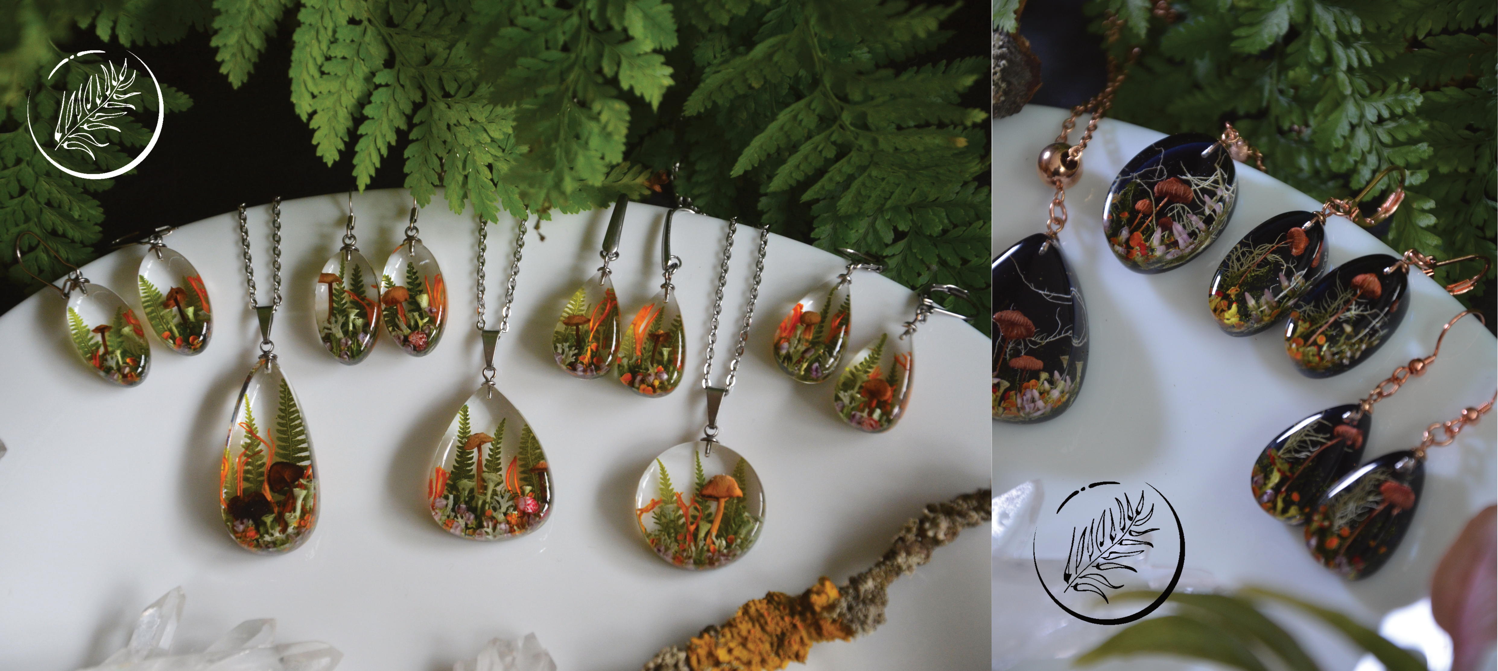
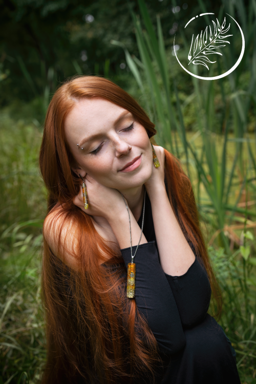
In this SciArt profile, we meet Gabriela Krejčová, a postdoctoral researcher at the University of South Bohemia, Czech Republic, who enjoys making nature-inspired jewellery.
Forest-inspired pendants and earrings with real mushrooms, ferns and lichens.
Can you tell us about your background and what you work on now?
My first scientific endeavour was carried out in the field of cancer immunotherapy. I found the metabolic changes of tumour cells particularly remarkable at the time. After completing my undergraduate studies, I started looking for a new laboratory where I could conduct my master’s thesis. I came across Dr. Adam Bajgar, who at the time was working on the metabolic polarization of Drosophila melanogaster immune cells during bacterial infection. Since it is well established that the metabolic setting of pro-inflammatory macrophages resembles in many aspects the metabolic changes occurring in some types of cancer cells, I changed my field of study because this topic represented a nice link to my previous research focus. During my PhD studies, I began to look into the signalling molecules released by the immune cells in response to their metabolic polarization, which subsequently mediate the inter-organ communication. I also become fascinated by the functional versatility of macrophages and their regulatory role in various stress conditions, which is my current focus.
A collection of pendants with real ferns.
Were you always going to be a scientist?
I would say I’ve always enjoyed unravelling the unknown, whether it was the mysteries of nature or Egyptian hieroglyphs. I remember wishing for a little spooky laboratory, and I cherish the memories of getting my own small kid’s microscope and exploring the world up close. Oddly enough, my dream job as a child was actually a fashion designer, which reflected my love for art.
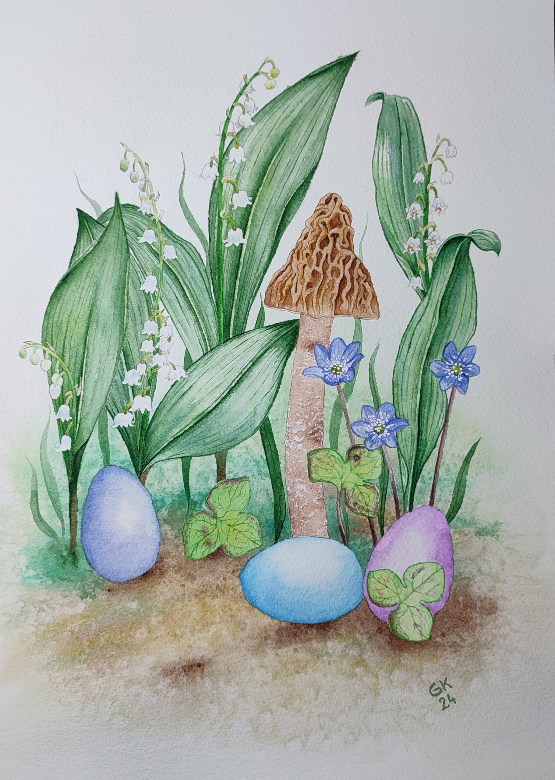
Spring-inspired watercolor painting of Verpa bohemica mushroom, lily of the valley (Convallaria majalis), Hepatica nobilis and Easter eggs.
And what about art – have you always enjoyed it?
By all means! I’ve always enjoyed all kinds of crafts and I’ve always had a desire to create pretty things – from drawings and paintings to jewellery and decorations such as traditional Easter eggs decorated with wax. Another passion of mine has always been dancing, so I found another way to express my urge for creating in designing and decorating dance costumes.
A set of earrings and a pendant with false chanterelle mushrooms (Hygrophoropsis aurantiaca), pink chervil, violet beautyberry, fern and lichens.
How do you make your jewellery?
I make my jewellery exclusively from products of nature and clear epoxy resin. So first, I forage for all sorts of flowers, mushrooms, lichens, mosses, ferns or berries to create tiny microworld compositions. Before casting, which is usually a multi-step process, all materials must be dried in a special way to retain their original colours and shapes. After demoulding, all must be sanded and polished, which is the most time-consuming part. Then I attach the jewellery findings and the piece is finally finished. The whole process takes approximately two weeks.
Sphere-shaped pieces are the most time consuming type of pendants I create.
What or who are your most important artistic influences?
My biggest muse is definitely nature itself. My goal is to preserve the beauty and diversity of shapes, structures, patterns and colours that nature has already created, and perhaps just slightly transform these pieces into small compositions and make them wearable. In this way, I would like to give people a piece of unspoiled nature that they can keep constantly with them.
I create also taxidermy jewellery with real beetles that I buy already preserved at insect sales exhibition.
Does your science influence your art at all, or are they separate worlds?
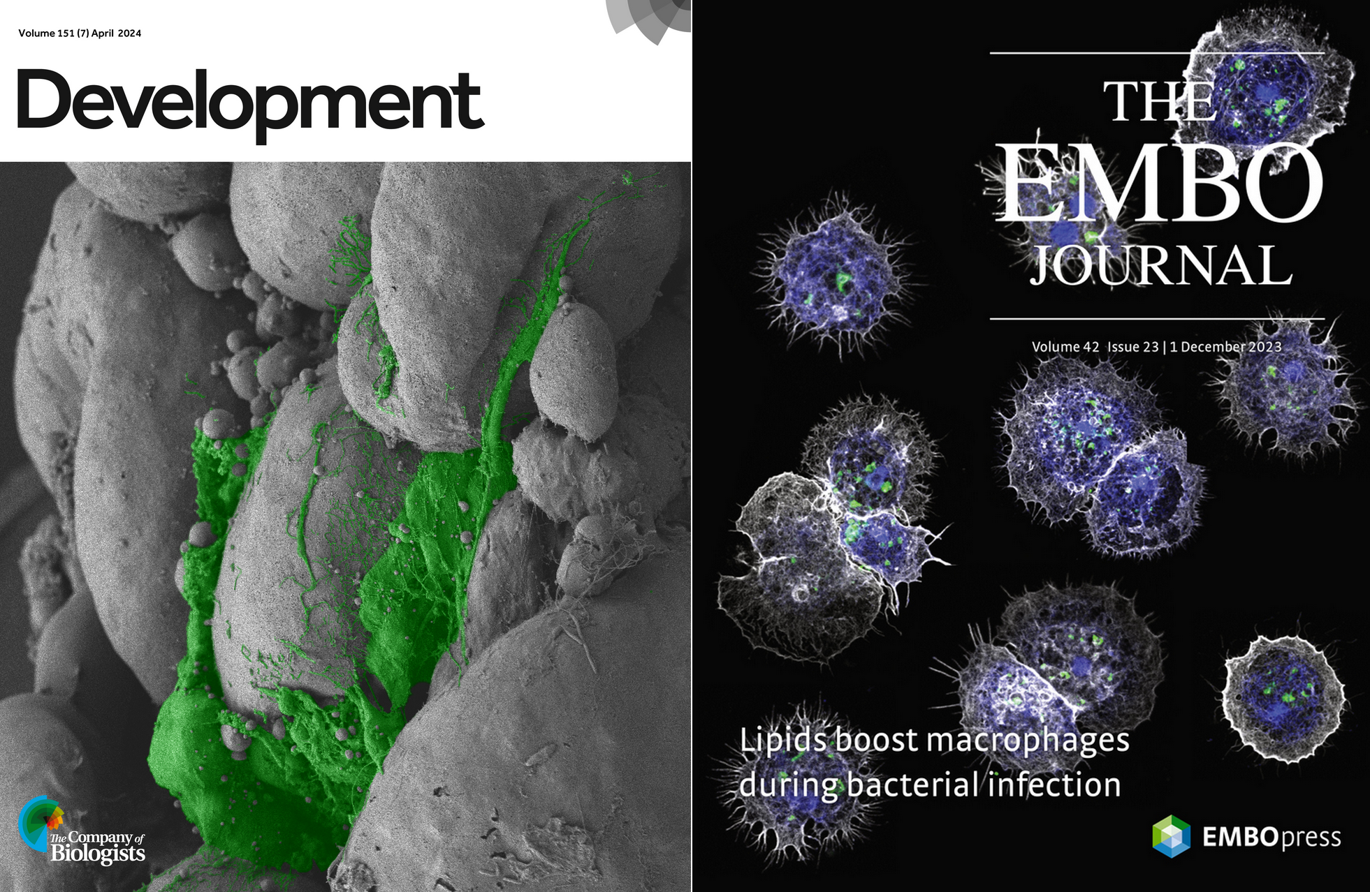
I would say it’s rather the opposite – my art influences my scientific outputs, or at least I hope so. I firmly believe that scientific imaging techniques provide ample room for artistic expression, and I hope that this is sometimes reflected in my scientific output. I especially enjoy the visualization of macrophages by confocal and electron microscopy, and the beauty of immune cells brought me much joy during my PhD studies.
Journal Covers for Development and The EMBO Journal.
What are you thinking of working on next?
It is now spring season in the Czech Republic, so everything is thriving and blooming. For me, it is the time of year when I need to stock up for the upcoming year so I have enough material to make jewellery in the colder months. Therefore, I have a lot of collecting, foraging and mushrooming ahead of me, which means a lot of quiet and fulfilling time spent in nature.
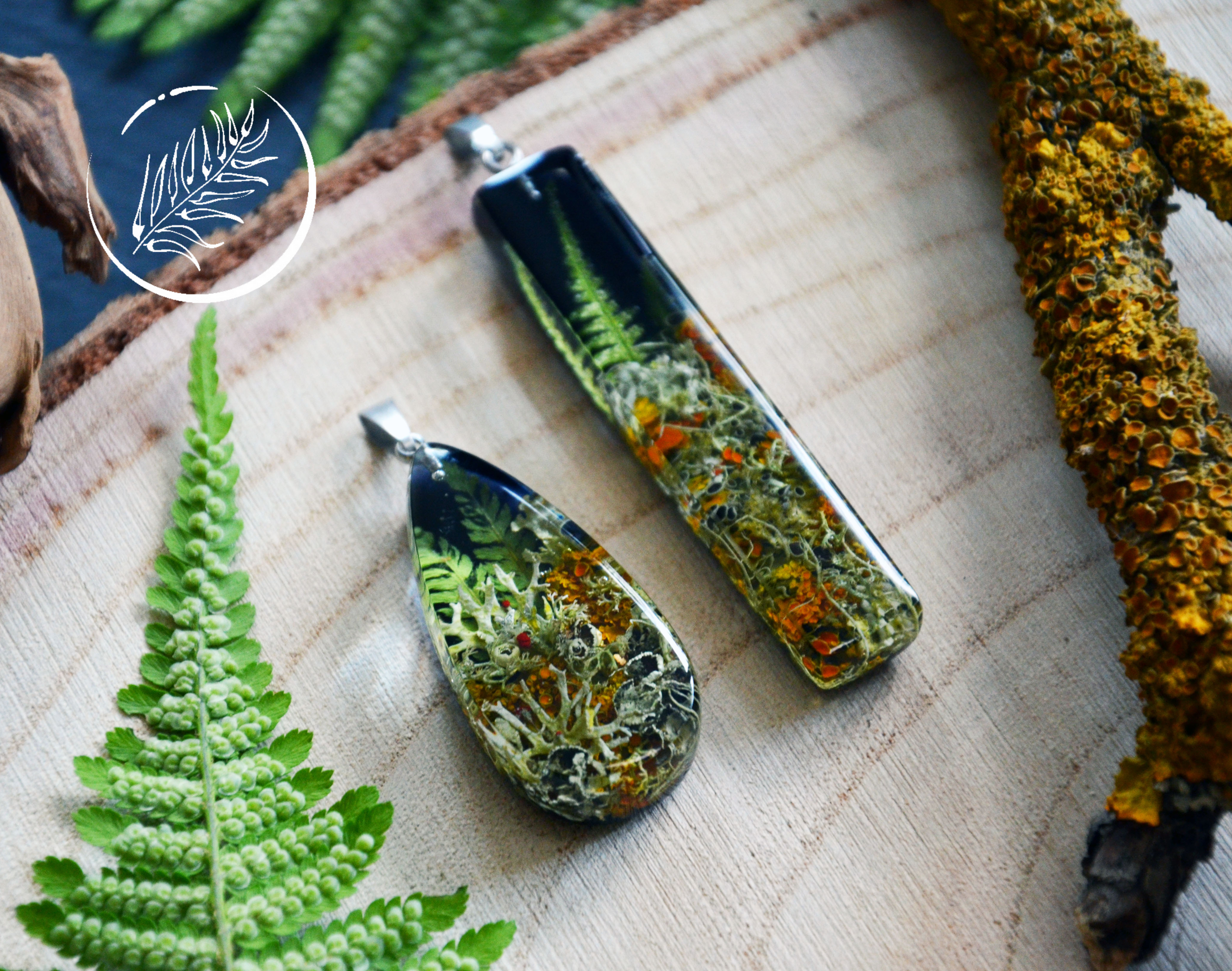
Pendants with many types of colourful lichens and fern
On the topic of mechanics and morphogenesis chaired by Development Editor, James Wells (Cincinnati Children’s Hospital Medical Center)
Wednesday 19 June – 15:00 BST
Clémentine Villeneuve (Max Planck Institute for Molecular Biomedicine) ‘Tissue-scale mechanics control stem cell fate and positioning during epithelial development’
Louis Prahl (University of Pennsylvania) ‘Branching, crowding, and packing: engineering the developing kidney epithelium’
Kyojiro Ikeda (University of Vienna) ‘Nanometric 3D printing: sculpting bristles by dynamic microvilli’
At the speakers’ discretion, the webinar will be recorded for viewing on demand. To see the other webinars scheduled in our series, and to catch up on previous talks, please visit: thenode.biologists.com/devpres
Neurofly2024 is the main conference on neurobiology of Drosophila, taking place every 2 years, and has been rotating across Europe for the last 40 years. The Company of Biologists Travel Bursaries will cover the registration fees of PhD students, enabling a wider participation in the Neurofly.
We have now advertised the bursaries, please visit:
The 20th Biennial European Drosophila Research Conference (Neurofly) will take place from 2 to 6 September 2024 at the beautiful University of Birmingham campus, in UK.
Neurofly 2024 will allow the opportunity for researchers from across the globe to meet to discuss their latest research and observations principally on the neurobiology of Drosophila but also including studies using other invertebrate model organisms. Drosophila offers major advantages for neurobiological research due to the wealth of genetic tools to observe and manipulate the nervous system. We aim to provide a venue for wide-ranging discussions and interactions between junior and senior researchers in an inclusive interdisciplinary environment that will allow the exchange of results, ideas and new concepts. A wide range of sessions will include Developmental and cellular neuroscience, Brain homeostasis and metabolism, Brain disease, injury and ageing, Gene expression and molecular neuroscience, Neural circuits and behaviour, Plasticity and remodelling.
We have lined up eight wonderful plenary speakers and the rest of talks will be selected from abstract submissions, allowing exciting, emerging research to be presented by early career and established researchers. We will also have a practical demo workshop on bridging connectomics and transcriptomics. And fun activities, including a trip to Stratford and the gala conference dinner.
Registration early bird and abstract submission deadline is 16 June 2024.
Please be aware that lecture theatre capacity is limited: please register with time to avoid disappointments.
A recent paper “Neural crest origin of sympathetic neurons at the dawn of vertebrates” challenges the prevailing dogma that the sympathetic ganglia arose only in jawed vertebrates. Instead, based on the findings in the sea lamprey, the authors suggest a late-developing rudimentary sympathetic nervous system may be present in the earliest jawless vertebrates. First author Brittany Edens and corresponding author Marianne Bronner tell us the story behind the paper.
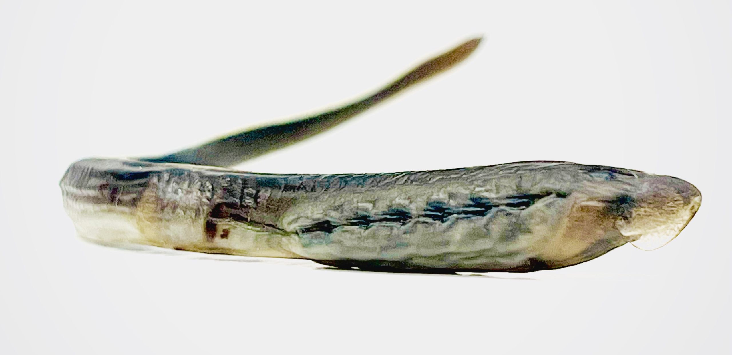
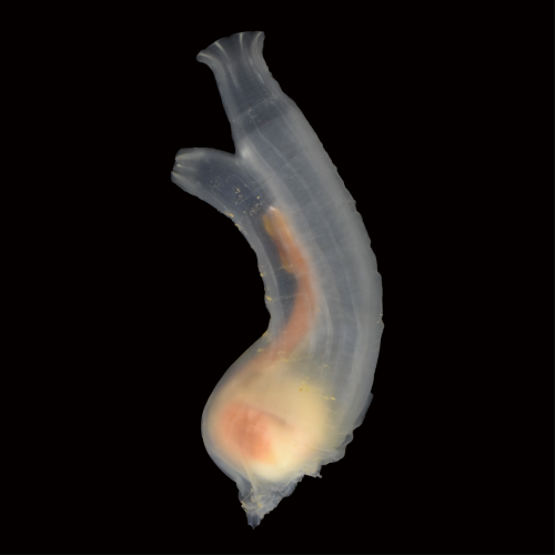
Wild-caught mature ammocete sourced from the Great Lakes.
How did the project start?
Marianne: Brittany was doing some staining of lamprey larvae with different antibody markers with the goal of defining different types of neurons in the developing enteric nervous system. We got together to look over the data and realized that some of the neuronal staining was not in the gut but dorsal to the gut in a position that was appropriate for the sympathetic nervous system. This was a surprise since lamprey are not supposed to have a sympathetic nervous system. So we started looking for more markers to test this possibility more rigorously.
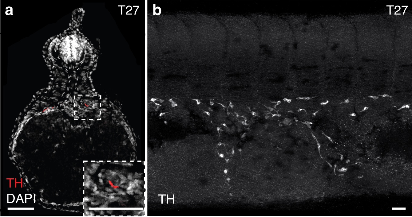
Detection of sympathoblasts in late embryonic lamprey: The catecholamine biosynthetic processing enzyme tyrosine hydroxylase (TH) is detected by immunohistochemistry at T27 in (a) transverse sections and (b) lateral whole-mount. TH+ sympathoblasts are localized dorsal to the yolk tube and flank the midline in bilateral streams. Scale bars=50mm (a) and 10mm (b).
Why do you think it’s been previously thought that jawless vertebrates lacked the sympathetic nervous system?
Marianne: That’s easy to answer. We think people (including ourselves) were initially looking at the wrong time. In higher vertebrates, the sympathetic nervous system develops rather early in development, initiating when neural crest cells begin to coalesce around the dorsal aorta. We actually looked at comparable stages in lamprey and did not see the markers characteristic of sympathetic neurons co-expressed. However, when we looked at larvae at about 1 month of development, we observed not only sympathetic marker genes but also the transcription factors known to be involved in their specification. Thus, there was a heterochrony in terms of the time of differentiation.
Why did you choose the lamprey to answer your questions?
Marianne: Lamprey are jawless vertebrates and have an important phylogenetic position at the base of the vertebrate tree of life. Lamprey fossils from the Cambrian period resemble modern lamprey in morphology. While we have no access to a “vertebrate ancestor” and lamprey have continued to evolve, they still are the closest approximation to what we think the ancestor may have looked liked.
Can you summarise the key findings of the paper in a paragraph?
Marianne: In gnathostomes (jawed vertebrates), the neural crest gives rise to a fate-restricted sympathoadrenal progenitor from which sympathetic neurons of the autonomic nervous system arise. A transcriptional program including Ascl1, Phox2b, and Hand2 specifies neural crest towards sympathoadrenal fates, and also promotes catecholaminergic identity (i.e., expression of tyrosine hydroxylase and dopamine beta-hydroxylase enzymes). Upon maturation, these neural crest-derived sympathetic neurons will express various pan-neuronal genes, as well as genes specific for catecholaminergic function. While the earliest vertebrates, which lacked jaws, were historically believed to lack sympathetic neurons within the trunk, we found evidence of these cells in the jawless vertebrate sea lamprey. We found that the same core transcription factors involved in sympathoadrenal specification were co-expressed in cells throughout the trunk in lamprey, as were the catecholamine pathway enzymes. Later in larval stages, these cells upregulated expression of pan-neuronal markers. Lineage tracing indicated a conserved origin in the trunk neural crest and finally, RNA-sequencing analysis suggested a transcriptional profile that was consistent with sympathetic neuron identity. Altogether our findings challenge the prevailing dogma that the sympathetic ganglia are a gnathostome innovation.
Were you surprised to find a rudimentary sympathetic nervous system in the lamprey?
Marianne: Yes indeed. We expected to see no sympathetic nervous system since that is what the literature says. It was a real surprise to see neurons in the right place with characteristics of sympathetic neurons.
How does the lamprey’s sympathetic nervous system differ from that in jawed vertebrates?
Marianne: There are many fewer neurons than seen in amniote embryos and no distinct ganglia. Just a few scattered cells all along the trunk region.
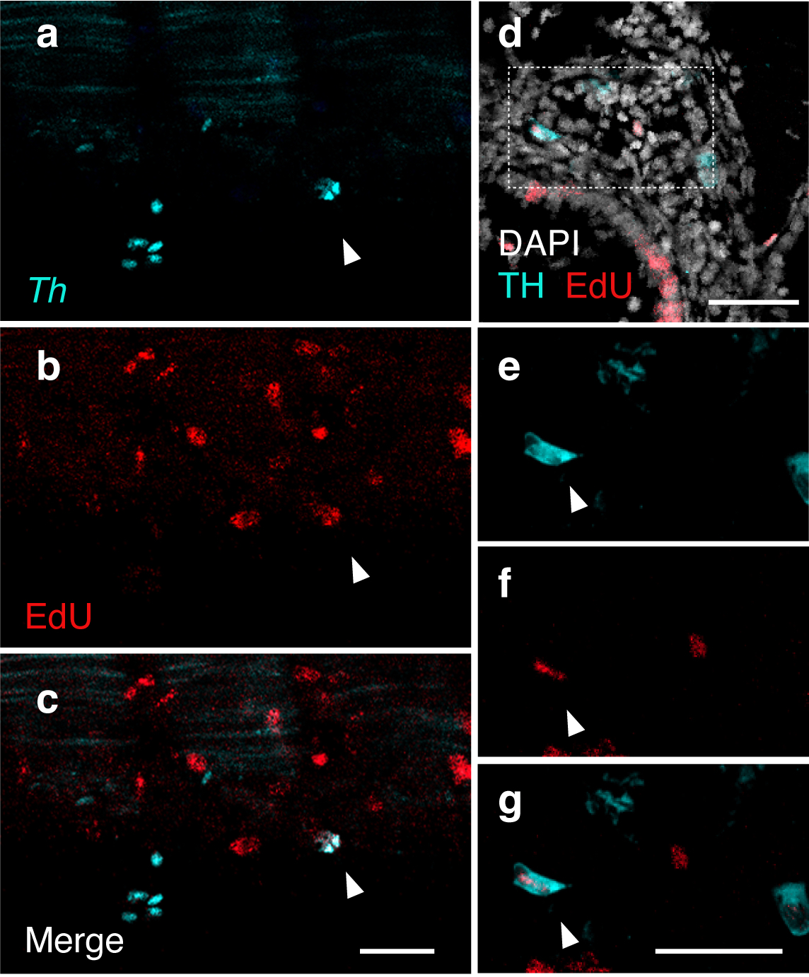
Ongoing proliferation of sympathoblasts in lamprey ammocetes: (a-c) HCR detection of Th (teal) and EdU staining (red) in ammocetes following an 8 hour EdU incubation. EdU detection in Th-expressing cells (indicated by arrowhead) reveals active division of sympathetic progenitors/neurons in lamprey trunk into ammocete stages. (d-g) Immunohistochemical detection of TH (teal) and EdU (red) co-expression in transverse sections of ammocetes following an 8 hour EdU incubation. Co-expression is denoted by arrowheads (n-p). DAPI is shown in white. Scale bars=50mm.
Brittany, were there any particular result or eureka moment that has stuck with you?
Brittany: Most of the experiments were performed on late-stage embryos and ammocetes that weren’t much larger, and as a result, a lot of our analyses documented sympathetic progenitors and immature neurons. To get a more mature population of sympathetic neurons for the final sequencing experiment, we actually had to source much larger, older ammocetes directly from the Great Lakes off-season. When they arrived at the lab, I was a bit shocked. They were so much larger than anything I was accustomed to working with, and I wasn’t sure if my tools were even appropriate for the dissections. The long and the short of it: the dissections were fine, but more importantly, the sympathetic trunks of these later-staged ammocetes were visibly discernible under the dissecting microscope. Of course we trusted our data from the late-stage embryos and the younger ammocetes, but I think it’s true that seeing is believing.
And the flipside: any moments of frustration or despair?
Brittany: When it comes to experiments and data, I try to keep a level head and clear perspective. As scientists, we are after the truth, and every clear result (even the ones we didn’t want or expect) gets us closer to the truth. The scientific process really does work, and having trust in that goes a long way on more challenging days.
What’s next for you, Brittany?
Brittany: Ultimately, I would like to be an independent investigator. I’m drawn to comparative embryology as a means to understand how peripheral sensory and autonomic neural systems first arose in vertebrates, and how genetic and environmental changes have driven diversification and adaptation of these systems over time. That’s a bit longer term, since I’ve just crossed the three-year mark as a postdoc, but in the meantime we have a collaboration with the Cai Lab at Caltech that I’ve been very excited about. We are looking to leverage their spatial barcoding technology, seqFISH, to better understand neuronal heterogeneity within the peripheral nervous system. Another endeavor I would like to mention is the work I’ve been doing with the support of the Caltech CTLO (Center for Teaching, Learning, and Outreach). One of my goals is to make hands-on science education more accessible to younger students, and with support from the CTLO and feedback from our local high school students, I have been developing grade-appropriate protocols and resources to introduce topics in embryology and neurobiology.
And Marianne, where will this story take the lab?
Marianne: We are continuing to work on many different neural crest derivatives in lamprey and would like to understand whether the gene regulatory circuits resulting in neural crest differentiation into things like peripheral neurons, craniofacial structures, etc. are conserved to the base of vertebrates. Right now, we are particularly interested in the enteric nervous system and how it has become elaborated.
I can’t believe it has been six months since I started this adventure as a group leader at the Center for Developmental Biology. Since then, few (but also lots of) things have happened.
One of the main things I would highlight is that the lab has grown. We are now four members!
Andrea Theodorou, who studied her BSc and MSc at Newcastle University, focusing on thyroid cancer and 3D in vitro models of Hepatocellular Carcinoma. She joins the lab as a Research Technician and is already immersing herself in Seville’s culture!
Irene Carrero Castro, who is completing her MSc in Omic Data Analysis and Systems Biology at the University of Seville. Her MSc thesis focuses on single-cell RNA-seq data analysis and she is being co-supervised by Dr. Fernando Casares and me.
Grace Wang, who is studying Computational and Applied Math, Data Science and Statistics at Rice University. Since 2022, she has been working on computational and mathematical projects together with Prof. Aryeh Warmflash and me, which hopefully we will be able to announce soon!
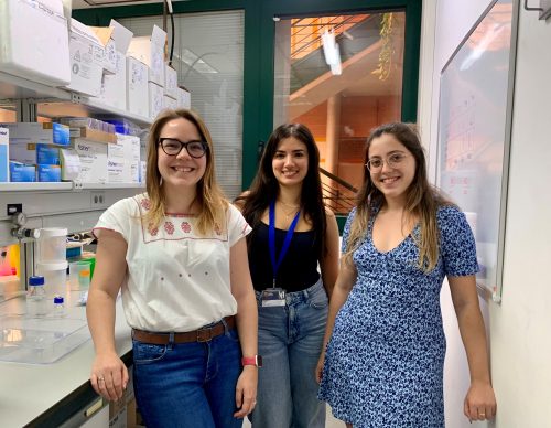
Last week we had a very special moment, as we took our first lab picture! Although we missed Grace, as she is located at Rice University, it feels great to be able to show the world the great team that is behind the scenes working on exciting projects. I feel very fortunate to work with these hardworking and brilliant scientists and even better people, and I can’t wait to see what we achieve together. If you want more details about our lab, please have a look at our (also recently finalized) website: https://systemsdevbiolab.com/
From left to right, Elena Camacho-Aguilar, Andrea Theodorou, and Irene Carrero Castro at the lab bench.
We also published my postdoc’s main piece of work on how combinatorial interpretation of BMP and Wnt signals controls cell fate decisions in early human development. Rice University wrote a press release with a nice summary of our work, but if you are interested in more details, you can find our publication here.
Apart from that, these months have been incredibly busy, and I have been trying to understand how to balance all the responsibilities as a new PI. I had heard and partially seen before how many hats one must wear as a PI, but I didn’t fully grasp the meaning of it until I started to experience it myself.
One of the hats that I am the most inexperienced with and that I am learning how to wear is the one involving bureaucratic processes. Although I thought I had seen it all after being an immigrant in a few countries, it seems like there are always things to learn :-) Jokes aside, last month, for example, I learned the steps needed for hiring people in the lab and helping them settle when the candidates are not from Spain. Luckily, with the advice of great colleagues, it all went smoothly, and if anyone is interested, I have made a step-by-step protocol for next time, which I am happy to share. The next bureaucratic step is to learn how to import reagents from abroad; wish me luck!
Balancing these bureaucratic tasks with other responsibilities has been a learning curve. To deal with long to-do lists, I am currently reading a very interesting book called Four Thousand Weeks. Unlike other productivity books, it encourages prioritizing tasks and accepting that time is limited, rather than trying to fit an endless to-do list into a finite day. As my to-do list grows exponentially, I am working on reflecting on what each task would entail before saying yes, even if it initially might seem exciting.
Overall, despite the challenges, I am thrilled with the progress and the new connections I am making. I hope to share some exciting experimental results in my next update. Talk to you soon!
This is part of the ‘Lab meeting’ series featuring developmental and stem cell biology labs around the world.
Where is the lab?
Our lab is in the center of Budapest, based in the Department of Anatomy, Histology and Embryology, Faculty of Medicine, at the Semmelweis University, Budapest, Hungary.
Our lab’s principal research goals are to understand the development of the enteric nervous system (ENS) and gut-associated lymphoid organogenesis, using the avian embryo as the model system. The avian embryo is a versatile embryologic model that we have leveraged to identify the role of multiple factors on the migration, proliferation, and differentiation of enteric neural crest-derived cells as they colonize the intestine to form the ENS. Abnormalities in this process lead to neurointestinal diseases like Hirschsprung disease, a congenital disorder in which ganglion cells fail to develop in the distal gut, leaving newborns with a bowel obstruction. We perform a variety of avian embryologic methodologies, including chimeras, parabiosis, gene overexpression and gene silencing studies using siRNA-RCAS viruses to examine the role of the extracellular environment in the embryonic gut that regulate the mechanisms of neural crest derived stem cell differentiation during intestinal morphogenesis.
Secondly, our lab focuses on mechanisms that underlie lymphoid organ formation. This work also uses the avian embryo as the model system, and addresses the cellular, molecular, immunological and morphological aspects of the primary and secondary lymphoid organ formation. Our aim is to discover how the avian lymphoid organs are built, and how immunosuppressive diseases affect their organization.
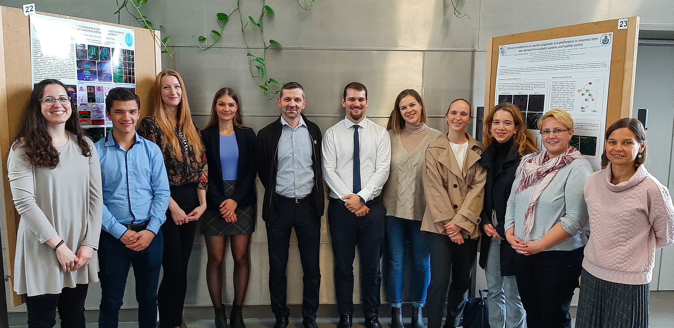
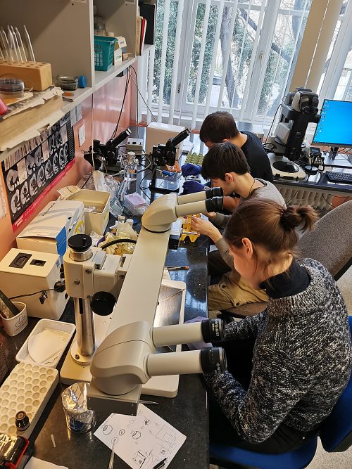
Lab group photo
Lab roll call
Nándor Nagy: As the head of the lab, my primary task is to ensure that everyone is enthusiastic about coming to the lab each morning. As a PI, I train the research staff to have the skills to execute research protocols competently. I am also a university professor responsible for leading the developmental biology course and teaching human anatomy classes for medical students.
Katalin Kocsis, associate professor: With decades of experience in teaching anatomy, histology, and embryology, I help students understand the complexity of the developing embryo. I am also providing the administrative background to our Developmental Biology course for medical students in both Hungarian and English programs.
Nóra Fejszák, post-doc: Using the avian embryo as a model system for early hematopoiesis, I investigate yolk sac stem cells in multiple avian species and their differentiation to tissue resident immune cells.
Viktória Halasy, PhD student: Currently I am writing my PhD thesis about the development of the extrinsic innervation of the colorectum.
Ádám Soós, PhD student: My main project is to establish the enteric neurosphere technique in the avian model system. I am responsible for the microscopy facility of our laboratory.
Emőke Szőcs, PhD student: I work towards understanding the inductive capacities of embryonic tissues and cell-cell interactions in forming primary lymphoid organs.
Zsanna Gecse, undergraduate student: As a third-year medical student, my task is to study the complexity of hindgut innervation, currently characterizing a new cell type in the mucosa of the avian hindgut.
Csenge Jurenka, undergraduate student: I joined the lab in the second year of medical school; I am trying to establish a novel Hirschsprung’s disease model to understand enteric neurocristopathies better.
Ábel Farkas, undergraduate student: I am a second-year veterinary medicine student. Since joining the lab, I started characterizing the development of the cloaca-associated lymphoid structure in domesticated birds.
Zsófia Bogya, undergraduate student: My student research project is to study the development of the dendritic cells in chicken primary lymphoid organs.
Réka Borbála Tóth, undergraduate student: I study bioengineering and try to understand the role of extracellular matrix proteins in the migration of enteric neural crest-derived cells.
Noémi Kegyes, lab assistant: I help with all ongoing projects in the lab, making sure everyone can continue their work with a smile on their face.
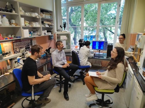
Ad-hoc lab meeting
Favourite technique, and why?
Nándor: My favorite techniques in studying the embryo are microsurgery and imaging. Avian embryos are accessible during all stages of embryogenesis and there is a large repertoire of methodologies, including tissue grafting, retroviral-mediated gene transfer, electroporation, and embryo culture I use to perturb and analyze gene function during development. Microscopy is the best way to show the complexity of the developing tissues and organs. Analysis of both live organ cultures and fixed tissue samples using transgenic lines or multiplex immunofluorescence, complemented by microscopy at different resolutions enables the examination of developing organism across various scales, from intracellular ultrastructures to complex multicellular tissues.
Apart from your own research, what are you most excited about in developmental and stem cell biology?
Nándor: I believe that being a stem cell and developmental biologist at this time is filled with excitement. While there has been significant progress in studying the embryo, the underlying biology and causes of many congenital diseases are still not well comprehended. By combining various techniques and resources, adopting a multidisciplinary approach that includes careful clinical phenotyping, genetics, developmental biology, and regenerative medicine, we have the potential to truly advance our understanding and stem cell treatment of congenital diseases within the next 5-10 years. Our goal is to be at the forefront of this important endeavor.
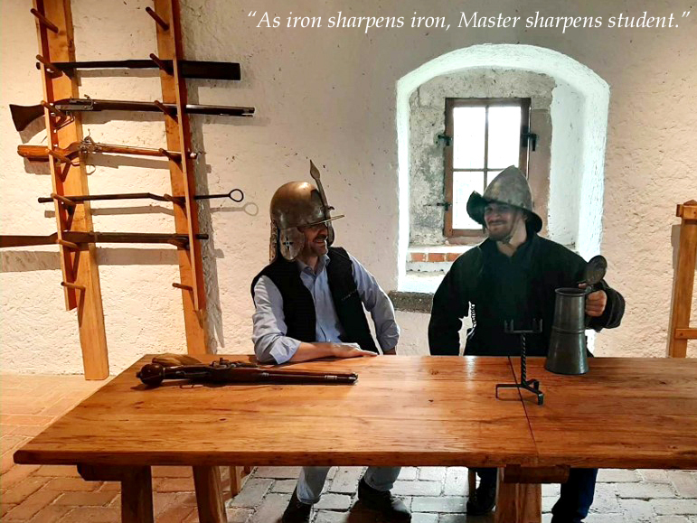
Our real teacher is the embryo
How do you approach managing your group and all the different tasks required in your job?
Nándor: I am privileged to lead this lab with highly motivated young scientists driven by their unique interests. Balancing between administrative and teaching tasks, I always enjoy working with my PhD and undergraduate students in the lab. Enthusiasm and fun are essential in science, and I believe lab members should be self-motivated, choosing projects that interest them. I dedicate time to help new students to acclimatize to the lab and encouraging them to find a topic to study and develop new ideas over time. Weekly lab meetings, joint lab meetings with other research groups and spontaneous discussions provide a strong foundation for our work and support the collaboration between the lab members. In addition, one-on-one meetings with lab members ensure their individual success, which I highly prioritize.
What is the best thing about where you work?
Nándor: The familiar atmosphere and numerous opportunities for scientific collaboration with other research labs in Hungary and internationally.
Katalin: In this lab one can always get the help and encouragement that is needed to be successful in the academic and educational fields.
Nóra: It is great to encounter the wonders and fragility of life almost every day through chicken embryos enclosed in eggs, and I have the opportunity to share this experience with a cohesive and supportive lab team in which new and exciting ideas are generated continuously.
Viktória: Our lab community exudes enthusiasm, cheerfulness, and unity, resembling a tight-knit group of friends, thanks to exceptional leadership. We prioritize teamwork and the sharing of thrilling new findings.
Ádám: The environment is very cozy, the lab members are really supportive. It’s a bit like a second family.
Emőke: Trying to understand the developing embryo is such an exciting task. We approach this with the most diverse methods, which makes day to day lab work so much fun – all while doing this with a great team.
Csenge: Whenever I am working in the lab, there is always someone to ask for help, get some advice from, or just have a chat with, and this gives me a lot of motivation.
Zsanna: Working in the lab is a fantastic opportunity for me to familiarize myself with laboratory techniques that I would not normally study and to deepen my knowledge in the field of embryology. I also love the fact that whenever I go there, I see the smiley faces of my coworkers who are always there to help me with any difficulty.
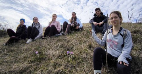
Field trip to Buda hill
What’s there to do outside of the lab?
Nándor: I love the vivid Budapest city and always enjoy its historical and multi-cultural hub character. I grew up in Transylvania (8 hours from Budapest), a historic eastern European region with truly wild mountain region, a land that is still rich in myths and legends. I love to go mountain hiking, visit castles and ruins, discover the various biodiversity, and spend time outdoors with my family. Budapest and Transylvania are the places to be!
Katalin: Living in the suburban part of the city with my family, we enjoy gardening around the house. Visiting the central part of the city is always like an interesting tour. Theatres, museums, the zoo – several exciting places to see frequently – not only in Budapest – but also in other parts of Hungary.
Nóra: Budapest is full of colorful life and offers a wide range of exciting indoor and outdoor activities. On workdays we sometimes go out for a coffee or an ice cream to a nearby park. During weekends I like to explore the hidden natural formations of forests around Budapest with my family. My favourite ones are the forests that belong to the floodplain of the river Danube.
Viktória: Beyond the lab, attending conferences and weekend trips always leads to memorable moments. During weekdays at night, we frequently chill out with activities like indoor rock climbing or ice skating in Városliget, the central park of Budapest.
Ádám: Our lab is close to the city center, making it convenient for us to unwind together outside of work. Whether it’s grabbing a drink at a local spot or engaging in activities like bouldering or hiking, there’s no shortage of options for us to bond and relax together.
Emőke: Budapest is a vibrant place; both city life enthusiasts and outdoorsy people can find activities to their taste. Just take a walk along the Danube in the evening, enjoy concerts, discover art galleries and museums, explore nature in the hills around Budapest, try kayaking on the river and have fun!
Csenge: There are some great bars near the lab, where we can celebrate after a successful conference, but Budapest itself gives great joy with its monumental buildings and wonderful scenery along the Danube.
Zsanna: I find Budapest the perfect place to be a university student, as the city is famous for its nightlife as well as its monumental historical heritage. Whether you are an extrovert who wants to meet friends every weekend at a new pub, or an introvert who wants to get lost in the museums, you will find your place here.
In the May Development presents… webinar, we celebrated the winners of Development’s 2023 Outstanding Paper Prize. Catch up on the recordings of the talks from the authors of the two winning papers.
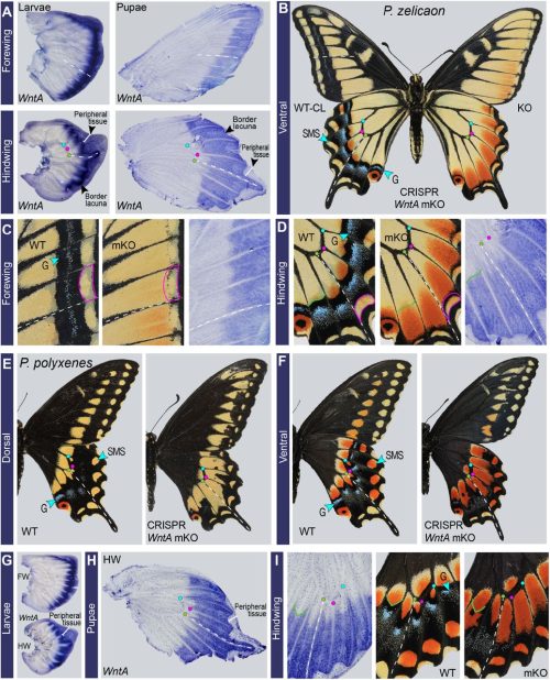
Ling Loh (The George Washington University) & Joe Hanly (The George Washington University and Duke University)
Do you know about Development’s ‘at a glance’ article types? These articles comprise a short text document highlighting the fundamental aspects of a developmental biology topic, which is accompanied by a large poster schematic that illustrates all you need to know ‘at a glance’.
Development has published a number of these articles over the years, ranging on topics from somitogenesis to gibberellin signaling, branching morphogenesis to the peripheral nervous system. To view the full collection, visit this page.
Some of the most recent, free-to-read articles are included below; to download the high-resolution image, click on the link in the ‘High-resolution poster’ or ‘Supplementary Information’ section of the online article. If you’d like your own physical copy of a poster to hang in your lab or office, look out for The Company of Biologists’ exhibit at the upcoming Society for Developmental Biology meeting (Atlanta, GA, USA) or the ISSCR annual meeting (Hamburg, Germany).
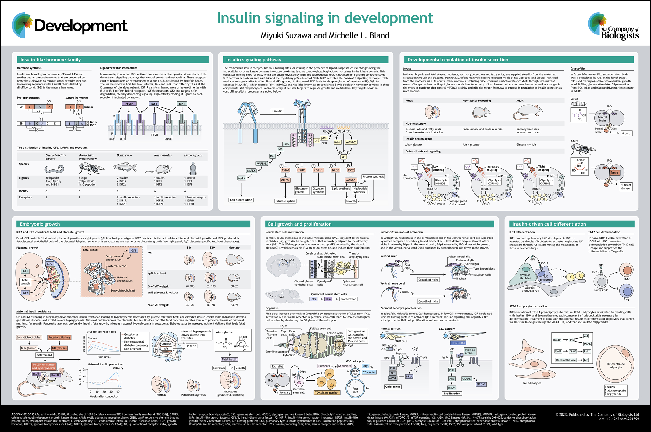
This Development at a glance article summarizes insulin hormone family signaling and highlights the roles of individual hormones in regulating growth, cell proliferation and differentiation.
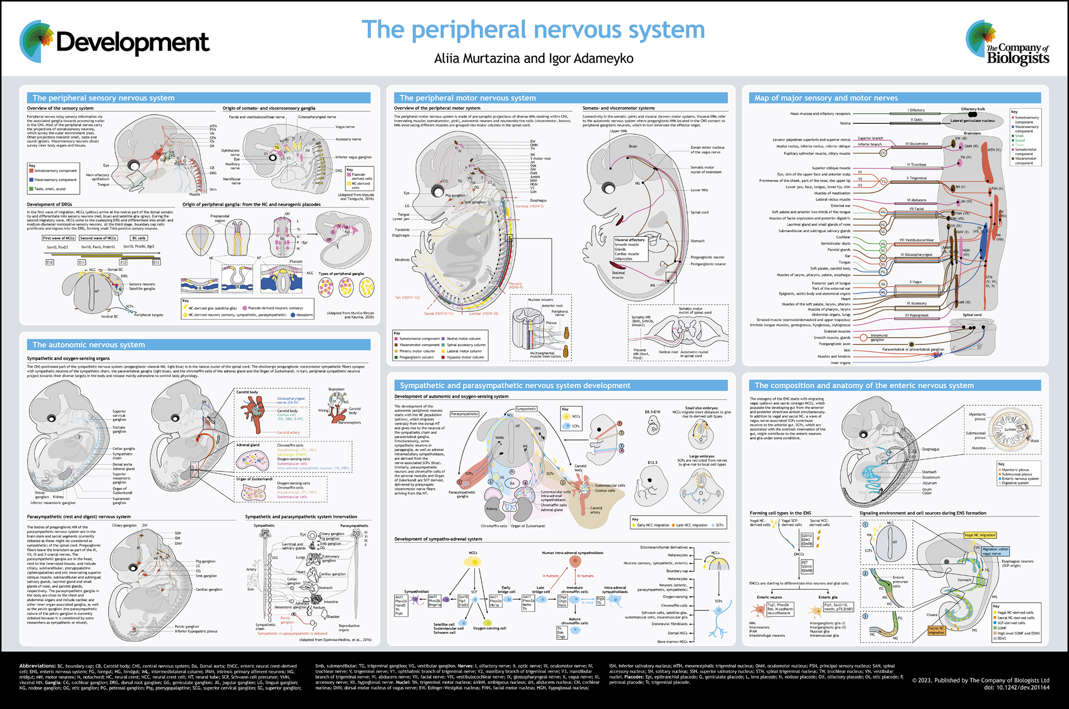
This Development at a Glance article provides up-to-date basic knowledge of the anatomical and functional organization of the peripheral nervous system (PNS), with additional insights into the development and cell-type heterogeneity underlying its different roles.
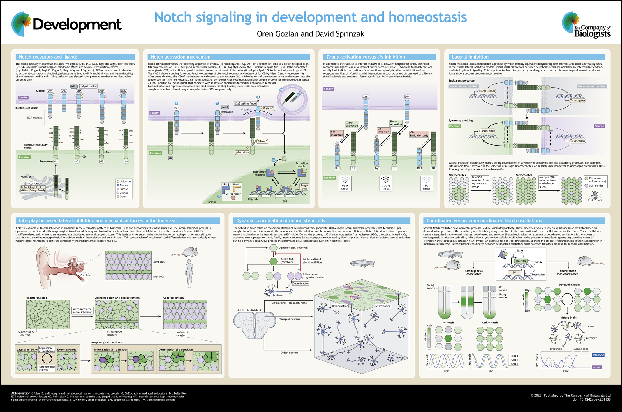
A brief overview of the Notch signaling pathway and its molecular activation mechanism, discussing different examples of Notch-mediated coordination of differentiation between neighboring cells during development and homeostasis.
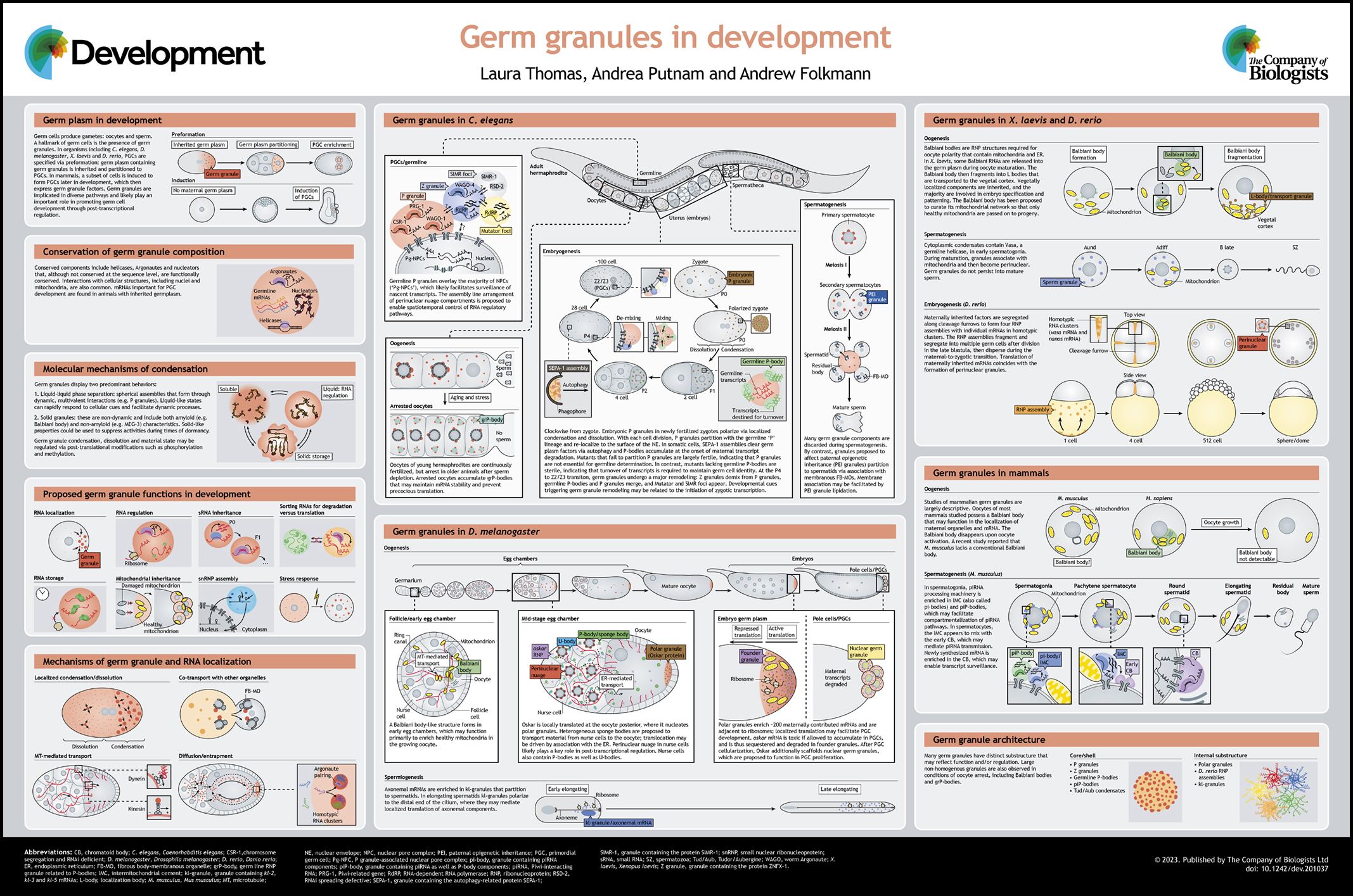
A survey of germ granules across organisms and developmental stages, highlighting emerging themes regarding granule regulation, dynamics and proposed functions.
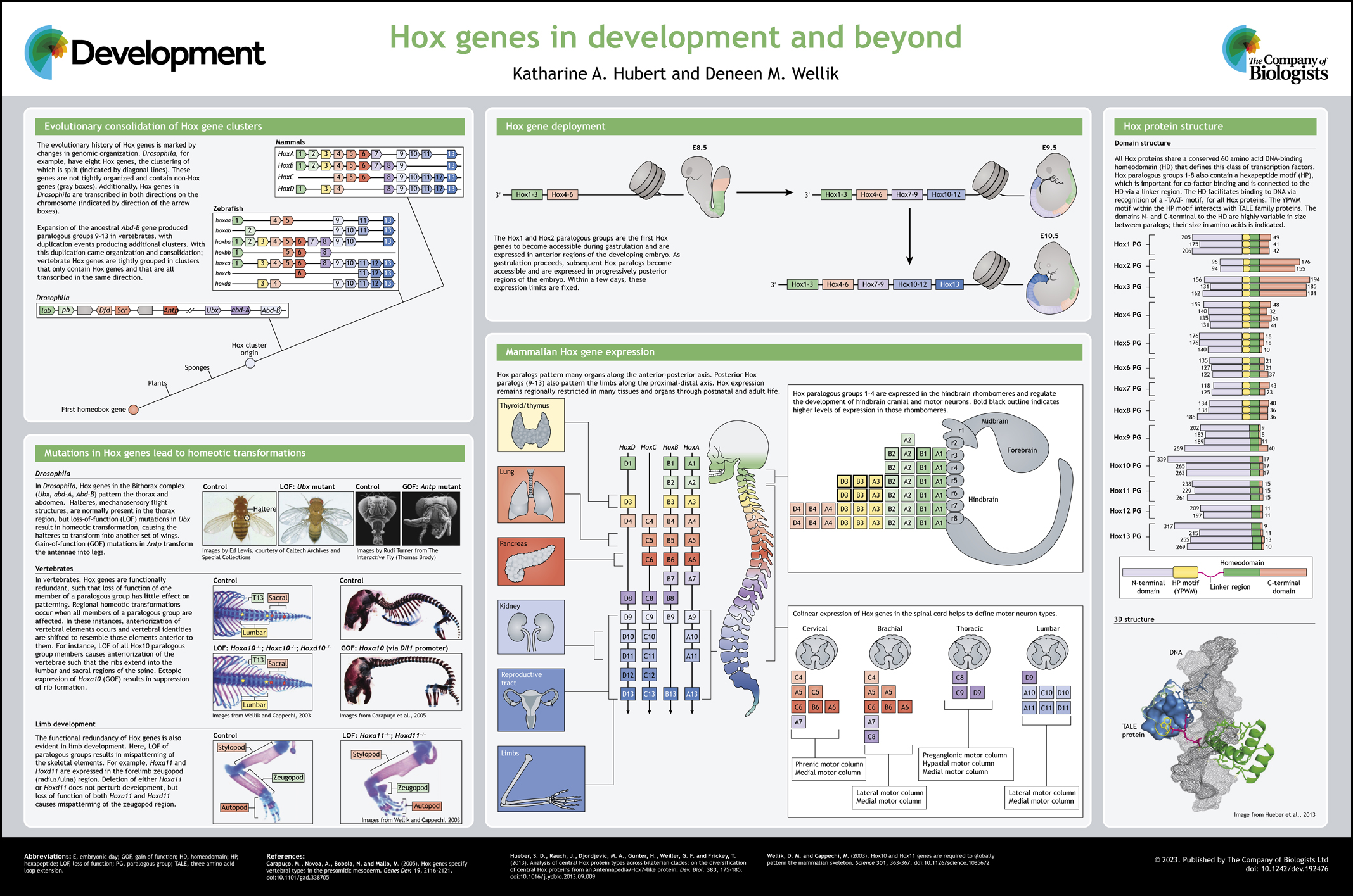
This Development at a Glance article provides an overview of the genomic organization, protein structure and regulation of Hox genes and our current understanding of their roles both during and after embryogenesis..
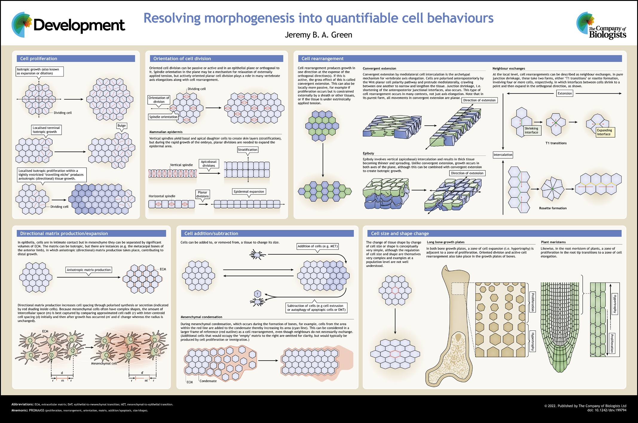
This Development at a Glance article describes how a cells’ limited repertoire of behaviours provide the basis for quantifying morphogenetic phenotypes.
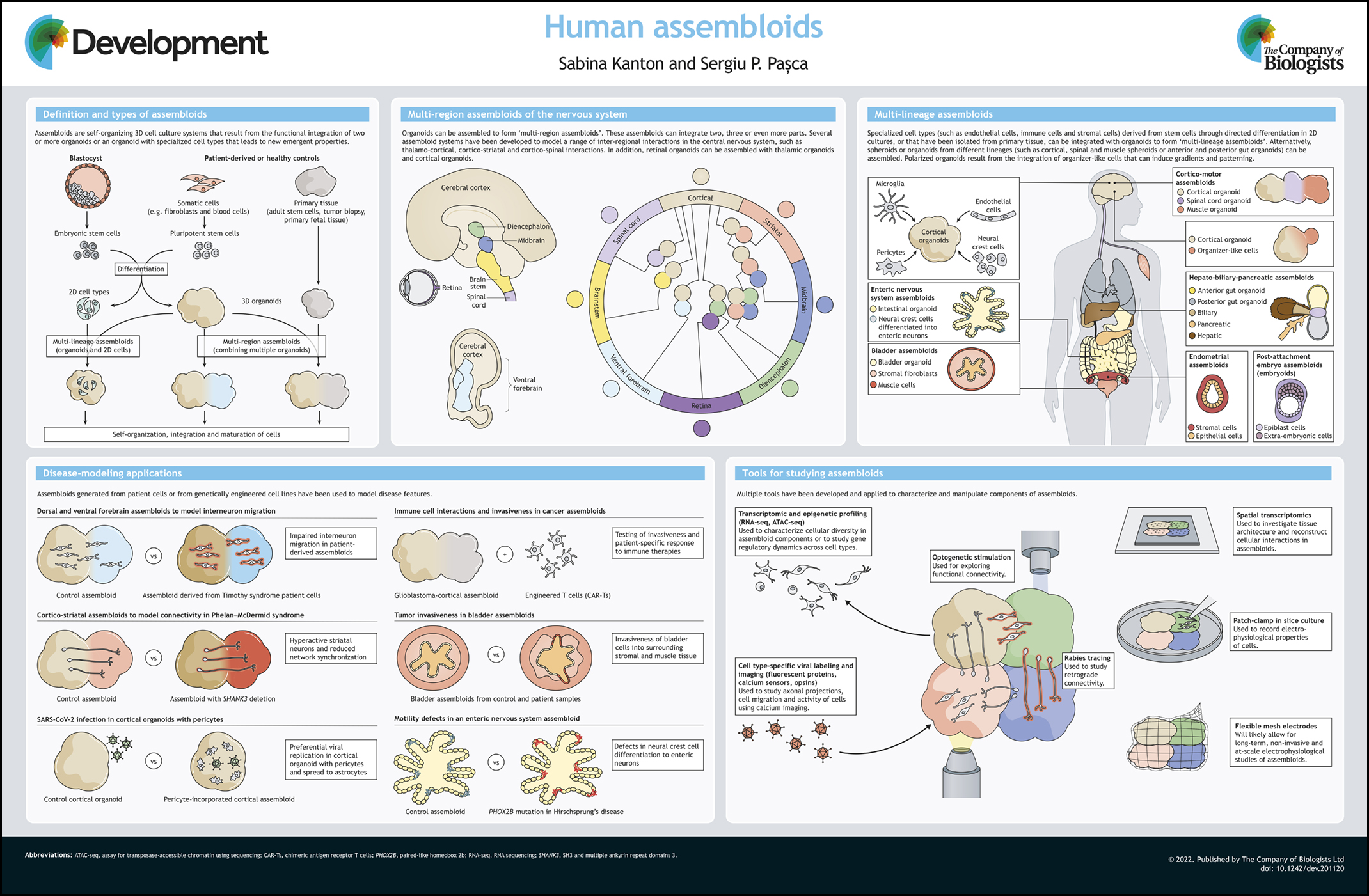
This Development at a Glance article gives an overview of the potential of assembloids – three-dimensional, self-organizing in vitro cell culture systems constructed by integrating organoids or organoids and other cell lineages.
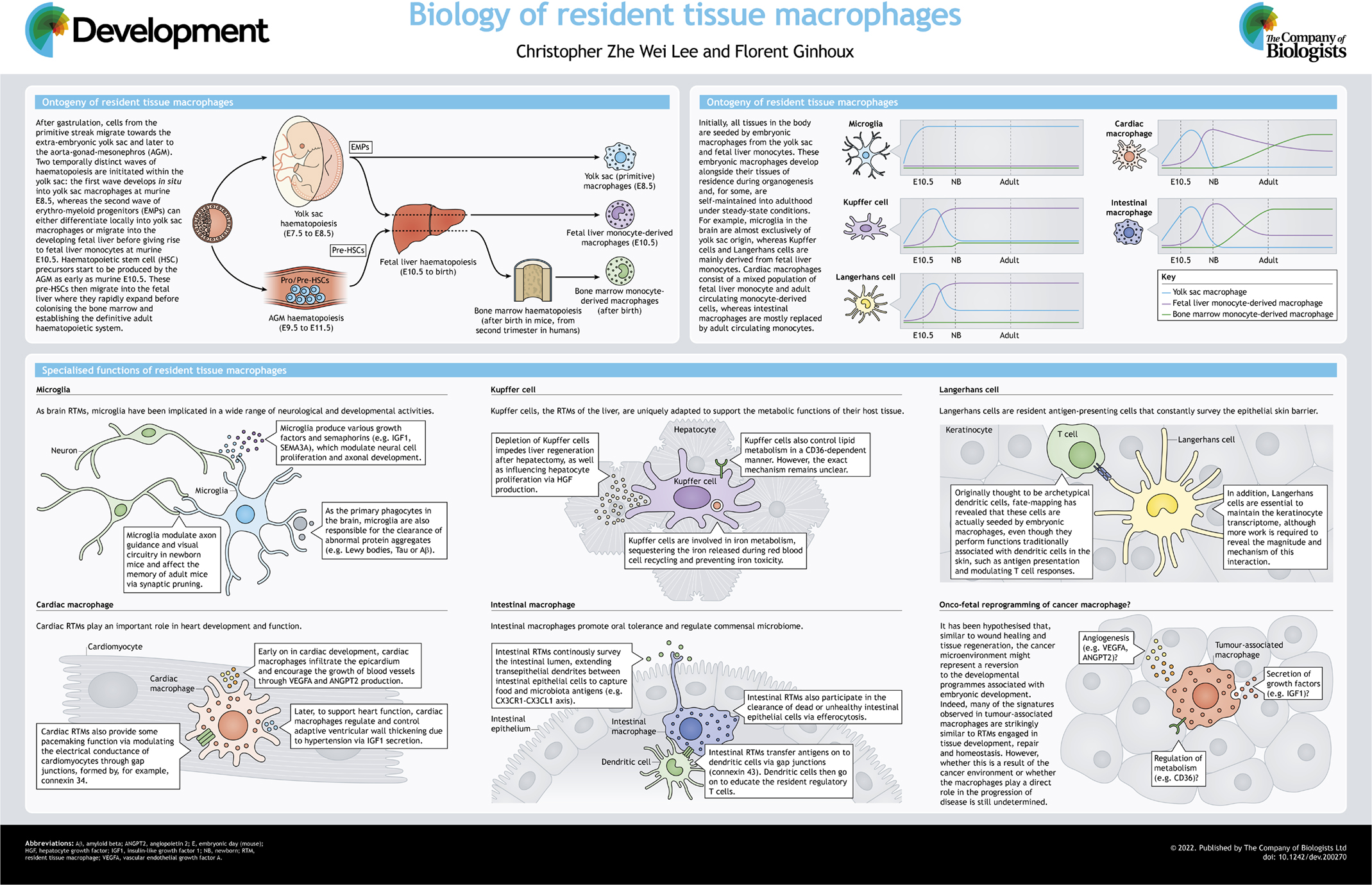
This Development at a Glance article gives an overview of the ontogeny, maintenance and unique tissue adaptions of macrophages, and highlights their role in development, homeostasis and dysfunction.
 (4 votes)
 (No Ratings Yet)