The community site for and by
developmental and stem cell biologists

Woods Hole images round 4- the winning movie

Posted by , on 30 September 2013

This year’s last Woods Hole competition had an exciting development- instead of choosing from 4 images, we asked the Node readers to choose from 4 stunning movies. It was probably hard to choose who to vote for, but in the end the beautiful movie of the ascidian metamorphosis was the big winner. The following collection of stills from the movie will be in the cover of a future issue of Development, while the movie itself will feature in the homepage of the journal.


Woods Hole movie- Ascidian metamorphosis


Congratulations to Matthew Clark, from the University of Oregon, for the winning movie.

The runners-up to this competition were Marina Venero Galanternik (University of Utah), Rodrigo G. Arzate-Mejía (Universidad Nacional Autonoma de Mexico), Jennifer McKey (Universite Montpellier) and William Munoz (The University of Texas MD Anderson Cancer Center) for their movie of Drosophila embryogenesis; Daniela Di Bella (Fundacion Instituto Leloir), Joyce Pieretti (University of Chicago), Saori Tani (Kobe University) and Manuela Truebano (Plymouth University) for their movie of cell divisions in C.elegans; Eduardo Zattara (University of Maryland, College Park) for his movie of zebrafish lateral line migration.

Thumbs up (2 votes)
Loading...

Tags: , ,
Categories: Images, News, Video

Navigate the archive

Use our Advanced Search tool to search and filter posts by date, category, tags and authors.

Stem Cells: An Insider’s Guide – the new book by Paul Knoepfler

Posted by , on 30 September 2013

coverI recently had the pleasure of interviewing Associate Professor and dedicated stem cell blogger Dr. Paul Knoepfler about his upcoming book “Stem Cells: An Insider’s Guide”. The book covers everything from defining stem cells and their clinical applications to stem cell regulation and even some stem cell urban legends. Here, Paul chats about his book, his own thoughts on the stem cell field, and possible approaches for cloning John Lennon…

Congratulations on your book, it’s a real stem cell tour-de-force. Can you tell me what motivated you to start writing this?

I’d been working on my stem cell blog for about three years and finding it really rewarding as an additional element to my professional life. It occurred to me that I could probably expand the audience further by putting some of my thoughts down in the context of an actual book. One of the things I did was to go on Amazon and look at the different books that were out there on stem cells. I found that there is this big gap: some are probably over-simplified, but most of them are way too scientifically dense, half a sentence on half a page, that sort of thing. I couldn’t really find the kind of book that I would have liked to read, or that a friend of mine who is not a scientist but is interested in stem cells would want to read. I tried to write that book, and hopefully I was successful.


So your book is aimed at both scientists and non-scientists?

I’m hoping to have a book that scientists will enjoy but also a wider audience can comfortably read and understand. I also want to challenge people to think, but I don’t want people to feel like they are reading a textbook. I aim to be in the middle, where people are challenged, but not overwhelmed.


Paul Headshot2013.resizeI think you do challenge people, partially because you present many different views, not just your own.

I went into this project trying to write a book that has a unique voice. Like you said, I do try to include other people’s opinions, but I didn’t shy away from including my own. And so that is a little different, or actually really different, from what we are formally taught in science. I intentionally said in the book that these are actual opinions, not necessarily facts. I think that’s helpful because it gets people’s attention, even if they don’t necessarily agree with me. Maybe it makes them think in a new way, makes them talk about it and gets a discussion going. I also wanted to get some humour in there, because even if it is a little edgy or risky, it’s also kind of fun.


You are also a cancer survivor. Was there a more personal motivation for demystifying the stem cell hype? 

Yeah, that definitely played a role in all of this. Having cancer has really opened my eyes to the fact that there is another side to this: the patient side. I think for me, having been a patient, it helped me to empathize and connect more with patients in the stem cell arena, because they are very vulnerable. They are often facing very devastating diseases and injuries and they don’t feel like conventional medicine has anything to offer them. So I try, by my own experience as a cancer patient, and also as a stem cell scientist, to help them in whatever way I can. But also I empathize with the situation they are in, because often times they’re willing to take more risks than I am comfortable with. They have a different perspective and I try to respect that. I try to take that patient perspective with me and put it into the book. I don’t think it is good to just write a book about stem cells without including things that are important for the patient. Having been a patient myself, it helps me to be more conscious of those issues.


You talk in the book about stem cell tourism and the problems it creates for patients and for the field. Whose responsibility do you think it is to crack down on stem cell tourism? 

That’s a great question, and I don’t necessarily have all the answers. I don’t know if people are aware but there is a sort of ad hoc group of stem cell scientists and bioethicists around the world who have been activists on this issue. And I include myself in that group, because we as individuals are putting ourselves at risk, promoting evidence-based medicine in the stem cell field and encouraging patients to ask more questions before jumping into a risky situation. Many of us, including myself, have experienced being threatened with litigation and serious retaliation in other ways. So I don’t think that model is really going to work in the long run because it puts too much risk and responsibility on individuals as human beings. And I am not saying that there isn’t a role for individuals, but I think that somehow we need to have a higher level response to stem cell tourism. While down here in the trenches there isn’t a whole lot of concrete data on stem cell tourism, there is definitely a feeling that the problem is accelerating, so I don’t think that just a group of ad hoc individuals is going to be able manage it. The FDA, at least in the jurisdiction of the US, has a responsibility to work on these issues. But it starts becoming very complicated when you are dealing with international companies, because the FDA doesn’t have jurisdiction outside the US.

sc bus

So then it really comes down to education. You’ve clearly tried to address this with your book, but is there a greater need to educate not just patients but doctors as well?

That is definitely something that I have advocated for. I proposed earlier this year for medical schools to start having a formal stem cell training program for physicians (see http://www.ncbi.nlm.nih.gov/pubmed/23477401). If you have a gastrointestinal problem, you want someone who has years of training dealing with gastrointestinal problems. It’s the same kind of thing with stem cells. I want patients to raise their expectations and to ask more questions. So I think you’re totally right, education might be in the long run the most effective way to go, so that patients and potential patients who think that they might want to get a stem cell transplant actually have resources that they can easily obtain. So that they can think “Ok, I should be asking this question, I should be asking that question, I should be skeptical about this” and so on. I am a real believer in outreach and education.


In your book you list over 10 different diseases that may be amenable to stem cell therapies. How close do you think we are to any one of those?

I don’t think we are as close as I would hope we are, and I don’t think we are as close as many other people hope we are. I wish it were different but the unfortunate reality is that if you want to do this kind of science properly, and I really think we need to do that, it’s a relatively slow process. You have to accept that there are going to be setbacks, and some things aren’t going to work, and other things will work. I am cautiously optimistic that within a decade, macular degeneration might be one of the first diseases where we see a substantial impact in a significant number of patients using stem cell technology. I have some optimism, and to me the basic idea behind that approach seems very straightforward and logical. And so I would say I am pretty hopeful on that. I think we are going to see some progress on diabetes too. I definitely wouldn’t use anything close to the word ‘cure’, but then again I think that you can see a pathway to helping patients via stem cell technology. It might take a decade, but you can see a roadmap where you can regenerate either endogenous beta cells that were dormant, or put in a new mini-pancreas from stem cell-derived beta cells. I believe there are definitely reasons for hope.

stembucks

You also encourage us to “get your geek on” and think about far-out applications of stem cells. Of all the ones that you discuss in your book, which would you most like to see happen?

I would love to see whole organs becoming available. Because nowadays, for example, if you get really serious liver disease, in the end there is a very good chance that is going to kill you even if the rest of your body is healthy. So I think this idea that you can just go get a new liver, or a new pancreas, or regrow your hand; I think that’s both really cool and practically speaking could have a spectacular impact on human health. I also like space related stuff. I was surprised to find that NASA actually funds a lot of research for stem cells in space. That is one of my own stem cell geeky fascinations. I think that is kind of cool.


I would like to see dinosaurs…

I have mixed feelings on cloning, I have actually come out against some of that stuff. I think it is going to be a tough one technologically speaking, but it’s very fascinating and it does capture peoples’ imagination that they might see a woolly mammoth. I am not so sure about human cloning: some guy recently bought John Lennon’s old tooth, because he wanted to clone John Lennon. I guess he was hoping to get some DNA or something. There are a lot of weird, potentially dangerous, yet fascinating ideas out there.


You have been named one of the Top 50 most influential people in the stem cell field. Where do you think the influence comes from, and how do you intend to use it?

When I started my career I would have hoped that my influence was purely scientific, but I think realistically that ranking in the top 50 must have had a lot to do with my blog and me being somehow a leader in stem cell social media. I know from the people who confidentially contact me that a lot of the other top 50 and other stem cell bigwigs do read my blog very regularly. It doesn’t mean that they agree with me, and they may even think I am totally crazy on some topics, but they come and read it so I think that is where that influence comes from in a large part.

My hope is to use my influence in a positive way: one of my biggest goals is to get people communicating more, people who wouldn’t normally talk to each other. And also to get patients connected up with resources. And I have definitely encouraged patients to try to find researchers’ emails in articles: the researcher may not respond to you, but then they might. That is my biggest goal, to get people more interconnected and educated.


alphabet soup

Stem Cells: An Insider’s Guide is available now via World Scientific and Amazon.com 

Thumbs up (7 votes)
Loading...

Tags: , ,
Categories: Book Reviews, Interview

And in the beginning was histone 1

Posted by , on 30 September 2013

A team at IRB Barcelona identifies an essential protein for embryonic viability during the first cell divisions in the fly Drosophila.

This protein, called dBigH1, which is a variant of histone 1, could also be associated with fertility issues.


A zygote is the first cell of a new individual that comes about as the result of the fusion of an ovule with a spermatozoid. The DNA of the zygote holds all the information required to generate an adult organism. However, in the first stages of life, during the so-called embryogenesis, the genome of this zygote is repressed and does not exert any activity.

In the fly Drosophila melanogaster, the genomes of the zygote are repressed until the thirteenth division, after which the embryo starts to express its own genes. Headed by Ferran Azorín, also CSIC Research professor, the Chromatin Structure and Function group at the IRB Barcelona has identified a protein in Drosophila that keeps the zygotic genome inactive until the correct moment. This function is vital for embryo life because without dBigH1 the genome is switched on too early and the embryos die. The results are published in Developmental Cell, the most important journal of the Cell group devoted to development.

This is the first time that scientists have described a specific function of histone 1 during embryogenesis. Although this protein is present in the first embryonic stages of humans and mice, nothing is known about its function.

“The fact that now we have also detected this protein in Drosophila has allowed us to study its vital activity during early stages of embryonic development more quickly and efficiently,” explains Salvador Pérez-Montero, PhD student and first author of the study, and Albert Carbonell, postdoctoral researcher who joined the project a year ago. “If this same function is conserved in humans, its alteration could be related to gestational disorders or early miscarriage,” says the head of the group Ferran Azorín.

The scientist goes on to explain that “they are not disorders —in the true sense— that are commonly treated and, in fact, problems during gestation can arise for many different reasons.”
 
 
Future studies on infertility

The protein dBigH1 could also be related to male and female fertility. In this study the scientists have revealed that this molecule plays a fundamental role in fly embryogenesis, but they are now focusing on defining the function of this protein in germinal cells.

The so-called germline comprises the sex cells, namely the cells that give rise to ovules and spermatozoids, and thus the very cells responsible for passing down genetic information from one generation to another. In the Drosophila embryo, even in the first divisions about 40 germline cells separate and differentiate and all of them express the protein dBigH1.

The scientists already have the first functional results, which point to dBigH1 regulating sperm production in males and ovule production in females. “When this gene is removed, this process is totally disrupted,” explain the researchers.

The next paper is expected to reveal whether there is indeed a relationship between the protein dBigH1 and individual fertility, and if so, the potential biomedical applications of this new discovery.
 
 
Reference article:

The Embryonic Linker Histone H1 Variant of Drosophila, dBigH1, Regulates Zygotic Genome Activation Salvador Pérez-Montero, Albert Carbonell, Tomás Morán, Alejandro Vaquero, and Fernando Azorín.
Developmental Cell (2013),

This article was first published on the 30th of September 2013 in the news section of the IRB Barcelona website


Thumbs up (1 votes)
Loading...

Tags: , , , , ,
Categories: Research

Biology Bytes: A Stem Cell-Focused Book for the Public

Posted by , on 22 September 2013

I recently published a book, called Biology Bytes: Digestible Essays on Stem Cells and Modern Medicine, which is on stem cells and other medical-related topics, and thought it would be of interest to readers at The Node. A large part of the book is just on stem cells — specifically, an entire chapter (74 pages) is on the many different stem cell types and relevant cutting-edge research (including a general introduction to stem cells), and another chapter is dedicated to bioengineering organs, regeneration in the salamander, and the amazing life-cycle of the jellyfish Turritopsis nutricula. The book is geared towards the general public, so it is perfect for introducing someone to these fields.

Here are some more details on the book:
“In Biology Bytes: Digestible Essays on Stem Cells and Modern Medicine, author Dr. Teisha J. Rowland discusses the history and latest scientific advancements in these fields of science, and many more. With a specific focus on issues that we increasingly encounter in the modern world around us, Dr. Rowland explores cutting-edge science through essays that can be easily digested: complex scientific concepts are broken down into key points based on the latest discoveries, technical jargon is clearly explained, and the impacts of these discoveries on our lives are explored. This book includes comprehensible explorations of a wide range of topics, including different types of stem cells and treatments they may be used in, the development and impact of in vitro fertilization (a technique responsible for over 1% of U.S. births today), how and why GMOs are made, the creation of vaccines to fight cancer, and fascinating food science behind delectable drinks such as beer, wine, and tea.”

The book is available as a Kindle eBook or paperback at Amazon.com. Feel free to spread the word to anyone you think may be interested.

Also, for those who may be interested, I recently published a second similar book that is focused on animals, called Biology Bytes: Digestible Essays on Animals Both Commonplace and Bizarre, which is also available through Amazon.com.

Thumbs up (1 votes)
Loading...

Tags: , ,
Categories: Book Reviews

Stem cell success is team work!

Posted by , on 17 September 2013


93773 Figure 3 cropped


Stem cell beauty…yes they are so beautiful, they have amazing properties, they bring a lot of hope for future therapies…well, yes they are the stars!

However, like in everyday life, “the star” is not the only one to be the key of its own success, success is teamwork! And for stem cells, the supportive team is the stem cell niche, also called microenvironment.

Studying stem cell niches is key to understanding stem cell biology since the niche directly influences stem cell fate choices, such as proliferation and/or differentiation.

Several factors regulate stem cells within the niche: interactions between stem cells and their neighboring cells but also interactions between stem cells and the surrounding matrix components or nutritional inputs (growth factors for example).  In addition, the physiochemical surrounding (pH, temperature,…) is very important within the niche.

This month in Development , Gancz and Gilboa show that the nutritional input conveyed by the insulin signalling pathway plays key roles in the development of the ovarian niche-stem cell units in Drosophila melanogaster. In this picture, we can observe Drosophila melanogaster ovaries at LL3 stage of development. When the insulin receptor is overexpressed (on the right panel), the ovary is much bigger than normal (on the left). Also, there are more terminal filaments cells (in pink), a major component of the germ stem cell niche.

Interestingly, their study shows that in addition of strongly influencing the development of the germ stem cell niche (terminal filament cells and intermingled cells), insulin signalling also regulates the number of germ stem cell precursors (ancestors of mature ovarian stem cells, in blue in the picture). So, insulin signalling contributes to ensure that germ stem cells and their niche develop coordinately.

Extensively describing niche components and understanding how they influence stem cell decisions is a major challenge in stem cell biology. Indeed, reproducing stem cell-niche interactions in petri dishes would give scientists the keys to instruct stem cells in the directions they are interested in!


D. Gancz, L. Gilboa, Insulin and Target of rapamycin signaling orchestrate the development of ovarian niche-stem cell units in Drosophila. Development,  (Sep 11, 2013). doi:10.1242/dev.093773


Thumbs up (2 votes)
Loading...

Tags: , ,
Categories: Research

In Development this week (Vol. 140, Issue 19)

Posted by , on 17 September 2013

Here are the highlights from the new issue of Development:


Roadmap for human trophoblast model

F1.smallCorrect trophoblast development is essential for placenta function and embryogenesis. In humans, the early stages of this process can only be modelled in vitro using embryonic stem cells (ESCs); however, owing to the failure to identify the stepwise progression that occurs during differentiation, the identity of the resulting cells is not clear. Now, on p. 3965, Mana Parast and colleagues narrow this gap by directly comparing various stages of in vitro trophoblast differentiation with in vivo cell types in the human placenta. The authors find that in vitro BMP4-driven trophoblast differentiation progresses through a p63+/KRT7+ stage that is akin to cytotrophoblast (CTB) cells in vivo. The CTB cells are the precursors of the terminally differentiated syncytiotrophoblast (STB) and extravillous trophoblast (EVT) cells. As expected, ESC-derived CTB-like cells can be differentiated further into functionally mature KLF4+ STB and HLA-G+ EVT cells. The authors propose that BMP4-driven differentiation of human ESCs is an accurate and informative model of human trophoblast development.


Plant germ cells count on chromatin

F1.small-1Unlike animals, plants do not set aside germ cells during embryogenesis. Instead, the precursors of these cells, called spore mother cells (SMCs), are generated via a somatic-to-reproductive transition that occurs later in life. Although epigenetic remodelling has been largely studied in the post-meiotic phase of germline development, it is unknown whether pre-meiotic events contribute to cellular reprogramming in the reproductive lineage. Now, on p. 4008, Célia Baroux and colleagues investigate these dynamic changes and uncover widespread chromatin reprogramming during the slow meiotic S-phase that accompanies specification of the female Arabidopsis SMC. As in animal primordial germ cells, the authors observe increased nuclear size, reduction in heterochromatin, depletion of linker histones, chromatin decondensation, changes in histone modifications and core histone variants in the female SMCs. The authors propose a bi-phasic chromatin reprogramming process that is necessary for proper somatic-to-reproductive cell fate transition and thus competency to establish the pluripotent, female gametophyte.


Vascular remodelling goes with the flow

F1.small-2Mechanical forces such as blood flow play a key role in regulating vascular remodelling and angiogenesis. Vessel diameter must be tightly controlled to establish correct hierarchical vascular architecture, but how this is achieved is unclear. Now, on p. 4041, Mary Dickinson and colleagues report that vessel diameter in the mouse embryonic yolk sac is regulated by blood flow via two distinct mechanisms: vessel fusion and endothelial cell migration. Using live confocal imaging of dual Flk1-reporting whole embryos, the authors observe that vessel fusion only occurs in areas of relatively high flow, and that this leads to a rapid increase in vessel diameter. In addition, high flow also facilitates the preferential recruitment of endothelial cells from smaller capillaries, which further increases vessel diameter. Analyses using the Mlc2a mutant, in which blood flow is compromised, supports these findings. This study highlights the importance of mechanical forces in regulating individual cell behaviour as well as more complex processes such as the establishment of correct vascular architecture.


Mechanical forces fly high in wing morphogenesis

Mechanical forces regulate cellular behaviour to guide correct tissue size and shape. Distribution of stress can affect individual cell morphology, but how global forces are balanced across the whole tissue is largely unknown. In this issue, two papers examine how mechanical stress influences cell shape, and how these forces converge to regulate tissue morphogenesis in the developing Drosophila wing.

F1.small-3On p. 4051, Thomas Lecuit and colleagues investigate the global pattern of stress in the developing wing disc. They find that cell shape is governed by stretching at the periphery and compression at the interior, which may arise from increased proliferation at the centre of the wing. Cells at the periphery counteract this stress by polarising Myosin II tangent to the wing pouch, and by switching their cell division axis to align with the direction of stress. To support these findings, the authors inactivate Hippo signalling to mimic the effect of overproliferation. They observe a loss of the directionally dependent stress effect and a reduced apical surface in the mutant clones compared with their wild-type counterparts, which results in distortion of the bordering cells. These results suggest that the pressure exerted by proliferating cells influences global mechanical stress, which in turn regulates tissue morphogenesis at the periphery of the wing.

F2.smallThe role of mechanical stress in regulating cell shape and tissue morphogenesis is further examined by Shuji Ishihara and Kaoru Sugimura (p. 4091), who investigate the formation of hexagonal cell geometry in the phase II pupal wing. The authors use live imaging and Bayesian inference to map global mechanical forces in response to defined perturbations. They show that the direction of stress originates primarily from the attachment site to the wing hinge, and that this results in a redistribution of myosin, which enables greater tension at the cell junction as a means to resist tissue stretch. This in turn provides directional information for the cells, which ultimately results in the hexagonal configuration and a structurally balanced wing.


Distinct SCL isoforms regulate HSC emergence

F1.small-4The emergence of haematopoietic stem cells (HSCs) from the aorta-gonad-mesenephros (AGM) region requires the coordinated activity of multiple transcriptional networks. The transcription factor stem cell leukemia (SCL) is necessary for this process; however, multiple scl isoforms exist and the exact stage at which each is required is unknown. In this issue (p. 3977), Zilong Wen and colleagues deconstruct the stage-specific requirement of scl isoforms scl-α and scl-β during the emergence of HSCs from the ventral wall of the dorsal aorta in zebrafish, which is equivalent to the AGM. Using dual fluorescent reporters coupled with in vivo time-lapse imaging, the authors show that scl-β is expressed in the haemogenic endothelial cells, whereas scl-α is expressed after the endothelial-to-haematopoietic transition has occurred. In loss-of-function studies, the authors show that sclβ is necessary for the specification of the haemogenic endothelial cells, whereas scl-α acts to maintain the newly born HSCs. This study represents an important step in teasing apart the transcriptional complexity that governs HSC development.


PLUS:


The developmental origins of adipose tissue

adiposeThe formation and maintenance of adipose tissue is essential to many biological processes and when perturbed leads to significant diseases. Here, Jon Graff and colleagues highlight recent efforts to unveil adipose developmental cues, adipose stem cell biology and the regulators of adipose tissue homeostasis and dynamism. See the Review on p. 3939


The regulation of Hox gene expression during animal development

HoxHox genes encode a family of transcriptional regulators that elicit distinct developmental programmes along the head-to-tail axis of animals. Here, Mallo and Alonso examine the spectrum of molecular mechanisms that control Hox gene expression in model vertebrates and invertebrates.  See the Review article on p. 3951

Developing insights into cardiac regeneration

Heart
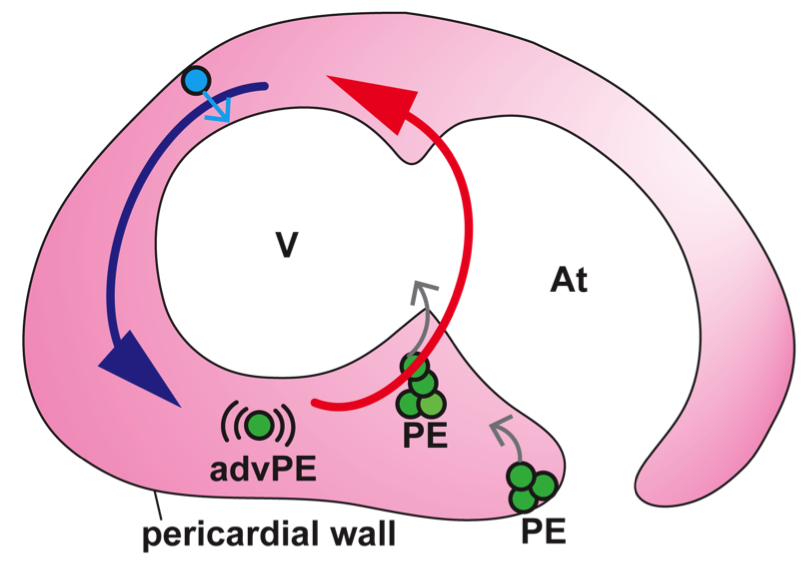
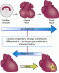
The recent EMBO/EMBL-sponsored symposium ‘Cardiac Biology: From Development to Regeneration’ gathered cardiovascular scientists from across the globe to discuss the latest advances in our understanding of the development and growth of the heart, and application of these advances to improving the limited innate regenerative capacity of the mammalian heart. Christoffels and Pu summarize some of the exciting results and themes that emerged from the meeting. See the Meeting Review on p. 3933


Thumbs up (No Ratings Yet)
Loading...

Categories: Events, Research

(Developmental) Biology around the internet- September 2013

Posted by , on 16 September 2013

Here is our monthly round-up of some of the interesting content that we spotted around the internet:


News & Research

– Nature reported this month that stem cells have been used to generate minibrains in vitro. You can read an excellent article by Ed Yong on this work .

– The hilarious 2013 IgNobel Prizes that ‘first make people laugh and then make them think’ have been revealed. Check out the list of winners!

– A paper was published this month reporting the first case of successful in vivo cellular reprogramming.

– How good is your stem cell knowledge? Take the Knoepfler lab 2013 stem cell quiz!

– And if you are keen on making a poster to communicate your research to the general public, here are some tips from the British Science Association.


Weird & Wonderful 

– We found two great science-inspired art websites. We found a website dedicated to art in a petri dish, as well as an artist who creates beautiful microscopy-inspired glass sculptures.

World Cell Race 2013 is now accepting applications! Do your in vitro cells have what it takes to be a winner?

– Science-inspired arts and crafts make another appearance this month, with this pattern to knit an axolotl as well as a brain-inspired hat.

– Check out the amazing camouflage of this moth! Not so successful in Spring though!


Beautiful & Interesting images

Cell Press has a great gallery of embryogenesis images that you should definitely have a look at.

– This beautiful image is a map of the scientific collaborations across the world. You can read an explanation of how the map was generated in this blog.

– And if you are a microscopist, you might want to have a look at these Victorian mounters for microscopy slides.


Videos worth watching

– This cool animation shows metamorphosis from tadpole to frog.

– The New York Times made this short video explaining what 3D bioprinting is, including footage of a working bioprinter!

– and we found a few science raps on the internet, including this rather catchy one from Stanford University about meiosis:


All the content on this post and more (including coming meetings and registration deadlines) was tweeted from the Node twitter account. If you don’t want to wait for the monthly posts, follow us on Twitter!


Thumbs up (2 votes)
Loading...

Tags: , , , , , , , ,
Categories: Images, News, Video

Proepicardial cells go for a swim: how fluid flows guide epicardial progenitors to the heart

Posted by , on 12 September 2013

The epicardium, the layer of cells covering the myocardium, plays an essential role in heart maturation and formation of the heart valves and coronary vasculature. It derives from the proepicardium (PE), a cluster of cells emerging from the inner lining of the pericardial wall, in which the heart is located. During development, PE cells need to be transferred to the surface of the myocardium, in order to form the epicardial layer. It is largely unknown how this process is regulated. Interestingly, this event takes place at a developmental stage at which the primitive heart has started to beat. Indeed, the heart is the first organ to acquire its function, and the role of blood flow in shaping the developing heart is well established, but the role of fluid forces generated in the pericardial cavity surrounding the heart is unknown.

Prior to our study, epicardium formation had been analyzed thoroughly, mainly by electron microscopy, on fixed samples in many different species. These analyses led to the proposal that epicardial precursors could be transferred to the myocardium by two different mechanisms: (1) the formation of a transient bridge between the PE and the myocardium that allows PE cell transfer or (2) the release of PE cell cysts into the pericardial cavity followed by their progressive adhesion to the myocardial surface. Some observations suggested that these mechanisms might be species-specific, while others suggested both mechanisms could work in parallel.

In order to shed light on how epicardial progenitors reach the heart, we set a multidisciplinary approach combining the expertise of the Mercader group in epicardial development (CNIC, Madrid) and the Vermot group in flow forces measurements (IGBMC, Illkirch). Using a novel transgenic reporter line, which marks epicardial precursor cells, combined with high-speed imaging, we found that epicardial precursors delaminate from the PE and are released into the pericardial cavity. There, PE cells and cell clusters circle around the ventricle for a certain length of time until finally adhering permanently to the surface of the heart. In vivo imaging also led to the identification of novel sources of epicardial precursors cells, which were not PE-derived but instead detached from the cranial pericardial mesothelium and were transferred directly to the myocardial surface. It is tempting to speculate that this could potentially represent a different type of epicardial progenitor type.


Advection of PE cells around the ventricle followed by attachment to the myocardium. Imaging at high temporal resolution was used to record a pair of proepicardial (PE) cells advected within the pericardial cavity during heart contraction. As the heart beats, the 2 advected PE cells (AC; white circle) can be seen circling around the ventricle. The second part of the movie, acquired 60 mins after the first, shows the same cell pair attached to the ventricular myocardium. A, atrium; AC, advected cluster; V, ventricle. Reproduced with kind permission from Current Biology.


Thus, transfer of PE cells to the myocardium through an intermediate step of release into the cavity seems to be the predominant mechanism operating in the zebrafish. This prompted our next question: Is the release of PE cells into the cavity dependent on the heartbeat? Blocking cardiac contraction genetically or chemically revealed that in the absence of a beating heart, PE formation as well as epicardial progenitor release was blocked. Together with the observed motion of PE cells within the cavity, this suggested to us that the heartbeat generates a pericardial fluid flow, which in turn directs on PE cells towards the myocardial surface. In order to measure the fluid forces exerted on PE cells we used focused light as a tweezer (optical tweezers). This technology has been developed with the help of Sébastien Harlepp, a physicist specialized in single particle force measurements at the IPCMS of Cronenbourg. We quickly realized that direct PE cell tweezing worked amazingly well to study pericardial flow forces. We could measure very small forces variations generated in the different places of the cavity and were able to show that PE cells are exposed to different fluid forces at different regions in the cavity. More fascinatingly still, sites of high forces correlated with sites of PE formation and sites of low forces, with sites of adhesion to the myocardium.

Thus, cardiac development and function is not only coupled by the blood flow inside the heart, but also by the pericardial fluid advections outside the heart, which play an important role in epicardium morphogenesis. Our next burning question is to unravel the molecular mechanism triggered by the pericardial fluid flow in PE cells driving their detachment from the pericardial wall and release into the cavity, as well as to understand the mechanisms through which the myocardium can trap the PE cells that are “swimming” in the cavity.

 Epicardium formation in zebrafish

 In vivo imaging of epicardium formation in the zebrafish reveals that the beating heart triggers pericardial fluid flow forces, which are necessary to transfer epicardial precursors from the pericardial wall to the myocardium. Blue and Red arrows represent the fluid flow direction and forces. Fluid flow forces are high (red) close to the atrioventricular boundary and lower (blue) around the outer curvature of the ventricle (V). Green circles represent proepicardial (PE) cells, which get released into the cavity (grey arrows). Blue cell represent a precursor derived from the cranial pericardium, which gets directly adhered to the myocardium (grey arrow). An advected PE cell (advPE) is shown, “swimming” in the pericardial cavity (pink). At, atrium. 


References

Peralta, M., Steed, E., Harlepp, S., Gonzalez-Rosa, J. M., Monduc, F., Ariza-Cosano, A., Cortes, A., Rayon, T., Gomez-Skarmeta, J. L., Zapata, A. et al. (2013) ‘Heartbeat-Driven Pericardiac Fluid Forces Contribute to Epicardium Morphogenesis’, Curr Biol. Epub 213/08/21


This post was written following an invitation by the Node team

Thumbs up (5 votes)
Loading...

Tags: , ,
Categories: Research

Antibody reporting in developmental biology journals

Posted by , on 12 September 2013

We are trying to encourage scientists and journals to work together to improve reporting of antibody experiments which is often poor. See our comment article;

http://f1000research.com/articles/2-153/v2

We would really appreciate any information of which DB journals have existing antibody reporting guidelines (so we can credit them) and encourage editors of journals who do not have them to consider adding them.


Andy Chalmers.

Thumbs up (1 votes)
Loading...

Categories: Research

An interview with Alejandro Sánchez Alvarado

Posted by , on 11 September 2013

Alejandro Sánchez Alvarado is an investigator at the Stowers Institute and Howard Hughes Medical Institute, and has worked for many years on regeneration in a little flatworm- the planaria. The Node interviewed Alejandro at the 2013 International Society of Developmental Biology meeting, and asked him about his career, his work on regeneration, and his role as co-director of the Woods Hole embryology course.


Alejandro Alvarado photo 3When did you first become interested in Biology?

My interest in Biology arose as the result of a phenomenal high school teacher named Maldonado, who had a very unique method of teaching. We walked into his classroom on the first day of Biology classes and he asked us: ‘What is the minimum number of characters that you need to make a language?’. My friend Nestor said one letter: ‘a’ could be one word, ‘aa’ could be two words, etc. And Maldonado said ‘well, that is how life works. Only it uses 4 letters, ACGT’. And he then started explaining DNA and other such concepts. He was very good at challenging us. Once, after he described how DNA is a double helix, he asked us if we could think of a way it could copy itself. My friend Francisco was very close, and suggested a conservative rule. Following this Maldonado explained Meselson and Stahl’s experiment [showing that DNA replication is semi-conservative], and this is when I got really hooked- I understood that you could arrive at all these fundamental conclusions without even seeing the molecules. I wanted to know more about this awesome molecular biology. He had a fundamental effect on how I did things down the road, because I was on my way to medical school, the only way to do Biology in Latin America at the time, and he opened my eyes to the possibility of molecular biology.


You are originally from Venezuela, but you moved to the US to study molecular Biology. Why did you decide to move abroad?

In Venezuela, the only way to study biological sciences was to go to medical school. But Maldonado inspired me to study molecular biology. I went to embassies in Caracas to check university catalogues- I considered going to France, England and even Russia, but I realized that there were many more opportunities in the United States, and I looked for universities where I could get a degree in molecular biology. I spoke no English at the time, and of the 4 universities that offered a molecular biology course I ended up choosing Vanderbilt, in Nashville Tennessee, primarily because nobody spoke Spanish there and I wanted to learn English. It was a little crazy, but when you are young everything looks like an adventure and I really wanted to study molecular biology.


You didn’t do a PhD straight after your undergraduate studies, and instead you decided to take some time off and travel around South America. Why did you do this?

I went back to Venezuela assuming that I would be able to practice as a molecular biologist at a research institution, but the economic situation at the time did not allow it. So I decided to do something that I had always wanted to: travel around South America. The trip was supposed to take 3 months, but it took almost 8 months to complete! I ran out of money in Brazil, then I got robbed, loosing everything I might be able to sell to make ends meet. From that point onwards I was at the mercy of people who helped me get all the way back home, and it was a long trajectory. By the time I arrived back in Caracas again, everyone thought I was gone, because I had not been in touch for months. My dad could hardly recognize me as I had lost so much weight. It was a terrific adventure! And it did teach me something: things are never as bad as they look.

Because of this adventure I couldn’t go to graduate school at Washington University in Saint Louis as I had planned. Instead I went to Cincinnati to be a technician and try to go to graduate school. There I met some terrific people, such as Jeff Robinson, Tom Doetschman and Arnold Schwartz who took me under their wing and gave me opportunities, so everything just worked out. But that hiatus… I would do it again for sure. The trip convinced me that what I really wanted to do was science- the best thing I could do for my fellow human beings was to give back by trying to expand knowledge. That trip also reinforced my desire to do basic research. Translational research is fine, but I don’t want to map already discovered continents- I want to find new ones.


You work on regeneration. Why is regeneration your continent to be discovered?

I came in to the field of developmental biology by accident. When I was a graduate student I joined the lab of Jeff Robbins to work with embryonic stem (ES) cells. I became absolutely hooked by the notion that these cells were capable of giving rise to everything in the mouse. I wanted to understand how genomic output and plasticity were regulated, and hence the early decision steps that those cells took. However, these processes were hard to study in vivo in mice, so I decided that I was going to work on animals that grow outside of the mother.

Later I was offered a staff position by Don Brown at the Carnegie Institution. I was not prepared to run my own lab- I barely knew any developmental biology! But I thought that if I went to an embryology department and was surrounded by developmental biologists I would learn. When trying to decided what to do for my NRSA (research fellowship) there was an article from an Indian researcher (Mohanty-Hejmadi) describing an unusual species of frog that could undergo a phenomenal homeotic transformation when exposed to a particular chemical: the tails would regenerate as limbs. Malcolm Maden showed the same thing in Rana temporaria, a species I had access to. I thought this was an amazing process. So I wrote my NRSA proposal on this topic, and began to compare normal tadpole tail regeneration with this homeotic transformation to find out what genes were being up or down regulated.

The whole idea was plasticity. When I began to study regeneration I realized that most people thought that regeneration was an epiphenomenon and therefore not worth studying- if you study embryogenesis, you understand regeneration. But I was never quite convinced of that. In regeneration, you have a situation where cells already know what they are: they are blood vessels, blood, connective tissue. You amputate them, and that amputation somehow resets the state of the tissue so that new tissues are made, and are functionally integrated into the pre-existing tissue. This does not happen in embryogenesis. I was completely hooked.


You mentioned how you started working with frogs, but you eventually moved on to the planarian. Why?

After I finished my screen, I had a beautiful collection of up and down regulated genes. The only way I could address the function of these molecules was to perturb them, but there was no way to do this in frogs at the time. I had to make a really hard choice: I could develop gene perturbation methodologies for the tadpole, but would have to be in this crazy species, Rana temporaria, that can only mate once a year; or I could take a bigger step back and find a new system to study regeneration that I could try to manipulate molecularly.

I took the Woods Hole Embryology Course in 1995 because I wanted to expand my understanding of the model systems available. I went back in 1996 to do some independent research, cutting everything that came out from the sea to see what regenerated. I had previously thought that regeneration was a rare event, but I realized that almost everything that I cut could regenerate! Then, also at Woods Hole, I found a book from 1901 called ‘Regeneration’ written by Thomas Hunt Morgan, who had actually worked on planarians (11-12 publications) before he moved a lot of his efforts to Drosophila. I read the book from cover to cover. I couldn’t believe it- why wasn’t anybody studying this phenomenal problem? This was the chance encounter that lead me to think that planarians might be a good system to study regeneration.


I heard the rumour that the planarians that established your current colony were collected from a fountain in Barcelona…

This was another of these chance encounters. I went to a British Society of Developmental Biology meeting in York and I concluded my presentation by saying ‘I am done with the frogs, I want to start working on planarians’. Someone in the audience mentioned that they had a postdoc in his lab who worked on planarians, and wanted to return to the US. That is how I met Phil [Newmark]. I invited him to come to my lab, and he brought with him a seed population of the Mediterranean species that we work with. We were just getting RNAi to work when all the planarians died: because of water conditions and so on- the usual things that happen when you are trying to establish a system.

Phil was ready to give up, but I said ‘Phil, we are going to Spain. It is rainy season, and you know these planarians come from some abandoned fountain. We’ll fly on Thursday, on Friday we’ll collect these animals and we’ll be back Monday or Tuesday.’ And that is what we did. We hopped on a plane with a green cooler that I still have, full with all kinds of bottles and traps, enough to seed every fountain in this specific park in Barcelona: we knew the planarians could be obtained from one of the fountains, but we didn’t know which. When we landed in Barcelona the first thing that I did was to check if it had rained: we knew that the fountain was broken, and that the worms would only come out when it was filled with rain water. Luckily it had rained. From the airport, we asked the cab driver to take us to a butcher, where we bought some liver to use as bait. Planarians love liver, but chopping it up in our hotel room was a bloody business! The word must have gone around because people in the hotel and the taxi drivers looked at us in a funny way! We seeded all the fountains in the park, and eventually, on the last day, we found a fountain teeming with planarians! We brought them back on the ice cooler. So yes, our entire planarian colony, and the planarians that most American and international labs are using, came from that fountain.


Where do you think the field of regeneration is heading? What are the big questions that excite you?

To me regeneration today its what embryology was at the beginning of the twentieth century, when people looked at all these very different embryos and discovered their shared properties. First principles are being uncovered, some similarities and dissimilarities are arising, and there is a great deal of things that don’t fit together. What I am excited about is the distinct possibility that we will be able to begin asking questions across multiple phyla, and get to the bottom of whether or not regeneration is something that was invented multiple times and independently in evolution or whether it is a fundamental attribute of multicellular life. We are beginning to really gain the tools to ask these questions, and perturb the system in ways that will allow us to test hypotheses. I am really excited by that.

I am also excited about how studying regeneration might allow us to understand some aspects of embryogenesis, such as scale and proportion. If a salamander regenerated its hand to the size of a embryo hand, it would be a useless hand. Instead, the hand grows to the perfect size. How scale and proportion are maintained is something that we don’t understand, but it’s fundamental. There are a whole bunch of other intriguing questions, like how do these newly formed tissues functionally integrate with pre-existing tissues, and how is it decided which cells are killed and which ones survive – both in regeneration and in tissue homeostasis.

I think that regeneration can provide the context to understand biological attributes that may be responsible for malfunctions, pathologies in humans or other organisms. It really is a new continent, one of the last unexplored frontiers of developmental biology.


You have previously expressed your interest in the history of science. Is this interest in pre-molecular literature strictly historical or do you think it is beneficial for research to explore ‘old’ literature?

I think it is both. I like to understand the humans behind the science but I also like to understand the context in order to understand the genesis of ideas. For example: who had the first idea of what a stem cell is? How far in biological thinking does it go? That is a big draw for me, and usually this happened in the pre-molecular era. I like to go back and read as far as I can, trying to identify who may have formulated those ideas. And I think that is a great exercise intellectually, because it allows you to interrogate biases that we immediately accept. For example, the notion that stem cells were just naïve cells waiting to be induced. We have a bias introduced by this concept, whereas now we know stem cells are a highly specialized cell type that is fighting constantly not to differentiate.


You have been the co-director of the Woods Hole Embryology Course for the last two years. Do you have a vision for where you want the course to go? How do you see your role as the director?

I think both Richard [Beringher] and I took on this position because we really believe that despite what everybody is trying to tell us, developmental biology and embryology are not dying arts. The magic of the course is to let people who have never seen a sea urchin or an ascidian embryo before watch them unfold, from some nondescript cell into organisms, in all their glory. There is a great deal of inspiration in that. There are thousands of different species out there, doing truly remarkable things that we are not even aware of. We try to plant the seed, in the few brains we get every year, that there is merit in looking beyond what is readily accessible.

The particular vision that I have for the course is to try to bring molecular tools to organisms that have been under-studied. We bring in organisms and say “let’s make an RNA seq library from this embryo, and sequence it. Let’s get a transcriptome for this species this year, the next species next year. Let’s see what we can learn from these diverse organisms.” I think there is some merit in that because it will allow us to expand our understanding of the early developmental stages.

But I think the main vision for the course is to try to convey to the students the tremendous value of thinking about discovery. Most of the students are hearing what I hear from my colleagues- how difficult it is to get grants if our research is not translational. I know that these are the exigencies of the time, but that is not the only way things should happen. I want to be able to convey to the students that other people can mind the shop, but that they should be doing discovery research. There are many more labs studying C.elegans than there are cells in these worms! Why? Because there are remarkable amounts of Biology there to be discovered. I want to be able to encourage the students to think a little bit about this.

We hope that there is a palpable difference between how the students think about their biological problems before they come to the course and when they leave. I don’t want them to specialize too much- specialization is for insects, not for humans! We should have an expertise, but that should not dictate what we can do.


This is quite an important year for the Woods Hole Embryology course- it is its 120th anniversary. Are you doing anything to celebrate?

We are. The MBL is celebrating 125 years, and the course has been going for 120 years. There’s a mini-symposium on July 12th, where prior directors, faculty and students are giving presentations about their current science and how Woods Hole affected their way of thinking. I want the students to see the origins of the ideas of these incredibly smart, established people, who have contributed enormously to the field.

It will be an opportunity for the students to see how connected the community really is. The developmental biology community is a tight-knit community, and I think this is one of the reasons why it has been able to weather all sorts of challenges. I think we are about to witness a renaissance on the importance of this field, as our ability to interrogate more and more species, and more and more diversity becomes more accessible. It is just a matter of time. I am hoping that this embryology course will be continuing for another 120 years.


Thumbs up (26 votes)
Loading...

Tags: , ,
Categories: Interview