We aim to understand how human developmental cell fate decisions are controlled and exploit this knowledge for regenerative medicine and disease modelling applications. We are focusing particularly on defining how embryonic cells acquire an anteroposterior (A-P) axial identity and its effect on disease vulnerability and developmental potency/plasticity. To address our questions, we employ human pluripotent stem cell (hPSCs) differentiation as an in vitro model of early human embryonic development and utilise protocols we have recently established toward the production of human spinal cord progenitors and neural crest cells that correspond to distinct axial levels.
Current projects in our lab include:
1) Understanding how various cellular components of the trunk (e.g. motor neurons, paraxial mesoderm, trunk neural crest) are derived from multipotent axial progenitors.
2) Defining the molecular/signalling basis of A-P axial identity acquisition/maintenance in distinct neural cell types.
3) Deciphering the molecular/signalling logic of cell fate decisions in vagal neural crest derivatives focusing on the enteric nervous system and the treatment of enteric neuropathies such as Hirschsprung disease.
4) Determining how abnormal trunk neural crest development is linked to the initiation of neuroblastoma, the most common extra-cranial solid tumour of childhood.
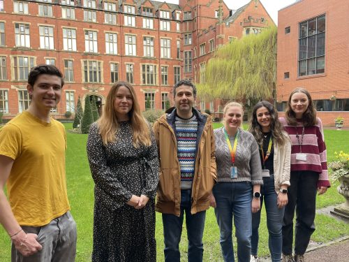
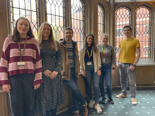
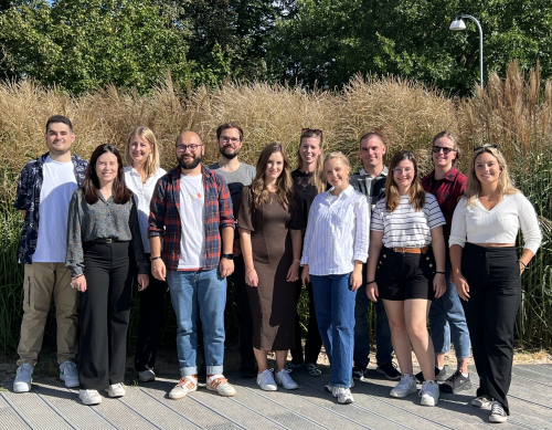
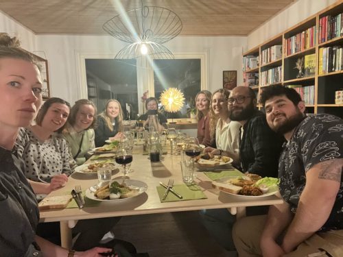
Lab roll call
James Birch: PhD student, studying the links between cell competition and neuroblastoma initiation.
Katy Boswell: PhD student, investigating the effect of chromosome 17q gain/MYCN overactivation on the enhancer landscape of hPSC-derived trunk neural crest cells.
Fay Cooper: Postdoc, studying determinants of axial identity in spinal cord progenitors and neural crest cells. She has also been involved in the development of a hPSC-based cell therapy for treating Hirschsprung disease. More recently, Fay has been examining the effect of DNA damage on trunk neural crest differentiation in hPSCs harbouring various neuroblastoma-associated aberrations.
Grace Gilbert: Joint PhD student (primary supervisor: Dr Dan Bose), dissecting the effect of Rubinstein Taybi Syndrome disease mutations on the epigenetics of neural crest/neuronal development.
Sude Uyulgan: PhD student, working on the study of axial identity acquisition by hPSC-derived lateral plate mesoderm and limb progenitors.
Favourite technique, and why?
Anestis: Immunostaining/fluorescence microscopy analysis of neuronal projections simply because it usually produces images of immense hypnotic beauty.
Apart from your own research, what are you most excited about in developmental and stem cell biology?
Anestis: I am very excited about the increasing number of complementary 2 -and 3-D hPSC differentiation models, which together with emerging cutting-edge technologies in microscopy, sequencing and bioengineering offer for the first-time unique opportunities for controlling and understanding human developmental processes. They can also facilitate examining older classic embryology experiments/ concepts under a new lens. I am very keen on using these approaches in the near future to address specifically our group’s questions.
What is the best thing about where you work?
Anestis: The University of Sheffield hosts a vibrant developmental/stem cell biology community, and I have met some fantastic colleagues, collaborators and friends here.
Sude: Supportive and friendly atmosphere, exciting research, and most importantly having Anestis as a supervisor!
James: The University of Sheffield is a great place to work. We have regular fascinating seminars and the university has many support groups from research on aging to data analysis (Sheffield R Users Group) to support you on your research journey. The university really cares what we think, I’ve seen management respond to student and staff surveys and to really be willing to put funding behind making changes where needed. I think all these contribute to making Sheffield an exciting and supportive working environment.
Fay: The people…. we have a really great dynamic in the lab at the moment which makes it a really enjoyable place to work.
What’s there to do outside of the lab?
Anestis: Sheffield is home to lots of pubs/breweries (it is currently the real ale capital of the world), restaurants, cinemas and concert venues so there are plenty of options. It is also close to the Peak District so stunning nature/landscapes are literally around the corner.
Sude: Hiking in the Peak District, great pie and good pubs.
James: Sheffield is a really vibrant and active city with loads to do. If you like music and street food we have loads of great music venues, pubs, clubs and food stalls all around the city. We’re also just a 20 minute drive from the peaks. One of my favourite hiking areas is the Edale valley, easily accessible by car and train. There are several great hiking routes and some of the highest peaks in the peak district, as well as a cozy pub (The Old Nags Head) for a drink and a great meal after a good walk.
Fay: Sheffield is the outdoor city… it is so close to the peak district which has some amazing walking, running and swimming spots. I spend as much time as I can enjoying this!
Browse through other ‘Lab meeting’ posts featuring developmental and stem cell biology labs around the world.
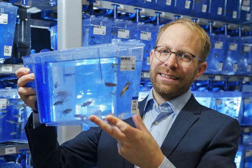
Daniel Wehner is Group Leader at the Max Planck Institute for the Science of Light and the Max-Planck-Zentrum für Physik und Medizin in Erlangen (Germany). At the 2024 joint German and Dutch developmental biology societies meeting in Osnabrück, Daniel was awarded the GfE Hilde Mangold Award for young investigators. After his award lecture at the conference, we chatted about his career background, his transition to becoming a PI, and his research into spinal cord regeneration in zebrafish.
(Image credit: Stephan Spangenberg)
Congratulations on winning the GfE Hilde Mangold Award! What does this award mean to you?
Receiving this award is of tremendous significance to me because it recognizes the efforts and contributions of our research team to the field. The German Society for Developmental Biology is my home turf, so I am deeply honoured to be recognized by this society for our work.
You received the GfE PhD prize back in 2015 as well!
Yes, this makes getting this Hilde Mangold Award even more impactful, as it validates that we are on the right path and encourages me to persist in my research direction. I believe our research has always been somewhat unconventional in that we never opt for the easiest route to tackle scientific challenges. For instance, our lab combines biochemical and physical methodologies to investigate spinal cord regeneration. It’s truly gratifying to see the efforts of our research group acknowledged. I extend my heartfelt thanks to all the members of my group and my past and present mentors who have supported us along the way.
Let’s go back to the beginning, when did you first become interested in science?
I have always had a keen interest in biology and in understanding the fundamental principles of life. However, it was during my final two years of high school in the biology course that my passion truly ignited. That was when I was introduced to the basic principles of cell biology, and I became captivated by the intricacies and sophistication that enable life. I went on to study (medical) biotechnology, yet I found myself unsatisfied with merely learning about established knowledge. The decision to pursue a PhD stemmed from my inherent drive to discover something new.
What did you work on during your PhD?
I completed my PhD under the supervision of Gilbert Weidinger, initially at the Technische Universität Dresden and later at Ulm University. Initially, my research focused on identifying new modulators of the Wnt pathway [1] [2]. Subsequently, I shifted my focus to investigating how Wnt signaling regulates fin regeneration. We adapted the TetON system to examine cell type-specific functions of Wnt signaling in a complex regenerating appendage [3]. This technological advancement led to the concept of Wnt pathway-controlled signaling centers that coordinate regeneration through secondary signals. [4] [5]. In another paper published in Development, we provided evidence that Notch signalling coordinates the proliferation and differentiation of progenitor cells — key events in tissue regeneration [6].
Then you moved to Edinburgh for your postdoc at the Becker lab. What did you work on?
Catherina and Thomas Becker have done pioneering work on spinal cord regeneration in zebrafish. It was an excellent synergy, bringing together the TetON system and their remarkable model system for central nervous system regeneration. It was during this time that we established the principle that after spinal cord injury, fibroblasts modulate the lesion site by depositing a complex extracellular matrix (ECM), but in strong contrast to mammals, in zebrafish this environment promotes regeneration [7].
Is that when you started to find your niche for your own research group?
Yes, I was intrigued by the interspecies difference in fibroblast behaviour because, in mammals, fibroblasts are typically viewed as inhibiting the regeneration of the central nervous system. For instance, research conducted by the Frisén and Göritz labs demonstrated that PDGFRβ-positive fibroblasts of perivascular origin contribute to the formation of inhibitory central nervous system scars [8] [9]. Therefore, the natural question to ask was: do these cells behave differently in response to spinal cord injury in zebrafish? To address this, we developed numerous genetic models to lineage-trace and cell type-specifically interfere with pdgfrb+ cell recruitment, or spatially-confined optogenetic depletion of pdgfrb+ cells. Our findings revealed that in zebrafish, pdgfrb+ fibroblasts are required for regeneration. We discovered that these fibroblasts possess the ability to downregulate specific inhibitory ECM molecules and upregulate regeneration-promoting matrix genes, facilitating axon regeneration. We further demonstrated that the unique biochemical composition of fibroblast-derived injury ECM in central nervous system lesions defines regenerative success, versus inhibitory scar formation in zebrafish and mammals. These findings resulted in the publication of our lab’s first two major papers [10] [11].
How was your experience transitioning to becoming a PI?
I must admit that the transition was not easy. There were moments when I considered leaving academia due to the lack of prospects. This is a challenge many colleagues face, particularly women in the field. As a father of two, I understand the difficulties and challenges of navigating a postdoc while caring for children, and I know firsthand the additional challenges young parents encounter. I consider myself very fortunate that things have ultimately worked out for me.
Following my postdoc in Edinburgh, I joined the Center for Regenerative Therapies at Technische Universität Dresden, working in Michael Brand’s lab with support from a return stipend from the German Research Foundation. Although I applied for an Emmy Noether Fellowship (similar to an ERC starting grant) and didn’t make the final cut, it was during this time that I crossed paths with Jochen Guck, a renowned researcher in tissue mechanics. We discovered excellent synergies by combining physics methodologies with molecular biology tools to investigate spinal cord regeneration in zebrafish. This led to an exciting opportunity to lead my own independent research group at the Max Planck Institute for the Science of Light.
Can you briefly talk about what your lab is working on?
My current research addresses the fundamental questions: i) How can regeneration-permissive lesion environments be established after central nervous system injury? ii) How can central nervous system axon regeneration be promoted in regeneration-limiting environments?
To achieve these goals, we are pursuing several approaches. i) We are investigating the intricate cell-cell interactions that dictate the biochemical and mechanical properties of the regeneration-permissive microenvironment in zebrafish, including the regulation of axon regeneration-permissive injury ECM. ii) Utilizing zebrafish as an in vivo platform, we are dissecting the regeneration-modulating properties of specific components of human scar tissue, along with elucidating their underlying mechanisms. iii) We are also in the process of developing humanized zebrafish models to explore novel strategies for spinal cord repair.
Do you have any advice for people thinking of starting their own lab?
Science thrives on passion and determination, existing within its own realm. Avoid comparing yourself to those outside of this world. Instead, dare to think innovatively and seek out environments that foster support, enabling you to embrace risks and confront challenges.
Speaking of supportive environments, how important do you think mentorship is in navigating an academic career?
A supportive mentor can profoundly influence career development. Navigating the academic path is challenging and fraught with obstacles. Having a great mentor who offers guidance on avoiding these pitfalls can be invaluable, sparing you from unnecessary setbacks.
I count myself fortunate to have benefitted from excellent mentorship. For those seeking PhD or postdoc positions, I recommend considering joining a lab led by a junior PI. Working with a junior PI provides valuable insight into the process of establishing a lab. Junior PIs often prioritize mentorship as it is a mutual responsibility.
My PhD supervisor, Gilbert, was a junior PI at the time. He taught me essential techniques such as microinjections, then entrusted me with the freedom to explore. This supportive environment was echoed during my time at Becker’s lab in Edinburgh, where guidance was readily available, yet I was encouraged to pursue independent research.
What’s your favourite technique? And what are you most excited about in your area?
My preferred technique is in situ hybridization—it’s remarkably reliable! Nevertheless, the method that currently captivates me the most is Brillouin microscopy. This non-invasive, label-free, all-optical approach enables the measurement of viscoelastic properties in vivo using light. Access to Brillouin microscopes is scarce, limited to only a handful of locations worldwide. Having the chance to employ this state-of-the-art technique at the Max Planck Institute in Erlangen to investigate the role of tissue mechanics in spinal cord regeneration is a true privilege. Achieving the initial results that contributed to our latest paper [11] was an overwhelming experience.
Finally, let’s get outside of the lab – what do you like to do in your free time?
Back then, I was deeply immersed in music, playing bass guitar in a band, and engaging in trekking, rock climbing, and mountaineering. However, with the arrival of my two daughters and the commencement of a new lab, I find myself with limited time for hobbies. Nonetheless, I consider science itself a passion—a hobby in its own right!
(Image credit: Stephan Spangenberg)
References
[1] Kagermeier-Schenk B, Wehner D, Ozhan-Kizil G, et al. Waif1/5T4 inhibits Wnt/β-catenin signaling and activates noncanonical Wnt pathways by modifying LRP6 subcellular localization. Dev Cell. 2011;21(6):1129-1143. doi:10.1016/j.devcel.2011.10.015
[2] Özhan G, Sezgin E, Wehner D, et al. Lypd6 enhances Wnt/β-catenin signaling by promoting Lrp6 phosphorylation in raft plasma membrane domains. Dev Cell. 2013;26(4):331-345. doi:10.1016/j.devcel.2013.07.020
[3] Wehner, D., Jahn, C., Weidinger, G. Use of the TetON System to Study Molecular Mechanisms of Zebrafish Regeneration. J. Vis. Exp.(100), e52756, doi:10.3791/52756 (2015)
[4] Wehner D, Cizelsky W, Vasudevaro MD, et al. Wnt/β-catenin signaling defines organizing centers that orchestrate growth and differentiation of the regenerating zebrafish caudal fin [published correction appears in Cell Rep. 2014 Feb 27;6(4):777-8]. Cell Rep. 2014;6(3):467-481. doi:10.1016/j.celrep.2013.12.036
[5] Wehner D, Weidinger G. Signaling networks organizing regenerative growth of the zebrafish fin. Trends Genet. 2015;31(6):336-343. doi:10.1016/j.tig.2015.03.012
[6] Bartholomäus Grotek, Daniel Wehner, Gilbert Weidinger; Notch signaling coordinates cellular proliferation with differentiation during zebrafish fin regeneration. Development 1 April 2013; 140 (7): 1412–1423. doi: 10.1242/dev.087452
[7] Wehner D, Tsarouchas TM, Michael A, et al. Wnt signaling controls pro-regenerative Collagen XII in functional spinal cord regeneration in zebrafish. Nat Commun. 2017;8(1):126. Published 2017 Jul 25. doi:10.1038/s41467-017-00143-0
[8] Dias DO, Kim H, Holl D, et al. Reducing Pericyte-Derived Scarring Promotes Recovery after Spinal Cord Injury. Cell. 2018;173(1):153-165.e22. doi:10.1016/j.cell.2018.02.004
[9] Göritz C, Dias DO, Tomilin N, Barbacid M, Shupliakov O, Frisén J. A pericyte origin of spinal cord scar tissue. Science. 2011;333(6039):238-242. doi:10.1126/science.1203165
[10] Tsata V, Möllmert S, Schweitzer C, et al. A switch in pdgfrb+ cell-derived ECM composition prevents inhibitory scarring and promotes axon regeneration in the zebrafish spinal cord. Dev Cell. 2021;56(4):509-524.e9. doi:10.1016/j.devcel.2020.12.009
[11] Kolb, J., Tsata, V., John, N. et al. Small leucine-rich proteoglycans inhibit CNS regeneration by modifying the structural and mechanical properties of the lesion environment. Nat Commun 14, 6814 (2023). https://doi.org/10.1038/s41467-023-42339-7
This is an Editorial in Volume 151, Issue 6 of Development, written by Alex Eve and Oliver Hobert.
This year, 2024, marks the 40th year of the discovery of the homeobox, a landmark discovery that fundamentally impacted multiple fields, ranging from genetics and genomics to developmental biology, neuroscience and evolution. The application of molecular biology techniques to analyze mutant animals, beginning in the 1980s, allowed two laboratories on two different continents to independently find that classic Drosophila mutants with characteristic developmental patterning defects carried sequence alterations in specific genetic loci which shared an ∼180 nucleotide sequence called the ‘homeobox’. This name arose from the intriguing phenotypic alterations observed in homeobox gene mutants, namely the transformation of the identity of some body part into that of another body part. Such identity transformations had long been known to exist in animals and plants, and had been dubbed ‘homeotic transformations’ by William Bateson, one of the fathers of modern-day genetics. The first Drosophila mutants with such a transformation were first identified by Calvin Bridges in Thomas Hunt Morgan’s legendary Fly Room at Columbia University, and further characterized by Ed Lewis at Caltech, whose work on homeobox genes earned him the Nobel Prize in 1995. After the molecular identification of homeobox genes in 1984 by Michael Levine and Bill McGinnis in Walter Gehring’s lab in Switzerland (McGinnis et al., 1984) and Matt Scott and Amy Weiner in Thomas Kaufman’s lab in the USA (Scott and Weiner, 1984), it quickly became clear that the homeobox encoded a DNA-binding domain, the so-called homeodomain. The monumental impact of defining the homeobox genes did not merely lie in understanding how specific developmental patterning events are genetically specified in Drosophila, but it rather lay in the realization that this domain is conserved throughout the animal kingdom and, eventually, that not only their sequence but also their function in developmental patterning is conserved. In fact, homeobox genes exist in unicellular organisms, but their number exploded with the advent of multicellularity, indicating that they have served as key drivers in the generation of the astounding complexity of animal body plans and cell types.
Another striking realization that came from studies across the animal kingdom was that homeobox genes form a part of a large family of genes encoding transcription factors, only a subset of which control developmental patterning along the anterior-posterior axis of a developing embryo (the genomically clustered HOX genes). Meanwhile, a much larger set of homeobox genes are involved in controlling the development of several other cell types, most notably and perhaps most predominantly so, the nervous system. Such neuronal functions of homeobox genes are deeply conserved in animals.
To celebrate this anniversary, Development has commissioned a series of review-type articles from leaders in the field demonstrating the impact that the homeobox discovery has had on different disciplines. This series, published throughout 2024, begins in this issue with two first-hand Perspectives that reflect on ‘perhaps the most exciting aspect of the homeotic gene story’ (Heasman et al., 1985): the first evidence that the Drosophila homeobox sequence is conserved in other animal species.
In the first article, Matthew Scott lays out the historical context for the discovery following studies of the Drosophila homeotic genes and describes from personal experience the work from Indiana University, USA, in identifying a conserved homeobox sequence (Scott, 2024). In its companion piece, Bill McGinnis and Michael Levine reminisce on their own ‘eureka moment’ from their studies in Basel, Switzerland, showing that the Drosophila homeobox sequence is conserved in other animals (McGinnis and Levine, 2024). Together, both Perspectives provide a fascinating and entertaining read, capturing the excitement of discovery and the maturation of competition into lifelong friendships.
To read the series as it grows, please visit the dedicated subject collection here: https://journals.biologists.com/dev/collection/10249/40-years-of-the-homeobox. To date, Development has published almost 5000 articles on homeobox genes and, of course, we welcome your future submissions detailing how this remarkable family functions in development, stem cells and regeneration.
We are located in Lund, a small city in Southern Sweden. The University was founded in 1666, and consists of many old beautiful buildings. The lab is affiliated with the Division of Pediatrics, and we’re part of the Lund Stem Cell Center.
We are intrigued by how we can utilize the setting of normal embryonic development to understand tumor initiation. Our focus lies primarily on how cancer forms neuroblastoma and paraganglioma develop during embryogenesis. These cancers arise in the adrenal gland and along (para)sympathetic ganglia, and originate from cells of the trunk neural crest stem cell population. Neuroblastoma is the most common tumor form in infants. It is believed to arise due to developmental defects during embryogenesis, but the precise origin and cause of initiation is unknown. Paragangliomas are on the other hand adult slow growing tumors, but also these are believed to be primed during development of the neural crest. We mainly use human patient-derived cells and chick embryos to dissect the role of neural crest specific genes in cancer initiation and progression. We develop techniques where we can analyze full-scale organogenesis and tumor formation in terms of the genomic, proteomic, and morphological landscape following genetic insults in the trunk neural crest stem cell population during embryonic development.
Lab roll call
Sinan Karakaya is a postdoc, exploring the role of a dysregulated hypoxic response in both normal development and the progression of tumors, specifically focusing on paraganglioma. His research investigates the role of HIF-2α in tumor initiation, development, and aggressiveness. Sinan is originally from Turkey, and did his PhD in Germany.
Perrine Burdeyron is a postdoc working on developing a model to study neuroblastoma by using human pluripotent stem cells with chick embryo as a host. This model allows us to study different genes involved in this childhood cancer. Perrine is originally from France.
Tom Gregor is a postdoc working on the role of HIF-2α during neural crest stem cell-derived childhood cancer progression. Tom is originally from Czech Republic.
Marina Mazariegos is working as a lab engineer, and arrived to the lab six weeks ago. She is involved in several projects with different people, as well as general management and organization of the lab. Marina is originally from Spain.
Niklas Engström is a lab engineer involved in a number of projects, as well as in maintaining the structure of the lab.
Elina Fredlund is a final year PhD candidate working to understand the initiation of neuroblastoma with main focus on an early development protein that has an important role in embryogenesis and a suppressive function in neuroblastoma.
Stina Andersson is a second year PhD student, and her work focuses on how different types of neuroblastoma cells respond to various cues from the embryonic microenvironment.
Zana Stirn is a master student. She is working together with our postdoc Perrine on the project to develop a human-chick chimera model, that will be used to manipulate different genes and pathways in neuroblastoma and consequently to get a better insight into the development and origin of this cancer. Zana is originally from Slovenia.
Pleun Jornick is a master student, fulfilling the last part of her master internship here. She studies the mechanisms of a tumor-suppressor gene in neuroblastoma. Pleun is originally from The Netherlands.
Özgür Rubar Altin is a medical doctor at the beginning of his career, and he is doing an internship in the lab. He is working on investigating the role of HIF-2α in chicken embryos and different tumor cells. Özgüris originally from Turkey.
Inés Sanz is a master student, investigating the role of how embryonic microenvironmental factors affect neuroblastoma cell behavior. Inés is originally from Spain
Favourite technique, and why?
Sofie Mohlin: There are so many techniques that we can use, each with its own advantages and beauty. But I’m going to go with a ‘simple’ on: microinjections and electroporation in chick embryos. It is such a basic technique that’s been around for ages, yet it’s the basis for so many experiments and essential knowledge we have on embryogenesis today. Unfortunately, I don’t spend a lot of time on hands-on lab work anymore, but when I do it’s always by the microscope doing injections. While it in one way requires focus and concentration, I tend to go into a meditative state where I can let go of stress and just enjoy the beauty of the embryos, one after one after one…
Apart from your own research, what are you most excited about in developmental and stem cell biology?
Sofie Mohlin: I think the process of (neural crest) stem cell lineage commitment is so delicate and exciting. With the recent in-depth sequencing techniques, we’ve come a long way in understanding the genetics behind, and as a result of, lineage commitment. However, there is so much left to understand about the biology surrounding lineage commitment.
How do you approach managing your group and all the different tasks required in your job?
Sofie Mohlin: My philosophy is that people are given full independence, but it comes with responsibility. In the lab we drive the projects forward together, but each individual project is one person’s pet project (or, someone’s baby, which is my favorite way of expressing it). However, I make sure I’m always available, as often as possible physically in the lab, otherwise being quick in answering questions via email/chat functions. We also have time-fixed regular lab meetings and one-on-one meetings with PhD students. Planning and prioritization are key, but to be honest it’s not always easy. With deadlines, grant applications and academic commitments, I often feel that time for actual science is sparse. I try to be open to the group when very intense periods occur so that they know why I might be more stressed and less available for a (hopefully short) while. I’m also working on being able to say no to things. There are so many things to do, and many of these are fun tasks. But also, with a limited number of hours per day, being able to prioritize and say no is crucial. At the end of the day, the time when I’m the happiest at work is after meetings with lab members, where we have discussed cool new data, experiments, and future plans, leaving with the feeling of exhilaration. This is an activity that I would never skip!
What is the best thing about where you work?
Sofie Mohlin: My group of course! I really enjoy discussing and doing science, as well as chatting about everyday life, with everyone, be it in the corridor, in the lab, in one-on-one meetings or lab meetings. I also work close with other great PIs, providing me with a fantastic peer community and surrounding.
Sinan Karakaya: The best part of working in our research lab is the collaborative environment fostered by our diverse team. Each member brings unique perspectives and expertise, creating an atmosphere conducive to innovative thinking and problem-solving. Additionally, the supportive and encouraging nature of Sofie and colleagues nurture professional growth and fosters a sense of camaraderie, making it an enriching and fulfilling experience to be part of our research endeavors.
Perrine Burdeyron: The best thing is the positive environment in the lab.
Marina Mazariegos: Definitely the work environment created by great people working for the good of the whole lab and not for themselves. And also, the prospect of achieving goals to contribute to progress in clinical research.
Elina Fredlund: The best things are my colleagues and the abundant opportunities available. Guidance is readily accessible upon request, as people are very friendly and supportive.
Stina Andersson: I love learning new things every day, and I really appreciate the collaborative atmosphere in our research group.
Zana Stirn: The environment! I really enjoy the dynamic of the group. We have many get togethers, workshops, potlucks, fikas. All of this contributes to an amazing environment, which makes coming to work even more enjoyable.
Pleun Jornick: The ambiance in the lab is amazing. It feels you can always go to anyone with questions and that your opinion is really appreciated. This makes me really motivated to give my all.
Özgür Rubar Altin: Excellent colleagues and scientific working environment.
What’s there to do outside of the lab?
Sofie Mohlin: Lund is a very picturesque city with lots of beautiful old buildings and surroundings, so just walking around town is nice. Ten minutes on the train then takes you to Malmö, a wonderful city with lots of culture, sightseeing, parks, food, and wine bars!
Sinan Karakaya: Outside of the lab, there are plenty of opportunities to spend your time indoors or outdoors. Besides being a scenic, historical town, Lund also offers variety of indoor and outdoor activities including museums, sports halls and trails for trekking and biking.
Perrine Burdeyron: During the winter you can enjoy ice skating and snow activities and during the summer you can enjoy the long days to go for hiking and visit all the national parks in the south. If you prefer the city, we are close to Copenhagen, the Danish capital.
Marina Mazariegos: It´s a small city but you can find a lot of activities and there´re a lot of nice places to go to relax with friends.
Elina Fredlund: Crossing the bridge, Copenhagen is just a 40 min train ride away. Söderåsen is a beautiful national forest that’s worth a visit during any season.
Stina Andersson: I enjoy spending my free time in nature or at dinner with friends and family.
Zana Stirn: Lund is like a big student campus, so the student life is very much alive. You can enjoy different cafes, bars, parks and a beautiful botanical garden. On the weekend, you can make a trip to Lomma beach or one of many natural parks in the area. For some bigger city vibes, Malmö and Copenhagen are only a short train ride away.
Pleun Jornick: As an international student in Sweden there is a lot to discover, but the tradition I cherish the most here is “fika”. A coffee break with a sweet pastry a few times a week is a celebration. There are a lot of different nice cafés in Lund, so the challenge is to try them all.
Özgür Rubar Altin: Lund has incredible nature that you can explore by bike. There is a nice football stadium where you can go to watch Lunds BK.
Browse through other ‘Lab meeting’ posts featuring developmental and stem cell biology labs around the world.
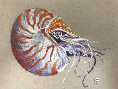
In this SciArt profile, we find out more about Brent Foster, whose artwork was selected as the Judge’s and People’s Choice in the Node-BSDB virtual art exhibition in the ‘Art by Scientists’ category back in September 2023. Brent is also one of the Node correspondents and has written a wide range of posts for our site. We caught up with Brent to find out more about his recent artistic projects and how he takes inspiration from the marine invertebrates he works with to create his drawings.
Chalk drawing of a nautilus.
Can you tell us about your background and what you work on now?
I have a rather eclectic range of science research experiences. I earned my bachelor’s degree in neuroscience from Brigham Young University in 2017, with minors in linguistics and creative writing. For two years, I explored human cognitive neuroscience methods — EEG, fMRI, MRS, TMS, and tVNS. Five years ago, I decided to try my hand at basic science research and was introduced to the world of marine invertebrates that challenges some of the basic principles of neuroscience that I used to take for granted.
Now, I’m working with comb jellies and trying to understand how nervous systems could have evolved in multicellular animals.
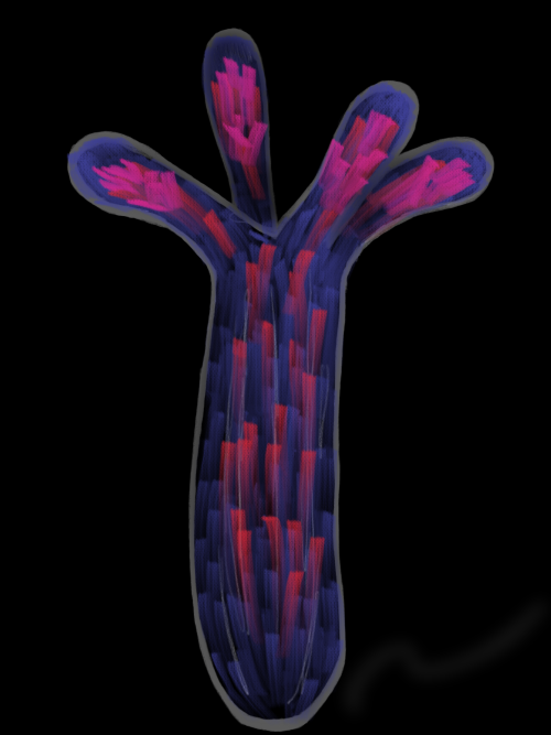
This is an artistic rendering of our recent paper‘s main result. We engineered Nematostella vectensis (otherwise known as the starlet sea anemone) to express a coral protein tagged with mCherry (red) that concentrates calcium (blue) in its tentacles (co-localization represented in pink).
Were you always going to be a scientist?
I’ve always been fascinated with the natural world and learning new things. As a kid, I was on a first-name basis with the librarian of our small local library and had read every book from their animal section. I used to distract my piano teacher with the latest factoids I had learned, and instead of practicing piano we would talk about things like giraffes and how weird their biology is. My dad was a high school biology teacher — I loved visiting his classroom and seeing his displays. One year he gifted me an old microscope with some slides of ants and bees. I’d sit at my desk and just stare through the oculars, mystified at being able to see the details of things that were so small.
So yes, I think I knew I was going to be a scientist. I just didn’t know what type of science I wanted to do.
And what about art – have you always enjoyed it?
I’m not formally trained in art, but I’ve always enjoyed drawing and painting.
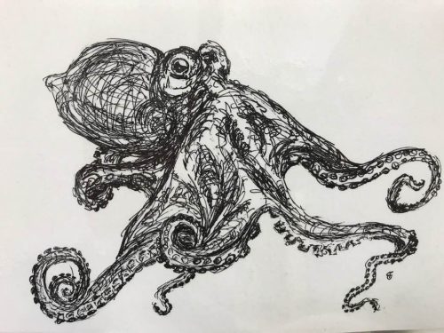
Living Water: Ink drawing of an octopus. When I see these creatures glide across a surface, I almost think of them as living water.
What or who are your most important artistic influences?
I haven’t really studied the great artists in any appreciable depth, but I’d say my favorite artists are Monet and Van Gough. There was a short period where I was fascinated with M.C. Escher, with his patterns that blended from mathematical precision to abstract imagination.
These artists were (and are) inspiring, but to be honest they’re intimidating — heroes on a pedestal looking down on us mere mortals. I’m sure they’ve influenced me in some ways, but I’m not sure it necessarily translates to my artwork. To me, my most important artistic influence was an art teacher during my elementary school years, Mrs. Marian Aranyos. She always inspired me to keep trying new things, and she made me feel like my art was something valuable, even when it wasn’t very good. She gave me space to experiment.
As for subject matter, I’d say nature has always been my greatest muse — especially animals. Nature, by far, is the most creative inventor.
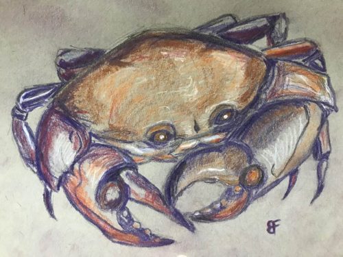
Crab – Chalk pastel drawing.
How do you make your art?
I generally prefer drawing (pencil, pen, or chalk pastel, mostly). I think pen drawings, especially, lend themselves to a sort of whimsy that can make challenging scientific concepts feel more tangible and relatable (or at least less scary). I’ve also dabbled with watercolor paints. Lately I’ve been playing around with an app called Sketchbook on my iPad. That’s opened up a whole new world of artistic possibilities.
Does your art influence your science at all, or are they separate worlds?
I don’t know if I’d say that my art is incorporated into my every-day science work. The type of art I create just isn’t precise enough to be technically relevant (although it helps me recognize the aesthetics of, say, micrographs). But my science definitely influences my art, especially since I started my current lab tech position. I’ve been inspired by the marine invertebrates I work with every day. They’re so beautiful and mysterious and remind me of what drew me to science as a kid. Now our home has a wall of chalk pastel drawings of crabs, jellyfish, anemones, nudibranchs — even an octopus wood cutout that my wife and I made together.
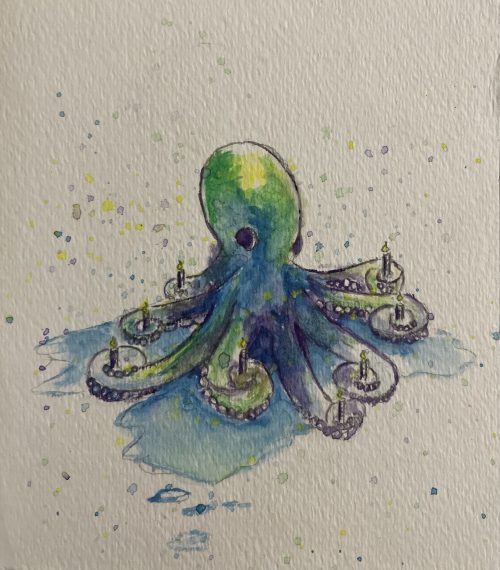
Octopus birthday – Watercolor birthday card I made for my wife, Alicia Boyd (she loves all things octopus).
What are you thinking of working on next?
I’m not sure what I’ll work on next. I tend to go through phases where I’ll create several pieces in the course of a few weeks, take a break, then try again when the inspiration strikes.
Just before Christmas, the Whitney Lab hosted an event to benefit the lab’s graduate students. I was able to share several pieces of art related to my research and used them to connect with our local community.
I’m playing around with the idea of using my art to tell some of the stories of science, geared especially for young kids (I think I have The Node’s “Show and Tell” series to thank for that inspiration). I created this science comic telling the story of a paper we published last month in iScience. I also drew an abstract representation of our main findings and submitted it to be considered for the issue’s cover image. It wasn’t selected, but it was a fun exercise.
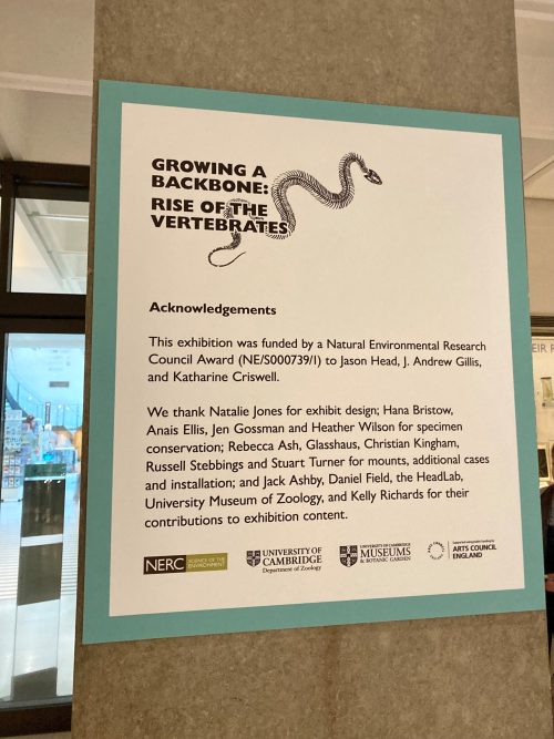
The people and the research behind the exhibition ‘Growing a backbone’
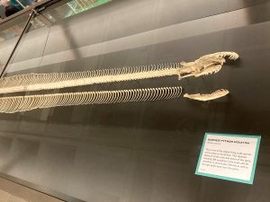
Where can you find a Victorian era ‘exploded skull’ and a 3.5-meter Burmese Python skeleton with every single one of its vertebrae and ribs laid out in a straight line? The answer is the University Museum of Zoology (Cambridge, UK), which recently opened an exhibition ‘Growing a backbone: Rise of the vertebrates’. One Sunday afternoon I decided to pop into the museum to have a look around.
Instead of having a separate space for the exhibition, the exhibits are dotted around the display cases in the museum. Trying to spot the ‘Growing a backbone’ display cards around the museum is like a treasure hunt. From skulls, jaws, teeth, spines, to limbs, visitors are taken along a journey to discover the evolution of the key features in vertebrates that make them unique.
Behind this exhibition is active research being conducted at the University of Cambridge on understanding vertebrate axial skeleton evolution. I reached out to Jason Head, Andrew Gillis and Kate Criswell, researchers working on the project, to find out more about their research on vertebrate evolution and the curation of the exhibition.
Evolution of the vertebrate axial skeleton
The project started with a conversation between Jason, Andrew and Kate. Jason is Professor of Vertebrate Evolution and Ecology and Curator of Vertebrate Palaeontology at the University Museum of Zoology, Cambridge. Before moving to Cambridge, Jason and his colleague Dave Polly had quantitatively analyzed snake skeletal morphology and found that Hox regionalization is maintained in snakes. Then, Jason met Andrew and Kate, who had been working with the little skate (Leucoraja erinacea) as a model system for vertebrate development. “We realize no one had looked in detail at axial skeletal evolution across jawed vertebrates,” said Jason, “and for a long time, people have paid little attention to the diversity of fishes.”
The three of them eventually obtained a grant from the Natural Environment Research Council (NERC), with the aim of integrating anatomical, developmental, and environmental data to reconstruct the evolutionary history of the vertebrate axial skeleton. The experimental aspect of the project was led by Kate, then a postdoc in Andrew’s lab, now Assistant Professor at Saint Francis University.
Kate looked at the different anatomical regions in vertebral columns of cartilaginous fishes (skates, sharks and stingrays) by studying skeletal anatomy and gene expression during development. “Fishes were thought to just have a trunk and a tail. I obtained CT scans of adult skates, created 3D models of each vertebra, and used morphometrics to quantify the shape change along the anterior-posterior axis.” She found that the skate do show some differences in shape in the vertebrae along the axial column, and the expression of certain Hox genes during development matches up with this adult pattern.
Looking beyond the little skate, the team investigated whether fish skeletons more broadly have axial regionalization, by looking at 90(!) fish species. Kate recounted, “a few times I got most of the way through segmenting or landmarking a vertebral column, only to find some defect in a vertebra (this happens a lot in fish!) and have to start from the beginning on another specimen of that same species.”
All the hard work was worth it eventually: the team discovered that the vertebral columns of many cartilaginous fishes have more regions than just a trunk and a tail. Kate said, “I started to get excited after running my analyses a few times on different fish, and consistently getting results suggesting that their vertebral columns were a lot more complicated than the textbooks had indicated. Fish are often treated as primitive or simple, but their anatomy can really surprise us when we look closely!”
Telling the story of vertebrate evolution
Evolutionary developmental biology is fascinating to many people, and creatures like skates and giant snakes often attract public attention. The ‘Growing a backbone’ exhibition in the museum is the perfect opportunity to showcase active research in vertebrate evolution.
Instead of creating the exhibition from scratch, Jason and his team made use of preexisting exhibits in the museum, by adding additional text about the development and evolution of different characteristics in vertebrates. The exhibition also included new specimens from the museum collection. One of Jason’s favorites is the Victorian era exploded human skulls, which were teaching utensils for medical students and zoologists in the mid to late 1800s. “The bones of the displayed skulls are pulled apart,” explained Jason. “The bones are on little armatures so that students could pull them back and forth to see the internal structure and the relationships of the bones.”
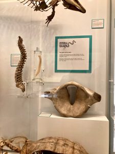
Due to my poor photo-taking skills, I could only capture half of the impressive 3.5m python skeleton with the individually laid out vertebrae and ribs.
Another highlight for Jason is the Burmese Python skeleton. “The Python was completely disarticulated. Our conservator, Natalie Jones, and I re-articulated the skull, and then we laid out the vertebral column and ordinated the ribs in a straight line. We even have one of the pelvic bones still preserved in the specimen.”
But not everything Jason wanted to include in the exhibit ended up being displayed— such as the skull of a sperm whale. “We have a display case about body size, explaining why vertebrates are basically animal giants, in part because we have evolved features allowing us to elevate our metabolism to be very plastic and eat anything that moves. But the sperm whale skull is rather large, so we didn’t really have a place for it.”
Writing museum display texts
Even though Jason has been working in museums for a long time, he acknowledged that it’s sometimes tricky to communicate complex concepts using very simple terminology. “I think it’s very hard to get consensus on how you should be targeting an exhibit for an audience. I tend to increase the expectation of audience comprehension. I think that a good exhibit challenges but doesn’t frustrate — you can introduce new terms and concepts, without blowing people’s minds to the point they can’t understand what you’re talking about. There’s always tension between the people working on the exhibit and the people who did the research. Compromise is how I’d say it works.”
One of Jason’s favourite displays is the case with different vertebral columns, showcasing the diversity of the spine, including the human vertebral column.
Embedding public outreach into the core of research
On the Sunday afternoon I visited the museum, the gallery was bustling with families and tourists. I was surprised to hear that the museum has not always been accessible by the public. Jason said that historically, the galleries were used for teaching Cambridge students. In recent years, the museum has made an effort to open up the galleries to the public, with temporary exhibitions such as ‘Growing a backbone’ to highlight findings from active research.
Jason thinks researchers should always try to incorporate outreach work as part of their grant proposal, which is what his team did in the NERC grant. “This is a great opportunity to educate the public, tell them the story of vertebrate evolution, and show them the homologies that they might not expect, for example, the fact that our teeth are basically modified scales!” said Jason. “It also gives funding agencies immediate deliverables, to show that our money is spent appropriately, and we can give back to the taxpayers who funded the research. As the exhibition will be here till September, we’ll also be looking at incorporating it into the museum’s public education and outreach programme.”
Filling in the gaps
We are still far from getting a complete picture about the evolution of vertebrate axial skeleton. “As a paleontologist,” said Jason, “I’m dealing with missing data from fossil records. It’s like you’re in a mansion at night: you have to figure out the entire layout of the mansion by looking in the dark with only a small box of matches.” That’s why Jason stressed the importance of expanding taxonomy sampling. “The field has been using the model taxon approach where you take one or two taxa from each major clade. We’ve got a decent sampling across the major divisions of vertebrates, but within the divisions, there’s not much we know. What we need to do is to continue to sample across phyla and look more into the diversity of vertebrates.”
“Most developmental biologists look at developmental processes and then think about what it means in terms of evolution. I’m a paleontologist. I go the other way,” said Jason. “I look at characteristics across phylogeny and then make inferences about the developmental mechanisms that might have led to that.” Kate, also a paleontologist by training studying an extinct group of fossil fishes called placoderms, has since expanded her research focus to include developmental biology: “I think palaeontology and developmental biology are complementary fields that help answer the same evolutionary questions using different approaches.”
Thank you to Jason and Kate for taking the time to explain their research and the process behind curating the exhibition. If you happen to be in Cambridge, UK, do pay a visit to the University Museum of Zoology. The ‘Growing a backbone: Rise of the Vertebrates’ exhibition is running from February to September 2024.
The road of academia is a pretty established ladder, PhD, then postdoc, then faculty. I am approaching the end of my PhD and the opportunities seemed endless. At least, when considering the current situation. There seems to be phenomenon that is now common in every scientific conference in the last couple years: many principal investigators coincidently appear to be looking for postdocs. Part of the reason could be due to recent reports have noted noting a 10% decrease in postdocs supported by NIH grants from 2020 to 2022, additionally, each of those years saw a 4% average decrease in postdocs across the science and health fields [1]. There are many reasons to blame for the decline in the interest of pursuing an academic postdoc, the main one being the cost of living surge in every state across the country. By April of last year, the prices of the essential needs had increased by 13% since 2021 while the NIH recommended minimum wage only increased a 4% in the same time frame [2,3]. For many, an academic postdoc is no longer financially viable and the biotech industry has not wasted the opportunity to keep up with their increasing demand which is projected to expand by an additional 15% by 2030 [4].
All these recent changes should point to an open post-doc market for someone looking to stay in academia. However, there is an additional demographic, that I happen to be part of, which makes up for over half of the postdocs in the United States who do not have the flexibility to shift to an industry position, which in many cases is a shorter term, riskier decision. Over a third of science and engineering doctorate degrees awarded in the US are to foreign nationals [5]. With another presidential election on the horizon, there is a potential risk to the 30,000 plus international postdoc visas that would need eventual renewing or to be newly awarded. The previous presidential administration cut down the number of green cards awarded by 18% across the board, and continued cuts is which is among the promises for the upcoming elections [6]. This limits academic institutions through increase in legal and processing fees, making international hires no longer viable. For those with the American dream willing to take a postdoc salary, the roadblocks might just get worse. Not only, the salaries will keep falling behind those offered in industry, but academic institutions may become more limited in the amount of help they can offer a postdoc in the efforts of finding a more stable migration status. As someone approaching the transition stage into a post PhD life, my options are were severely limited by the number of institutions that had their hands tied in terms of what they could offer, and with the elections just around the corner, stability is a privilege that is hard to come by.
It should be normal to have these discussions, I hope many are open to connect and try to point each other to possible opportunities and share what we are thinking for our next steps. Are you close to finishing your PhD? What are your options?
1- Gewin V. 2023, Postdoctoral researchers warn NIH that cost of living are gutting the workforce. Nature Career News https://www.nature.com/articles/d41586-023-02202-7#:~:text=The%20number%20of%20postdocs%20supported,respectively%2C%20between%202020%20and%202021.
2- Winters, M. and Cortes G. 2023. These 5 charts show how much 2 years of inflation have really cost you. CNBC Spend https://www.cnbc.com/2023/04/14/charts-how-much-inflation-increased-since-2021.html
3- 2022. Postdoc Salary in the US. PostdocInUSA https://postdocinusa.com/postdoc-salary-usa/
4- Ameco Research. 2023. Biotechnology Market is Anticipated to Reach USD 1,334 billion by 2030, Growing at a substantial CAGR of 15.5% from 2022 to 2030. LinkedIn Open Immersive Reader https://www.linkedin.com/pulse/biotechnology-market-anticipated-reach-usd-1334-billion/
5- National Science Board: Science & Engineering Indicators. 2020. Foreign-born students and workers in the US science and engineering enterprise. NSF https://www.nsf.gov/nsb/sei/one-pagers/Foreign-Born.pdf
6- Nowrasteh, A. 2021. President Trump reduced legal immigration. He did not reduce illegal immigration. Cato Institute https://www.cato.org/blog/president-trump-reduced-legal-immigration-he-did-not-reduce-illegal-immigration
Recently, Brent Foster and I published an article on non-model organism (NMO) research, where we interviewed several researchers working across the globe on the challenges, rewards, and the particular questions they investigate while working on (relatively) little-studied organisms. Although we aimed to provide a bird’s-eye view of NMO research in a pot pourri-style article, we found that they each of our interviewees had many interesting things to say that we thought it was a shame not to share with a wider audience. For this reason, we decided to publish the full interviews as well, which we will be doing over the next several weeks.
This interview is with Dr. Claudia Patricia Ornelas-García, investigator at the National Autonomous University of Mexico (Universidad Nacional Autónoma de México, UNAM), who works on the Mexican cavefish (Astyanax mexicanus) and other freshwater fish, looking at their systematics and speciation. The full transcript of the interview is below:
Can you summarise your work in a few sentences?
I work mainly with the systematics and speciation mechanisms in freshwater fish species. Since my PhD, I have worked with Astyanax, a very interesting genus because it is distributed from Argentina [all the way] to the Mexico-US border. During my PhD we reconstructed the evolutionary history of the genus in a particular region identified as Mesoamerica. But in general terms, what we were doing in that project was to analyse four molecular markers, three mitochondrial and one nuclear, to recover the systematics of the group. During the development of the project, I was very interested in the lacustrine systems of Central American Lakes in Nicaragua and Mexico. In these lacustrine systems there are a pair of morpho-species that were sympatric and we were very interested in the correlation between the morphology and the genetic differentiation, because when we were doing this systematic group, they at least seemed to be sharing some haplotypes in the mitochondria. We continued working on the ecological divergence, ecomorphological divergence, and some morphometrics in this pair of species, and more recently with RadSeq Data.
Years later, when I came to Mexico and established my own lab, we started working with cavefish. Actually, when I started, my first job was in Querétaro, very close to the caves in San Luis Potosí. I started working on the caves and I fell in love immediately because the environment is so amazing. It’s very particular. And the system is also very interesting. As you know, there are a lot of genomic resources available nowadays. In the beginning, when I started working with this group, there were maybe 10 or 11 cave populations already analysed from [a] phylogenetic perspective, but not from an evolutionary or developmental perspective. So in the beginning, I just wanted to include as many caves as we could, so we could test these hypotheses of how many times the fish has been able to adapt to the caves. There were several hypotheses, some of them say that it happened only once and there has been a lot of drift. The other says that there’s two independent lineages. I’m in that group that would suggest that there are two independent lineages that came to the caves and adapted to them. Actually, in our most recent paper, by a master’s student of mine, we assess this question, using not only the complete genomes that are already available, but also including some caves that were never analysed before. So we have a very, very exhaustive sampling. And in our results, we have at least three independent colonisation events of the caves, which for some is crazy, it’s not possible. But from our point of view, we are really relying on exhaustive sampling of the caves, and that is what we’re suggesting.
Nowadays, we are starting to move to some developmental analyses, because we were able to capture some fish from the caves, and we are reproducing them here in the lab. So far, we have been able to reproduce five different populations, different from the common ones like Pachón or Tinaja. We are reproducing Escondido, Arroyo, Tigre, Chica, and Pichijumo. Sabinos is common, but it’s less common than others. So far, we are trying to characterise some developmental features.
One of my students is working on the Rad-seq, and we are starting to work with RNA-seq, and we are a trying to compare the genetic convergences across different cavefish lineages, particularly including some less studied populations such as Escondido, which is from the [second] linage in the Guatemala region. We are trying to compare the differences during early development because we have realised that they have a very particular mutant in some visual pigments, so we are trying to match that variation with the phenotype. We are also exploring the phenotypic convergence with other cavefish in Mexico, like Prietella phreatophila, a catfish in the northern part of Mexico, and also with other families of cavefishes from the southern part of Mexico. We are trying to investigate them for convergences in phenotype, particularly in some processes related with asymmetry that has been reported in Astyanax and in P. phreatophila, and we are trying to check out if it’s consistent across cavefish living in Mexico or only in these two cavefish.
What kinds of asymmetry is this?
Astyanax is very interesting because in Pachón there has been described a directional asymmetry, as well as in Ictalurids [such as Prietella], and actually it’s in the same side, left turn of the head. Particularly in a cave called Chica there is one very well characterised Astyanax hybrid population (between the surface [fish] and the cavefish). In that population there is fluctuation asymmetry, [toward the] left or right, [similar to] chiclids, [that have] fluctuating asymmetry in the mouth of a scales-eating species. It’s interesting that when you analyse it in the hybrid population, the asymmetry is different in comparison to the other caves.
I understand that there are a lot more resources for you to study these types of species now. But obviously, it is generally a group of species that is not studied by an enormous number of people around the world. Are there particular challenges associated with that?
Yeah, I think one of the challenges is that there are a very restricted number of people working with these [species]. And in a way, it’s fascinating, because you will find something new for sure. But [from another point of view], in research groups [studying established organisms] it is easier.
We were trying to characterise the microbiome of the fish, we have a paper on that. And it was a little challenging, because there was not a lot of information already published on protocols or how to treat the data. Or when we are trying to set up [experiments], for example, for physiology or for another kind of ecological analysis, it’s sometimes difficult. But in a way, I think it’s very, very interesting. During my Bachelor’s, I was working with mice, the typical model in the lab, and somehow I think that the number of questions sometimes can be very restricted because there’s already so many studies in these animals, that it’s difficult to come up with something new.
You did touch upon this when you’re describing your work, but was there a specific thing that convinced you that these fish are what you want to work on?
I think the main reason was a because I have always been interested in mechanisms driving the evolution of morphology. When I started working on the lacustrine forms, it was like “Why do they have these different teeth, or different heads, or different body shapes?” and in the caves it’s dramatic, the change is impressive. Are these important morphological changes? When I started investigating the environment, it’s fascinating that they can survive under those conditions. That was one of the reasons.
Another important thing to highlight in Astyanax is that we have the Annual Meeting. It is very interesting because there are a lot of young people, together with senior researchers, and the community is very open. Because it’s not a model organism, [everyone is] really [willing] to talk about the system in a very open way, and include new researchers. Particularly for me, when I was finishing my PhD, this was a very dramatic point, because I saw a potential in the system that I can be included. And I have a lot of things in my favour, I am from Mexico, I can work in the field, I can do a lot of in situ experiments. Even nowadays, there are very few Mexicans working with Astyanax. It just happened that there were a lot of things that made me realise that there was a lot of potential in the [Astyanax] system for me.
Would you say there’s a particular question in your field that you find really fascinating? Or a finding or a result that you really weren’t expecting?
Yeah, as a systematic biologist, I have a very, very important question for me, which is how many species [the cavefish are]. This is an important question from two different perspectives. One is conservation. The other is from an evolutionary point of view, because for sure, they can hybridise, even lineages between the two branches can hybridise. So in a way, it’s very difficult to test for the biological concept of a species. Sometimes it’s difficult even for the developmental biologists, because they are really trying to understand the link between the gene and the morphology. We are trying to understand the mechanisms giving rise to this kind of systems, and how easy it is to speciate in this context. So the implicit question regarding these ecomorphs or ecotypes is, are they different species? And how significant is this for the evolutionary history of the model?
So in a way, it was very interesting for me at the beginning, because when I started going to these conferences on Astyanax, it was very easy for me to always think about the systematics, about the phylogeny. And sometimes [other groups] were interpreting some variation in the morphology through local adaptation. But actually, it’s related to the [evolutionary] history of the system, like the number of vertebrae, or various characteristics, actually, they were shared by ancestry, not by local adaptation. So I think that’s very, very important in these kinds of studies, trying to link between the genetics and the morphology. That’s one of the reasons I want to have a very good notion of how many times this [cave dwelling] model has evolved, because if it [evolved] only once, the interpretation of repeated evolution, or whatever we are asking, is different. The other question that is very fascinating for me is how their morphology can be so divergent, even under gene flow circumstances. How is this possible without any barriers [between the putative species]? That’s the reason we work with Astyanax.
How does data analysis and sharing between labs that use Astyanax and related species differ, in your opinion, compared to studies that use more established model organisms? You said, it’s a fairly open community. But you know, are there any major differences?
I must admit that I don’t know if I have enough experience on this question. All of my experience is with Astyanax. Even though during my Bachelor’s I was working with mice, I’m not familiar with other groups. In the lab where I was working, they were very specific in the questions that they were [asking], and it was not very easy to share information with other labs.
In my opinion, one of the things that the Astyanax model has is that [researchers working on it] are very open. For example, when we were trying to reproduce a fish, we were obtaining a lot of information [from other groups].
From what I know about Astyanax, that there are labs that work on it in the context of heart regeneration, because some have a non-regenerative heart, in contrast to the regenerative zebrafish heart. How large is the community that works on the more phylogenetic aspect of the species compared to those working on developmental or regenerative questions and is there any crosstalk?
Nowadays, a lot of people are trying to [investigate] this model [from] the eco-evo-devo perspective. They have realised [that it is important to distinguish between the different Astyanax lineages], because some of the results that they get are related with the lineage, and not with particularly with the environment. In these terms, there is a growing number of labs wanting to work in the field, know more about the ecology in situ, learn more about the behaviour, the physiological adaptations. Some are is trying to [replicate] experiments in the field, check if the same thing happens in the lab versus in natural conditions. In my opinion, the species gives a very particular opportunity, because it’s not like in other places where it’s very difficult to get access to the cave systems in the in the field. So nowadays, even though regulations are increasing, they are attainable. [Researchers] can [request] permits and get them in a year, which a reasonable time. So it’s more a question of what the interests of the researchers are, because they can really put their questions in different contexts, and navigate between eco, evo, and devo.
Is there anything else that you would like to add?
The problem with non-model organisms is the conservation situation. For sure, nobody will catch Mus musculus from the field, they already have so many reproductive lines in captivity that they don’t have to. The non-model organisms are in the opposite situation. Most of the labs [working on them] want to have more wild lines, more related with what is really happening in the field. And if you have 200 labs working [on cavefish], imagine the impact that we can have on the natural population. These cavefish are not really large [populations]. We published, just at the beginning of 2023, a paper [on] size estimation of the fish population in the caves, and it’s maybe around 1000 fish, or [a few thousands] of fish. It’s not really that large [a] number. And imagine, in the last 10 years, more or less, there have been around 200 fish extracted from the caves. So if you imagine a system that has to recover from 20% of the population being lost only because of scientific sampling, it’s a problematic situation.
When you try to make researchers aware of the situation, [they] really believe that the main extinction drivers of this kind of population are not related with our sampling. Most of us really believe that it’s all global warming, or local people extracting water for drink. I’m very surprised, because normally you have to fight this kind of attitude outside the scientific [world]. So in my position, I’m trying to make people aware of this, but it’s not easy. But none of the above is false. Global warming, and people extracting water for drink, are part of the problem. Thus, it’s important to be part of the solution, and maybe we should consider what we can do to solve the conservation situation.
Some of the most iconic populations, like Pachón, you can find thousands of papers published on it. And it’s in a little bit of a critical conservation situation right now. Many labs have been able to reproduce it. And most [subpopulations] can be easily [obtained] from [other] labs. But even nowadays, to avoid inbreeding, some labs could require to collect wild fish. The Guatemala region has conservation issues, because although they are not easily-accessible caves, most populations are very small, and not well-connected between them, because of the phreatic level of the water, which can lead to extinction more due to their demography than to other causes. So its conservation situation is very different. But in a way, we have the same situation in other places.
And it’s not only in Astyanax, it could happen in other non-model organisms too. For example, Axolotl is also an amazing model system. If 50 labs in the world start working with one particular population, there can really be an impact in the local population. That is why I think we need to be more aware of the impact and do our best to guarantee the prevalence of this model for future generations. [Because] what makes these organisms amazing also makes them vulnerable, in a way.
Understanding the effects of the environment on animal physiology and biomechanics is at the core of Journal of Experimental Biology. Environmental factors such as temperature, food availability, sound or the presence of predators can profoundly shape how an animal grows and matures into an adult. In this Special Issue, we take a close look at developmental plasticity, which is the influence of conditions experienced during early development on an animal’s phenotype. In her classic book of 20 years ago, ‘Developmental Plasticity and Evolution’, Mary Jane West-Eberhard proposed that ‘alternative phenotypes’ that arise in organisms under different early life conditions play a critical role in moulding animal evolution and diversification (West-Eberhard, 2003). The ensuing years have seen increasing attention on how developmental plasticity may contribute to evolution. Given this, coupled with the explosion of new information on the epigenetic mechanisms underlying developmental plasticity, the growing number of submissions to JEB in this area, and the fact that an earlier special issue on ‘Phenotypic Plasticity’ (Hoppeler et al., 2006) is now 18 years old, the time seemed right for a special issue on developmental plasticity. In the current issue, we have capitalized on the diversity of animal models under study, from worms to dung beetles and lizards to mice, to assemble a strong comparative approach to the topic. We also aimed to bring together researchers considering developmental plasticity from diverse angles, from molecular and cellular biology to whole animal physiology, ecology and evolution, to more fully understand and integrate new approaches and research findings.
Developmental plasticity is defined by the rearing environment, from nutrition to social conditions, which provides critical information that developing animals use to shape the maturation process and resultant adult behaviour and physiology. Such context-dependent plasticity during development is often considered to be both widespread and adaptive, although the extent to which this is the case remains unclear (Sánchez-Tójar et al., 2020). It is also important to recognize that conditions such as resource limitations or exposure to environmental contaminants can result in damaged phenotypes that are clearly not adaptive. In this Special Issue, Metcalfe (2024) discusses a third possibility – that variation in early conditions need not always result in obvious adult changes, but may alter developmental trajectories in ways that have more nuanced consequences over longer periods of time. Other articles in this Special Issue focus on identifying critical environmental factors that serve as cues for developmental adjustments, and how these, in turn, are transduced within the developing animal. For example, information transmission may be mediated by parental behaviour (e.g. Mariette, 2024) or indirectly via provisioning of the egg. Hotter temperatures, food scarcity or stress (e.g. from predators) experienced by a parent provide anticipatory cues to developing animals that may prepare them for similar stressors in later life. Food availability or nutrition, in particular, appears to be of fundamental importance in an animal’s developmental trajectory, to the point where we may ask whether it is a ‘master’ regulator of development. Understanding the mechanisms involved in nutritional effects on development is a critically important area for future research.
Cover: The male gazelle dung beetle (Digitonthophagus gazella) develops nutritionally plastic head horns. Males with access to low-quality nutrition during their larval stage develop into hornless adults (left). By contrast, males with access to a high-quality diet develop into large adults with exaggerated head horns (right; both images are on the same scale). Rohner et al. (jeb245976) review the many ways in which plasticity, symbionts and niche construction interact in shaping dung beetle development and evolution. Photo credit: Patrick Rohner. (No Ratings Yet) Loading...
 (No Ratings Yet)
 (1 votes)
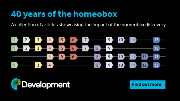

